1525
A comparison of pseudo continuous arterial spin labeling perfusion MRI (pCASL) and permeability imaging with dynamic contrast-enhanced MRI (DCE-MRI) in human rectal cancerYuichi Kumagae1, Yoshihiko Fukukurra1, Koji Takumi1, Hiroto Hakamada1, Tomoyuki Okuaki2, and Takashi Yoshiura1
1Department of Radiology, Kagoshima University Graduate School of Medical and Dental Sciences, Kagoshima, Japan, 2Philips Electronics, Tokyo, Japan
Synopsis
Our purpose was to investigate potential correlations between the blood flow (BF) measured by pCASL and dynamic contrast-enhanced (DCE) MRI-derived pharmacokinetic parameters in rectal cancer. There were significant positive correlations between BF and Ktrans (p = 0.006, r = 0.579) or Kep (p = 0.002, r = 0.644). These results suggested that pCASL may have the potential to be a noninvasive alternative to DCE MRI.
Introduction
Perfusion MRI has the potential to provide pathophysiological biomarkers for the evaluating, staging and therapy monitoring of rectal cancer. Pseudo continuous arterial spin labeling (pCASL) perfusion MRI is a noninvasive, nonradioactive and non-contrast-enhanced method capable of quantitatively measuring microvascular perfusion characteristics of tissues by tagging arterial water. The objective of this study was to investigate potential correlations between the blood flow (BF) measured by pCASL and dynamic contrast-enhanced (DCE) MRI-derived pharmacokinetic parameters in rectal cancer.Methods
Twenty-one consecutive patients with pathologically confirmed rectal cancer were recruited. A 3D pCASL and DCE MRI were performed on a clinical 3.0T Philips scanner. The acquisition of pCASL was performed by using 3D volume isotropic TSE imaging to obtain control and labeled images. The labeling plane was 80mm above the center of the image field of view. Labeling was applied for 3 seconds followed by a 1.6-second post labeling delay before image acquisition. The total scan time of one pCASL examination in one subject was 4min 7sec. DCE MRI were acquired after the bolus injection of 0.1 mmol/kg using power injector (3.0mL/s). Image acquisition was performed using a 3D-T 1fast field echo (T1-FFE) sequence. For T1 maps, precontrast 3DT1-FFE with dual flip angles (5o and 15o) was performed. BF on pCASL and DCE MRI-derived parameters including Ktrans (forward volume transfer constant), Kep(reverse reflux rate constant between extracellular space and plasma), Ve (the fractional volume of extracellularspace per unit volume of tissue), Vp (vascular plasma volume) and iAUC (initial area under the enhancement) within rectal cancers were calculated Spearman’s bivariate correlation was used to assess the relationship between BF and DCE MRI-derived parameters.Results
Perfusion result from one subject was presented in Figure 1. There were significant positive correlations between BF and Ktrans (p = 0.006, r = 0.579) or Kep (p = 0.002, r = 0.644). There were no significant correlations between BF and other DCE MRI-derived parameters (Ve, Vp or iAUC) (Table 1).Discussions
In this study, there was a significant positive correlation between BF and Ktrans. Several studies have shown that Ktrans is the most important pharmacokinetic parameter of DCE-MRI with regard to evaluation after CRT for rectal cancer 1,2. A high Ktrans before CRT indicates high permeability and vascularity of the tumor, which is believed to make the tumor more accessible to chemotherapy and less hypoxic, making the tumor more sensitive for radiation 3.Our result shows that BF could be useful for evaluation and response prediction after CRT for rectal cancer. There was significant positive correlation between BF and Kep. Kep has been shown to correlate well with micro vessel density, a marker of tumor angiogenesis and with T-stage. This could be explained by more extensive neoangiogenesis in more aggressive tumors, which causes greater accumulation of contrast in these tumors. Therefore, BF on pCASL may be proposed as an imaging biomarker for tumor aggressiveness.Conclusion
pCASL may have the potential to be a noninvasive alternative to DCE MRI.Acknowledgements
No acknowledgement found.References
1. Kim SH, Lee JM, Gupta SN, et al. Dynamic contrast-enhanced MRI to evaluate the therapeutic response to neoadjuvant chemoradiation therapy in locally advanced rectal cancer. J Magn Reson Imaging 2014; 40:730-737.2. Intven M, Reerink O, Philippens ME, et al. Dynamic contrast enhanced MR imaging for rectal cancer response assessment after neo-adjuvant chemoradiation. J Magn Reson Imaging 2015; 41: 1646-1653.
3. Tong T, Sun Y, Gollub MJ, et al. Dynamic contrast-enhanced MRI: use in predicting pathological complete response to neoadjuvant chemoradiation in locally advanced rectal cancer. J Magn Reson Imaging 2015; 42: 673-680.
Figures
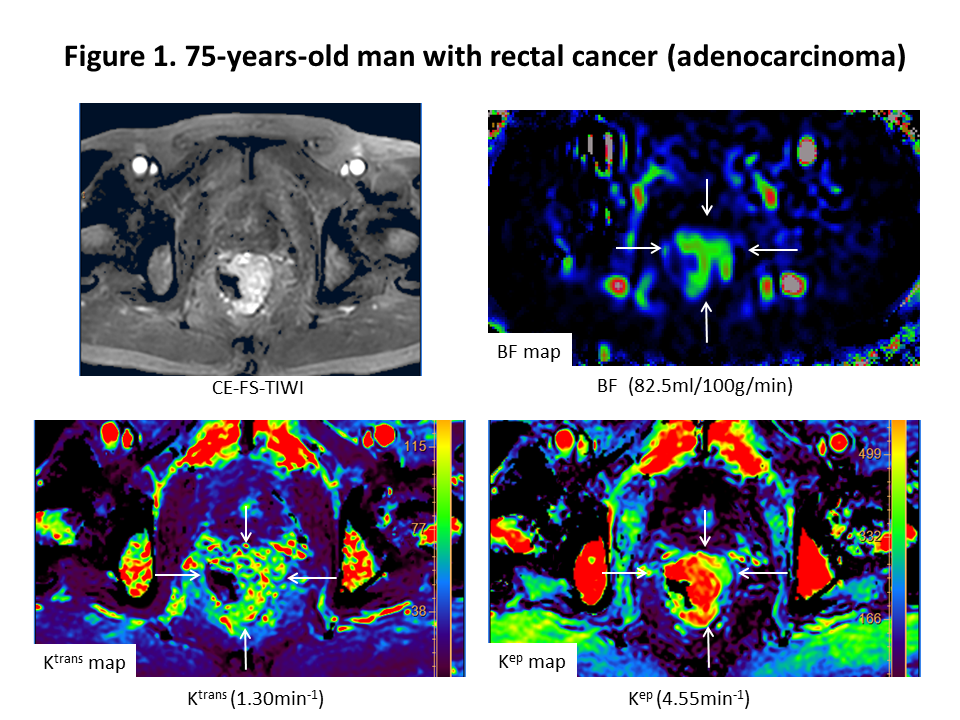
The BF map show higher BF in the carcinoma. Ktrans and Kep maps show higher values in the carcinoma.
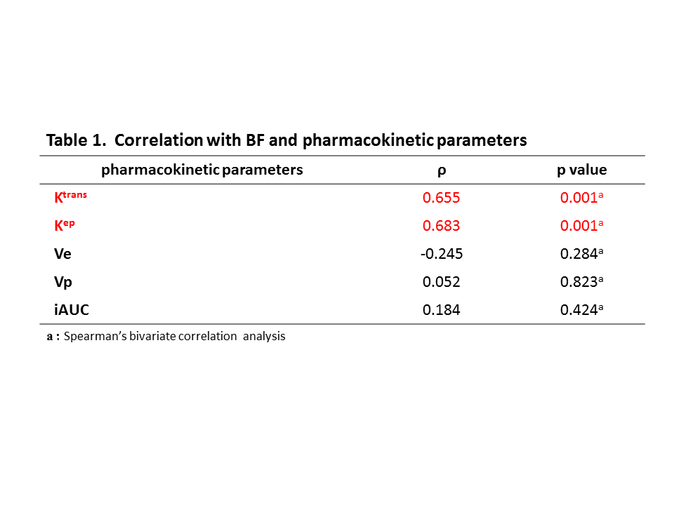
The table shows the results of correlation with BF and pharmacokinetic parameters. There were significantly positive correlations between BF and Ktrans, Kep, respectively. Although, there were no significantly correlations between BF and other pharmacokinetic parameters.