1502
Monitoring of Acute Thermal Coagulation in Muscle Using PSIF Sequence in MRI-Guided High-Intensity Focused Ultrasound Therapy1Shenzhen Institutes of Advanced Technology, Chinese Academy of Sciences, Shenzhen, GuangDong, China, 2University of Chinese Academy of Sciences, Beijing, China
Synopsis
MR-guided high intensity focused ultrasound (MRgHIFU) is a new noninvasive approach for thermal ablation of focal lesions with clinical applications in uterus, bone, prostate, brain, breast, and liver.Traditionally, the volume of tissue coagulation is evaluated through contrast enhanced T1-weighted images (CET1). However, there are several limitations for CET1 used for thermal lesion detection.
In this study, acute thermal damage following HIFU ablation in muscle was assessed using a PSIF images. this preclinical study demonstrates that PSIF sequence offers a good T2 contrast for visualizing acute thermal damage in muscle tissue during HIFU treatment, and has an obvious advantage in acquisition time, making PSIF a suitable sequence for real-time monitoring tissue changes during thermotherapy at high field system.
Introduction
MR-guided high intensity focused ultrasound (MRgHIFU) is a new noninvasive approach for thermal ablation of focal lesions with clinical applications in uterus, bone, prostate, brain, breast, and liver. Traditionally, the volume of tissue coagulation is evaluated through contrast enhanced T1-weighted images (CET1). However, there are several limitations for CET1 used for thermal lesion detection in focused ultrasound therapy [1].
Recently, a magnetization-prepared 3D T2-weighted MRI sequence was proposed to assess the acute thermal damage using T2-weighted image with background suppressed. However, the acquisition time is rather long for the sequence, and it is not suitable for real-time monitoring of the tissue change.
In this study, we investigated the feasibility to use a real-time heavily T2-weighted SSFP sequence (PSIF) at high field to assess the occurrence of acute thermal damage in tissue during HIFU therapy.
Materials and Methods
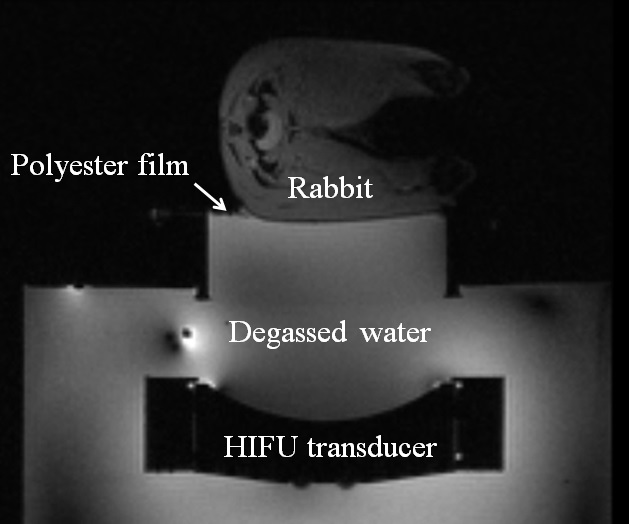
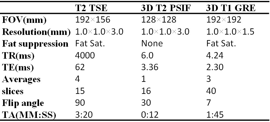
Animal experiments were performed in accordance with the approval of IRB. A male rabbit (weight 4.8 kg) was sonicated in the left thigh by HIFU. During the sonication, the rabbit was anaesthetized by isofluorane, and the vital parameters were monitored throughout the entire treatment, including respiration and rectal temperature. All experiments were conducted on 3T MR system (TIM Trio, Siemens, Germany). A customized MR compatible animal platform, which integrated animal table, water tank and HIFU transducer and positioning system, was used for rabbit experiment. The HIFU transducer (1.0MHz, Imasonics, France), whose active diameter and geometric focal distance were both 100mm, was fixed on the bottom of an acrylic tank filled with degassed water. A customized 6 channel coil was used for signal reception. The whole system configuration is shown in Figure 1. Before the sonication, a 3D T1-weighted GRE images were scanned for target localization. The positioning system moved the transducer so that the HIFU focal point was inside the target area. Reference T2-weighted TSE and PSIF were scanned. The focal area in left thigh was then sonicated with acoustic power ~120 W for 25 seconds. After sonication, T2-weighted TSE and PSIF were scanned for assessment of acute thermal damage for comparison. Contrast agent was administrated and a 3D T1-weighted GRE was scanned at the end of whole procedure. The parameters of the sequences mentioned above were summarized in Table 1.
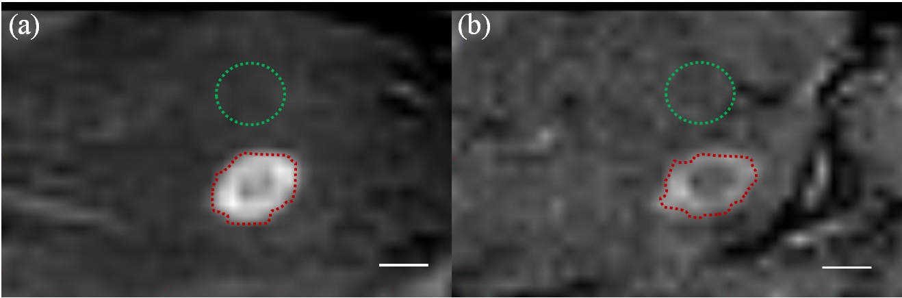
The animals were then sacrificed. The coagulated tissue was resected and fixed in formalin. The tissue damage was evaluated with the digitized H&E sections. The contrast-to-background ratios (CBR) between each lesion and nearby nontreated regions of interest (ROIs) in muscle were calculated from both T2-weighted TSE and PSIF images. The CBR time efficiency defined as CBR to square root time was also compared.$$CBR=\frac{mean_{lesion}-mean_{muscle}}{standard\quad deviation_{muscle}}$$$$CBR_{efficiency}=\frac{CBR}{\sqrt{t}}$$
Results
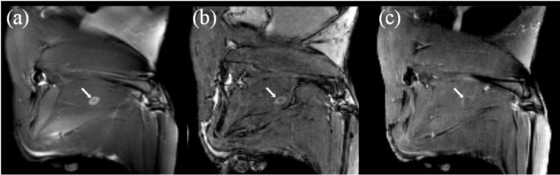
The Figure 2 depicts the appearance of the thermal damage regions on T2-weighted TSE, PSIF and contrast enhanced T1-weighted images. The T2-weighted TSE images and PSIF images offered the better visualization of the regions of tissue damage. The CBR and CBR time efficiency between regions of thermal damage and normal muscle were calculated for the 2D T2-weighted TSE, and PSIF images in Figure 3. The CBR for T2-weighted imaging was 20.15, significantly higher than the ratio for the PSIF sequence 5.88. However, considering the acquisition time, the CBR time efficiency for the PSIF sequence was 1.69 significantly higher than the T2-weighted imaging 1.42. The volume of the thermal damage regions was measured based on long axis and shot axis of ellipse area. The length of long axis was 18.1mm, 17.9mm and 18.2mm for T2-weighted TSE, PSIF and CET1 images, respectively. The length of shot axis was 7.21mm, 7.75mm and 6.34mm for T2-weighted TSE, PSIF and CET1 images, respectively. Lesion dimensions had good agreement with three different sequence images.Discussions and Conclusions
In this study, acute thermal damage following HIFU ablation in muscle was assessed using a PSIF images. The results demonstrate that the CBR time efficiency for PSIF was significantly higher than the tradional T2-weighted TSE sequence.
In conclusion, this preclinical study demonstrates that PSIF sequence offers a good T2 contrast for visualizing acute thermal damage in muscle tissue during HIFU treatment, and has an obvious advantage in acquisition time, making PSIF a suitable sequence for real-time monitoring tissue changes during thermotherapy at high field system.
Acknowledgements
No acknowledgement found.References
[1] Staruch RM, et al. 2017 Assessment of acute thermal damage volumes in muscle using magnetization-prepared 3D T2 -weighted imaging following MRI-guided high-intensity focused ultrasound therapy J. Magn. Reson. Imaging 46:354-364Figures