1501
Simultaneous displacement and T2 mapping of High-intensity focused ultrasound therapy1Shenzhen Institutes of Advanced Technology, Chinese Academy of Sciences, Shenzhen, China, 2Zhengzhou University, Zhengzhou, China, 3University of Chinese Academy of Sciences, Beijing, China
Synopsis
In this work, a hybrid ARFI sequence based on segmented SE-EPI is proposed to simultaneously monitor the displacement and T2 change of tissue during HIFU therapy. The reliability of this sequence was validated first. The quantified displacement and T2 show good consistence with the reference ARFI and SE results. The hybrid sequence was then applied before and after HIFU therapy to evaluate the treatment effects. With the occurrence of ablative lesion, T2 relaxation time decreased in the lesion center and increased in the boundary. While the displaced region (region with obvious displacement) and the maximal displacement at focus both enlarged. In general, this hybrid ARFI is a potentially useful HIFU monitoring method in clinical application.
Introduction
HIFU with the ability to treat deep tumor precisely and noninvasively is a fast developing therapeutic method. A variety of MRI parameters have been proved to be sensitive to HIFU effects1 , making it the first choice HIFU guidance modality. MR-ARFI is a powerful HIFU localization method, as it can encode the micro-scale tissue displacement into phase variation. Besides, it can also reflect the tissue stiffness change caused by HIFU necrosis2. Inflammatory edema induced by acute thermal injury in the boundary can be depicted by the T2 change of tissue. A hybrid ARFI and T2 relaxometry sequence based on segmented SE-EPI is proposed in this work, to simultaneously monitor the displacement and T2 change quantitatively after HIFU therapy.Materials and methods
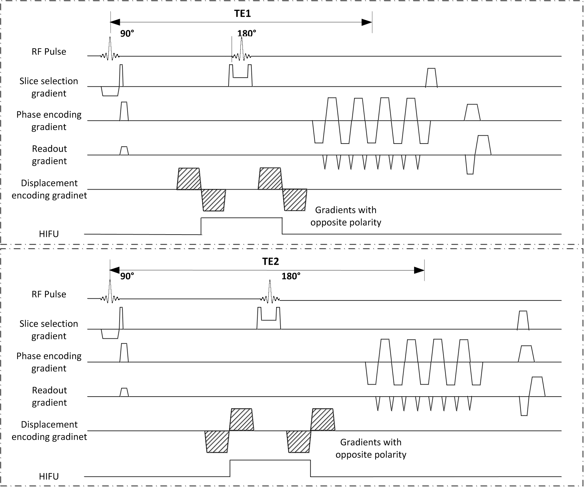
Sequence: The hybrid ARFI and T2 relaxometry sequence was illustrated in Figure 1. A pair of images were acquired with opposite polarity of displacement-encoding gradients and different TEs. Tissue displacement map was quantified based on two phase images3: $$∆x=(φ1-φ2)/(2γG_e τ)$$ Where $$$G_e $$$and $$$ τ $$$ were the amplitude and duration of displacement encoding gradient respectively. T2 map was calculated by fitting the mono-exponential decay of the magnitude signals4:$$T2=(TE_2-TE_1)/(ln(S(TE_1)/S(TE_2)))$$ Reference displacement map was acquired through traditional ARFI, TE = 35ms with the same displacement encoding configuration. Reference T2 mapping was based on single echo SE scans, with TE = 15, 30, 45, 60ms, TR = 5000ms.
Experiment: All experiments were conducted on 3T MR system (TIM Trio, Siemens, Germany). 1MHz HIFU transducer with 33W acoustic power was driven at pulse mode to induce local tissue displacement and synchronized to the motion encoding gradients (amplitude: 32mT/m, duration: 10ms). The porcine muscle sample was sonicated with continuous 33W acoustic power for 1min to produce thermal lesion. The hybrid sequence were scanned both before and after ablation to assess changes in tissue stiffness and T2 value. The imaging parameters were: TR = 600ms, TE1/TE2 = 35ms/55ms, Resolution = 1.2*1.2*3.0mm3, Matrix = 128*46, EPI factor = 9, BW = 814Hz/pixel. T2-TSE image was also acquired after HIFU ablation, with the parameters: TR/TE = 5000/97ms, matrix = 256*256, resolution = 0.6*0.6*3.0mm3.
Results
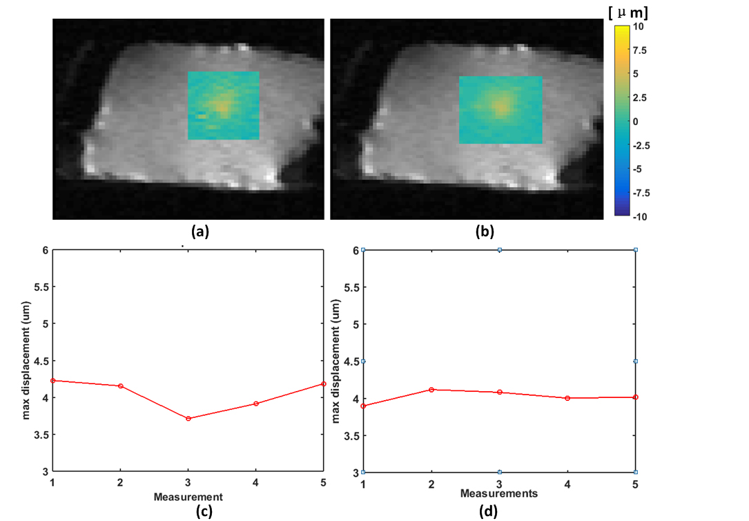
Figure 2 shows the displacement map and the maximal displacment at focus during five consective meaurments of hybrid and reference ARFI. The averaged maximum displacement was 4.04±0.22 μm for hybrid ARFI, and 4.02±0.08 μm for reference ARFI.
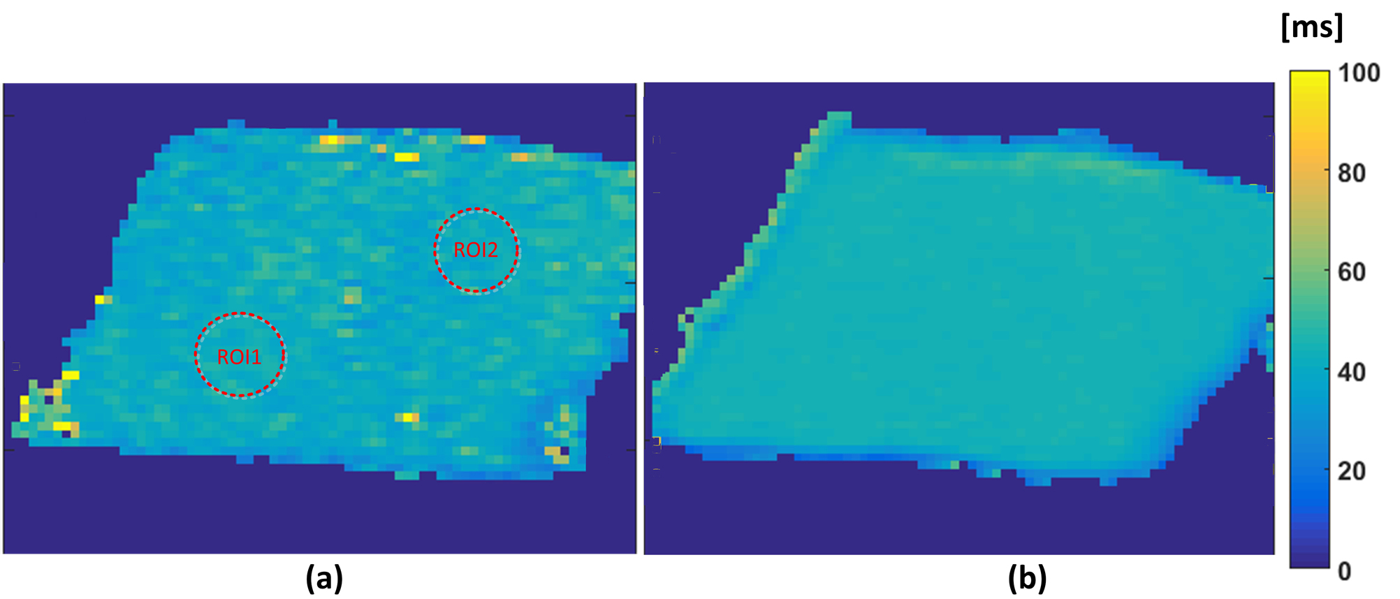
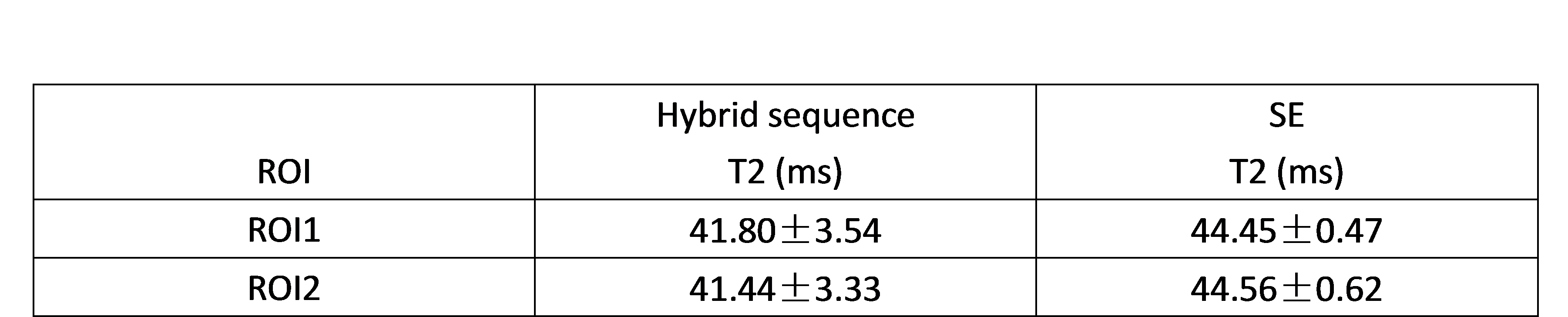
Figure 3 shows the T2 map quantified by hybrid ARFI and reference SE. The T2 map based on hybrid sequence was more noisy than that based on reference SE due to the relatively lower SNR. The T2 vaule in two selected region of interest (ROI) were assessed and compared in Table 1.
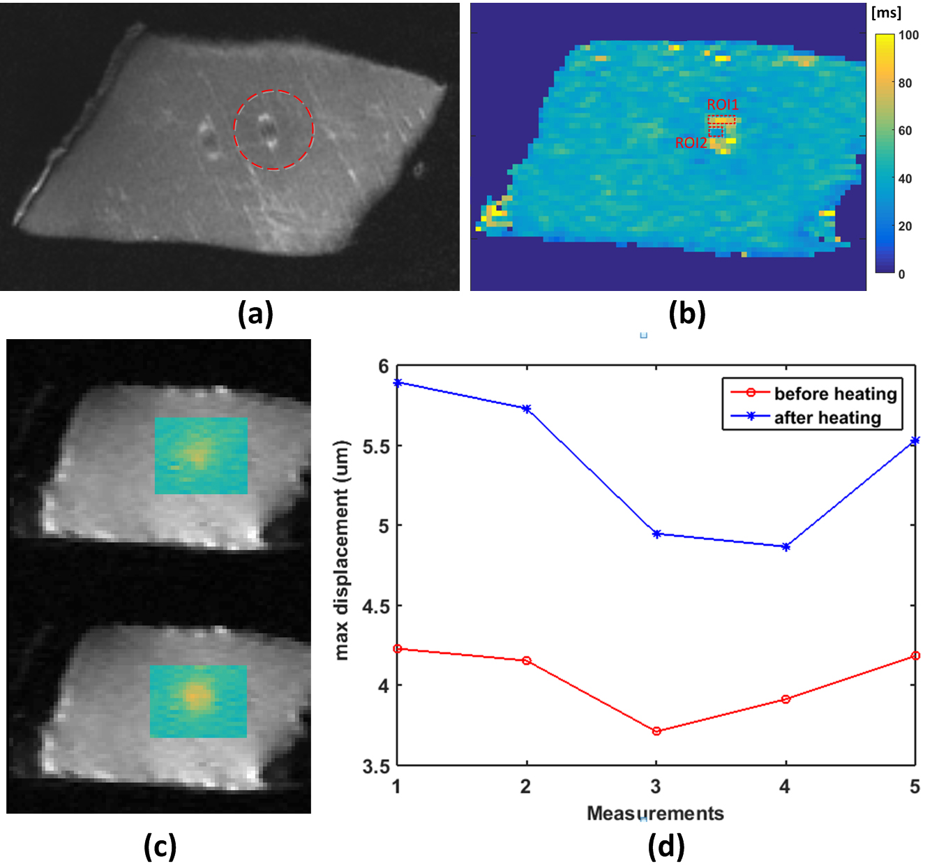
Figure 4 demonstrates the application of hybrid sequence to evaluate the HIFU ablation. The red dashed circle in figure 4(a) indicated the ablated region. The hyper-intensity in both figure 4(a) and (b) represent edema around ablated region. The T2 value in ROI1 was 72.29±9.97ms, while in the center region ROI2 was 36.56±3.78ms. The area of displaced region (region with obvious displacement) enlarged, and the maximal displacement increased from 4.04±0.22 μm to 5.39±0.46 μm, indicating the occurrence of necrosis3.
Discussion
It is reported that the cell response to HIFU ablation is complicated5. In the central zone, coagulative necrosis would occur, inducing change of tissue stiffness, which can be detected by MR-ARFI. While T2 at the periphery zone would significantly increase due to the inflammatory edema. To finely imaging the effects of HIFU ablation, a hybrid ARFI and T2 relaxometry sequence with the ability to obtain local displacement and T2 map simultaneously was proposed in this work.
The accuracy of the hybrid sequence was verified first. Excellent consistency between the hybrid sequence and the reference ARFI was found, except that the hybrid sequence demonstrated a slight higher standard deviation. This may due to the longer TE acquisition which had a lower SNR6. For T2 map, hybrid ARFI calculated a slight lower T2 with a larger standard deviation compare to SE, which may due to the signal attenuation caused by displacement encoding gradients. Nevertheless, the deviation was around 7%, making it still reliable.
Second, the hybrid sequence was applied to HIFU therapy monitoring. With the occurrence of ablated lesion, T2 decreased in the lesion center, and increased in the boundary areas due to edema. From the displacement aspect, it is observed that both the displaced region and the maximal displacement enlarged. It is possibly due to that the increase of absorption coefficient at the ablated region induced the increased acoustic radiation force.
Conclusion
In general, the proposed hybrid ARFI and T2 relaxometry sequence can detect changes of local tissue displacement and T2 value simultaneously, making it an attractive HIFU therapy monitoring method.Acknowledgements
This research was supported by the National Natural Science Foundation of China No. 81327801, 61302040,11504401, 81527901, 81301242, U1301258, National Key R&D Program No. 2016YFC0100100, Shenzhen Scienceand Technology Research Program No. JCYJ20150630114942317 and JCYJ20150521094519487References
1. Hectors S, Jacobs I, Moonen C, et al. MRI methods for the evaluation of high intensity focused ultrasound tumor treatment: Current status and future needs. Magnet Reson Med. 2015:302-317.
2. Bitton R, Kaye E, Dirbas F, et al. Toward MR-guided high intensity focused ultrasound for presurgical localization: focused ultrasound lesions in cadaveric breast tissue. Journal of magnetic resonance imaging : JMRI. 2012;35:1089-97.
3. McDannold N, Maier S. Magnetic resonance acoustic radiation force imaging. Med Phys. 2008;35:3748.
4. Baron P, Ries M, Deckers R, et al. In vivo T2 -based MR thermometry in adipose tissue layers for high-intensity focused ultrasound near-field monitoring. Magnet Reson Med. 2014;72:1057-64.
5. Chu K, Dupuy D. Thermal ablation of tumours: biological mechanisms and advances in therapy. Nat Rev Cancer. 2014;14:199-208.
6. Chen J, Watkins R, Pauly K, et al. Optimization of encoding gradients for MR‐ARFI. Magnet Reson Med. 2010;63:1050-8.
Figures