1492
Volumetric and rapid MR-acoustic radiation force imaging using simultaneous multi-slice imaging1IHU-LIRYC, PESSAC, France, 2Univ. Bordeaux, Centre de recherche Cardio-Thoracique de Bordeaux, Bordeaux, France, 3INSERM U1045, Bordeaux, France, 4Image Guided Therapy, Pessac, France, 5Center for Magnetic Resonance in Biology and Medicine - UMR 7339, Marseille, France, 6Siemens Healthcare, Saint-Denis, France, 7Siemens Healthcare, Erlangen, Germany
Synopsis
The local tissue displacement induced by acoustic radiation force impulses (ARFI) during MR guided HIFU can be used to localize the focal spot position before thermal ablation and to monitor qualitative changes in tissue elasticity during ablation. However current MR-sequence implementations lack of spatial coverage, for a temporal resolution in the order of the timescale (<1Hz) of displacement changes during sonication. To address this limitation, we developed a simultaneous multislice MR-ARFI sequence with a slice acceleration factor up to 3. Displacement estimations measured with accelerated sequences are compared to reference values using a non-accelerated sequence.
Introduction
MR-guided thermal therapies using high intensity focused ultrasound (MRgHIFU) are increasingly being applied in oncology and neurology as a fully non-invasive treatment. Displacement induced by the acoustic radiation force (MR-ARFI[1]) can be used to localize the focus position before thermal ablation and to monitor changes in tissue elasticity during ablation [2]. For the latter purpose, volumetric displacement monitoring with high temporal resolution is desirable. Recently, simultaneous multislice imaging (SMS) methods have been extensively developed to increase volume coverage at fixed duration and/or to reduce acquisition time[3]. In this work, we have investigated the benefits of SMS acquisitions for real-time monitoring of displacement by combining SMS with blipped-EPI acquisition and GRAPPA acceleration. Image reconstruction is performed online to visualize local displacement induced by HIFU in real-time. Validation was performed on an ex vivo swine muscle during MRgHIFU sonications.Methods
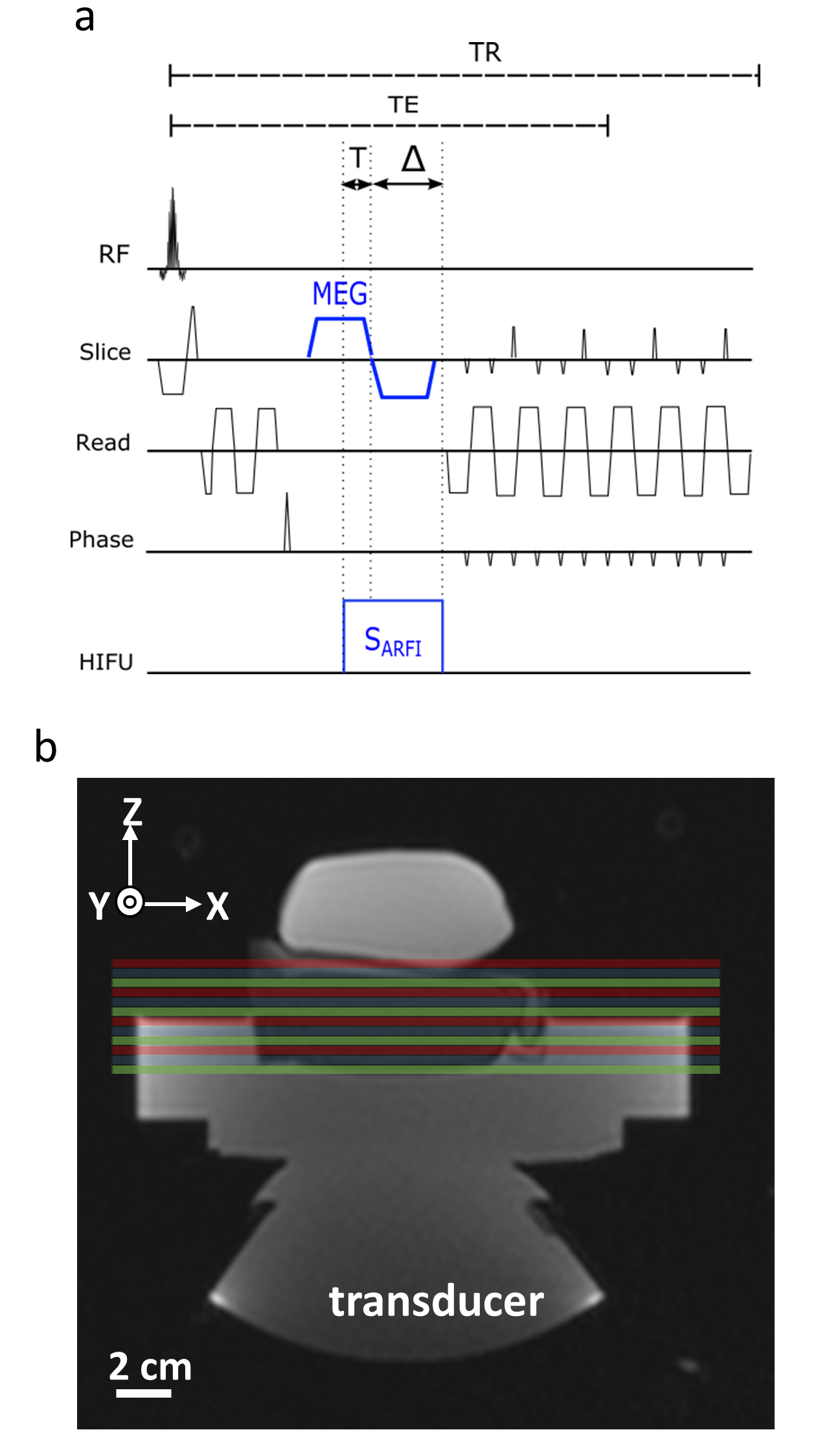
A single-shot gradient-echo echo-planar imaging (EPI) sequence was combined with simultaneous multi-slice and GRAPPA accelerations using the Blipped-Controlled Aliasing in Parallel Imaging (blipped-CAIPI)[4] technique. The prototype sequence was implemented at 1.5T (MAGNETOM Aera, Siemens Healthcare, Erlangen, Germany) and the vendor reconstruction was used. A bipolar Motion Encoding Gradient (MEG) was integrated before the EPI readout to encode micrometric tissue displacement induced by HIFU (see Figure 1a). Magnitude and phase images were reconstructed online and data were transferred online to a separate workstation (Thermoguide™, Image Guided Therapy SA, Pessac-France) for computation and visualization of displacement images. The displacement estimates were then computed as follows:
$$ΔD=\frac{φ-φ_{ref}}{γ.B_O.Δ.A}$$
Where $$$φ$$$ is a phase image when sonication is performed and $$$φ_{ref}$$$ is a reference phase image taken as the first dynamic in the time series, without sonication. γ is the gyromagnetic ratio, $$$B_0$$$ is the magnetic field. A=24 mT/m and Δ=5 ms are the MEG amplitude and duration, respectively. A delay T=3 ms was added from the center of MEG in order to take into account tissue viscoelastic properties. A temporal phase unwrap was performed between φ and $$$φ_{ref}$$$, before displacement computation. During displacement monitoring, 12 slices were acquired in coronal orientation using a slice interleaved reordering scheme (Figure 1b). The distance between adjacent slices was set to 0.3 mm resulting in a spatial coverage of 39.3 mm in the slice direction. Sequence parameters were: FOV=260x260 mm2, spatial resolution 2x2x3 mm3, TE/TR/FA = 36 ms/125 ms/60°, GRAPPA acceleration factor of 2, 50 % phase oversampling, 6/8 partial Fourier and bandwidth = 1955 Hz/pixel. The precision of the method was quantified for different Multiband factors (MB) by computing the standard deviation in displacement maps over the first 8 volumes in the time series, the 10th volume being acquired with a $$$S_{ARFI}$$$ HIFU pulse of 8 ms duration at 350 W acoustic. Displacement measurement datasets were acquired at the same location with 12, 6 and 4 sonication repetitions for MB=1, 2 and 3, respectively. In this study, displacement estimates without slice acceleration (i.e. MB=1) were taken as gold standard estimations of the displacement. Displacements measured with MB >1 were compared to this reference measurement in Bland-Altman plots.
Results
The temporal resolution for acquiring the 12 slices
were 1500 ms, 760 ms and 507 ms for MB=1,
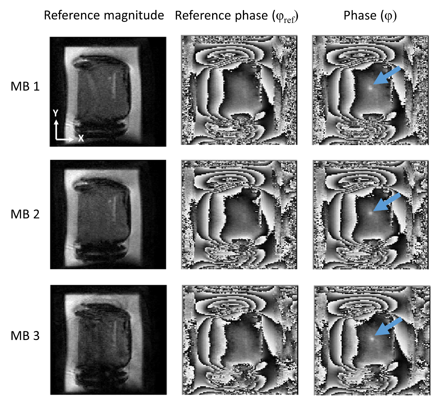
2 and 3, respectively. Figure 2 displays representative magnitude and phase
images for each MB factor. Blue arrows depict the position of the focus.
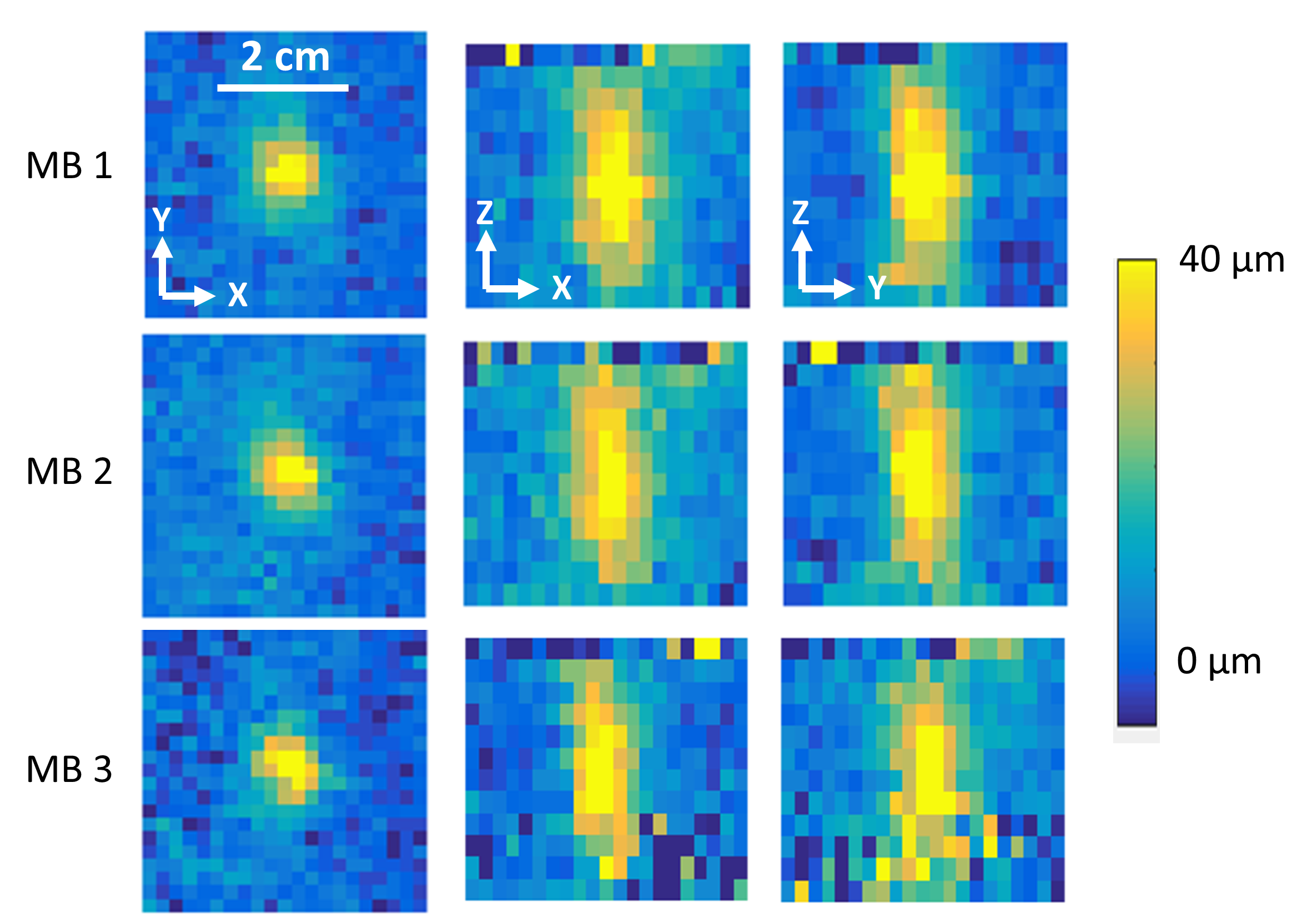
The spatially averaged temporal standard deviations of
displacement on the first 8 frames (without sonication) in a region of interest
(8x8x3 pixels centred on slice #6) centred on the ultrasound focus were 1.8,
1.9 and 2.1 µm. For MB=1, 2 and 3 the maximum displacements were 44, 47, 51 µm,
respectively.
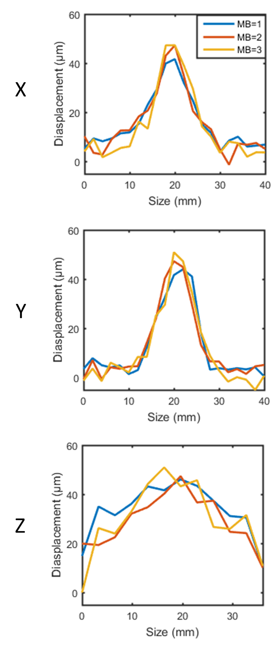
Figure 3 displays the displacement profiles at the
focus (21x21x12 pixels) in three orthogonal directions for MB =1, 2 and 3. The
displacement profile dimensions along X, Y and Z
axis (Figure 4), measured
at half the maximum displacement were [12, 8, 10], [10, 10, 8] and [33.75, 26.2,
26.2] cm, for MB=[1, 2, 3], respectively.
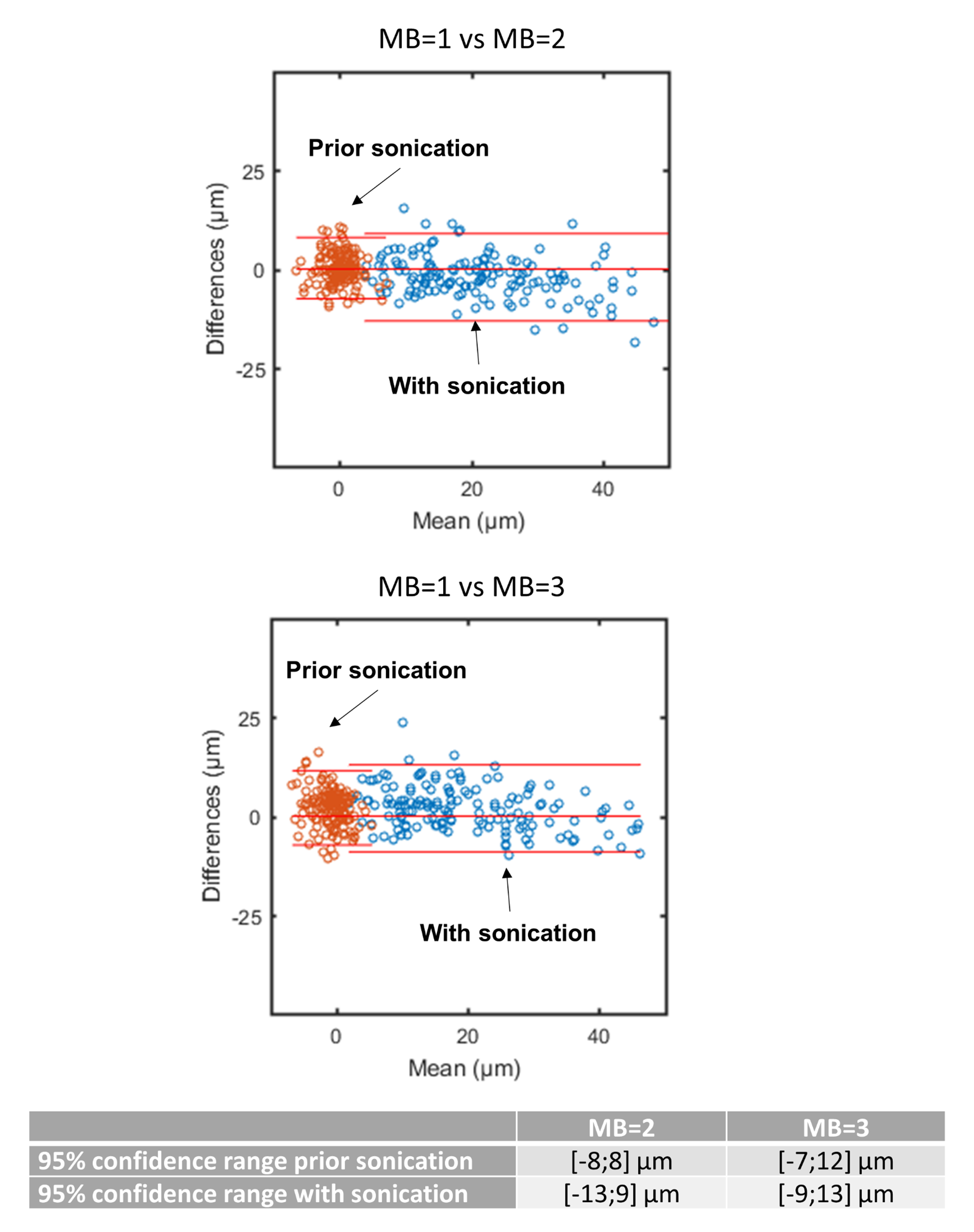
Bland-Altman
plots of the displacement estimation, in a ROI of 8x8x3 pixels centred
on slice #6, are shown in figure 5. Displacement estimations prior to
sonication and with sonication are displayed in orange and blue, respectively. The
Bland-Altman 95% confidence range remained under 13
µm for MB=2 and 3, respectively.Discussions and conclusions
Acknowledgements
This work received financial support from the French National Investments for the Future Programs: ANR-10-IAHU-04 (IHU Liryc) and Laboratory of Excellence ANR-10-LABX-57 (TRAIL), and the research programs ANR-11-TecSan-003-01 (TACIT) and Equipex ANR-11-EQPX-0030 (MUSIC).References
[1] N. McDannold et S. E. Maier, « Magnetic resonance acoustic radiation force imaging », Med. Phys., vol. 35, no 8, p. 3748, 2008.
[2] P. Bour et al., « Real-time monitoring of tissue displacement and temperature changes during MR-guided high intensity focused ultrasound », Magn. Reson. Med., 2017.
[3] M. Barth, F. Breuer, P. J. Koopmans, D. G. Norris, et B. A. Poser, « Simultaneous multislice (SMS) imaging techniques: SMS Imaging », Magn. Reson. Med., vol. 75, no 1, p. 63‑81, janv. 2016.
[4] K. Setsompop, B. A. Gagoski, J. R. Polimeni, T. Witzel, V. J. Wedeen, et L. L. Wald, « Blipped-controlled aliasing in parallel imaging for simultaneous multislice echo planar imaging with reduced g-factor penalty », Magn. Reson. Med., vol. 67, no 5, p. 1210‑1224, mai 2012.
[5] Souheil J Inati, Michael S Hansen, et and Peter Kellman, « A Fast Optimal Method for Coil Sensitivity Estimation and Adaptive Coil Combination for Complex Images ». [En ligne]. Disponible sur: http://cds.ismrm.org/protected/14MPresentations/abstracts/4407.pdf. [Consulté le: 26-avr-2017].
[6] D. L. Parker, A. Payne, N. Todd, et J. R. Hadley, « Phase reconstruction from multiple coil data using a virtual reference coil: Phase Reconstruction Using a Virtual Reference Coil », Magn. Reson. Med., vol. 72, no 2, p. 563‑569, août 2014.
Figures