1476
Evaluation of 2D simultaneous multi-slice EPI for high resolution thermometry in the brain at 3T.1IHU Liryc, Electrophysiology and Heart Modeling Institute, Fondation Bordeaux Université, Bordeaux, France, 2Univ. Bordeaux, Centre de recherche Cardio-Thoracique de Bordeaux, U1045, Bordeaux, France, 3INSERM, Centre de recherche Cardio-Thoracique de Bordeaux, U1045, Bordeaux, France, 4Image Guided Therapy SA, Bordeaux, France, 5CENIR, Centre de NeuroImagerie de Recherche, Paris, France, 6ICM, Inserm U 1127, CNRS UMR 7225, Sorbonne Universités, UPMC Université Paris 06 UMR S 1127, Institut du Cerveau et de la Moelle épinière, Paris, France, 7Institut Langevin Ondes et Images, ESPCI ParisTech, CNRS 7587, UMRS 979 INSERM, Paris, France, 8Centre de Résonance Magnétique des Systèmes Biologiques, UMR5536, CNRS, Univ. Bordeaux, Bordeaux, France
Synopsis
MR-guided HIFU in the brain currently lacks from insufficient spatial and temporal monitoring of the effect of ultrasound. In this study, we combine simultaneous multi-slice (SMS) echo planar imaging (EPI) technique with in-plane parallel imaging to achieve high spatial resolution with large volume coverage and/or short acquisition time during temperature mapping at 3T. The sequence was tested in vivo in a human brain with different multiband (MB) factors. SMS reconstruction and temperature mapping were computed using the Gadgetron framework. Then, validation was performed on an ex vivo chicken muscle during HIFU sonication to validate the method.
INTRODUCTION
MR-guided HIFU in the brain (noninvasive focal ablation for the treatment of essential tremor [1] or neuromodulation [2]) currently lacks from insufficient spatial and temporal monitoring of the effect of ultrasound. In this study, we combine simultaneous multi-slice (SMS) echo planar imaging (EPI) technique with in-plane parallel imaging [3] to achieve high spatial resolution with large volume coverage and/or short acquisition time during temperature mapping. Online scanner SMS implementations have now reached a sufficient level of maturity and such sequences could easily be used to quickly expand temperature monitoring for therapeutic application. In this context, a 2D SMS-EPI sequence was optimized at 3T to achieve a 1.7 mm3 isotropic resolution while covering the whole-brain. The sequence was tested in vivo in a human brain with different multiband (MB) factors. SMS reconstruction and temperature mapping were computed using the Gadgetron framework. Then, validation was performed on an ex vivo chicken muscle during HIFU sonication to validate the method.METHODS
Acquisition was performed at 3.0T on clinical imaging system (MAGNETOM Prisma, Siemens Healthcare, Erlangen, Germany). Two acquisitions with different MB factors (MB 1, 2) were compared using the single-shot gradient echo blipped-CAIPI SMS-EPI using the CMRR distributed gradient echo sequence. Acquisition parameters were FA = 60°, GRAPPA acceleration factor of 2, 6/8 partial Fourier.
Volunteer: the study was approved by the Institutional Review Board and subject gave written informed consent to be included into the study. 36 temperature slices were acquired sequentially in transversal orientation using a standard 20 channels head coil. Other acquisition parameters were TE/TR=16.8 ms/2423 ms for MB=1, 22.8 ms/1330ms for MB=2 (with 50 % phase oversampling), FOV=220x220 mm2, leading to an isotropic spatial resolution of 1.7 mm3, bandwidth = 2440 Hz/pixel.
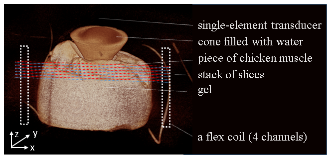
HIFU ablation device: A single-element transducer [4] (H115, Sonic Concepts, Bothel, USA) was operated at 850kHz. MR temperature mapping was performed, using one 4-elements flex antenna positioned around the sample (Figure 4), during a 5 s HIFU pulse for MB=1 and 2, in two consecutive acquisitions. Twelve slices were acquired using an interleaved pattern in coronal orientation every second. TE/TR=22 ms/842 ms for MB=1, 22 ms/437 ms for MB=2. FOV=200x200 mm2, spatial resolution 1.8 mm3 isotropic, pixel bandwidth = 1750 Hz/pixel, 50 % phase oversampling.
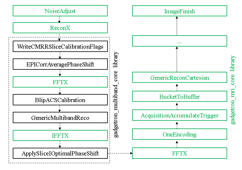
Image reconstruction: The raw data files were converted into ISMRM Raw Data format [5]. SMS-EPI reconstruction was performed offline in the Gadgetron framework [6] (Figure 1). Slice aliased images were unfolded using a standard slice-GRAPPA algorithm (5x5 kernel). Then, the Gadgetron implementation of GRAPPA was applied to correct for phase-encoding undersampling.
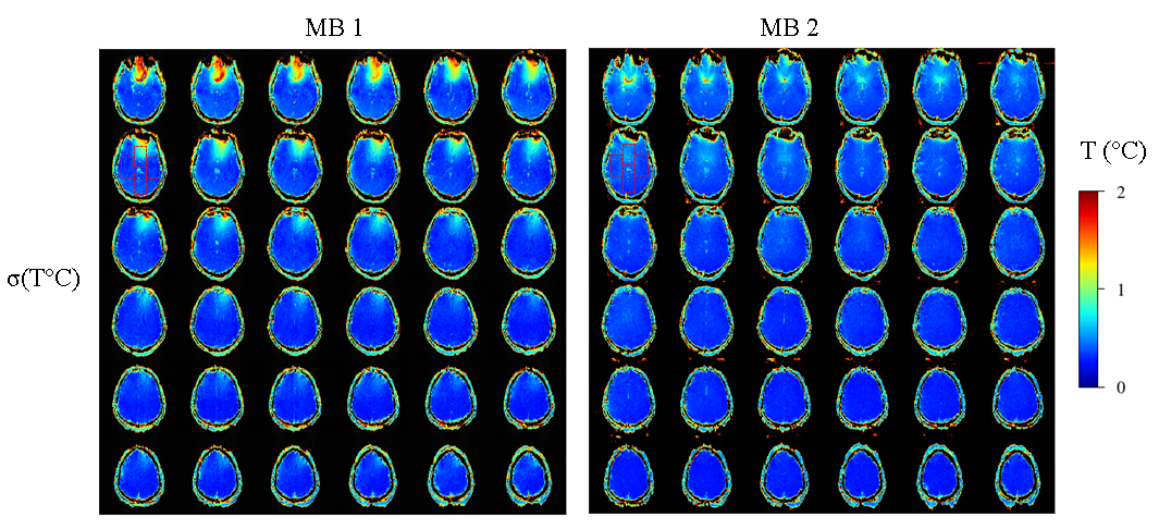
Thermometry: Temperature maps were calculated using the proton resonance frequency shift using phase subtraction. To assess the precision of MR-thermometry, the temporal standard deviation σ(T) and temporal mean μ(T) of temperature were computed in each pixel over 99 consecutive repetitions and the distribution of σ(T) and μ(T) values were analyzed on a four arbitrary drawn ROI (~15000 voxels each). Neither spatial nor temporal filtering were applied.
RESULTS
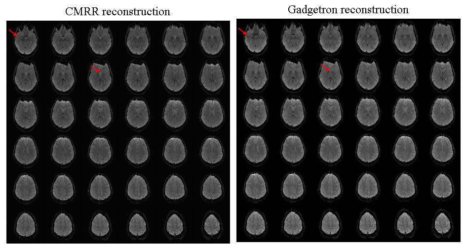
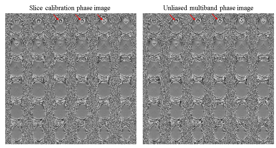
Volunteer study: The MB acquisition was 45% faster than conventional EPI-Grappa acquisition. Resulting magnitude and phase images showed acceptable quality (Fig. 2 and Fig. 3). The spatially averaged temporal standard deviation and mean of temperature in the four regions of interest (Fig.4) were (σ(T), μ(T)) = (0.5. ± 1, -0.2 ± 1.4) °C for MB=1 and (0.4 ± 0.8, -0.3 ± 1.2 ) °C for MB=2.
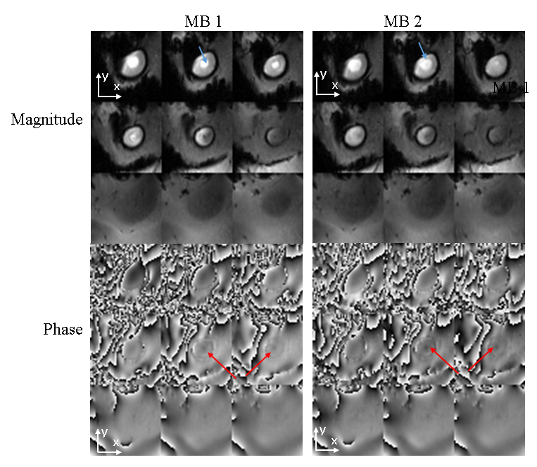
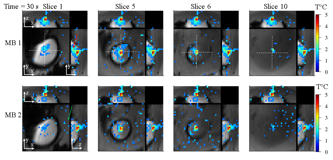
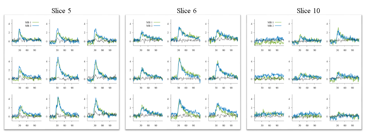
HIFU study: Figure 6 displays representative magnitude and phase images for each MB factor during heating. Red arrows depict the position of the focus. Four channels were used to reconstruct the data and showed acceptable image quality. Figure 7 displays the temperature profiles at the focus (4x3x7 pixels) in three orthogonal directions for MB =1, 2. Figure 8 shows temperature evolution in time at the focus in slice #5,#6,#10. No false-positive temperature spots due to potential signal leakage between simultaneously excited slices were observed under the tested conditions.
DISCUSSION AND CONCLUSION
Using this initial implementation, no difference between conventional slices and MB slices were found on the phase images, opening the way to accurate pseudo volumetric and rapid MR thermometry in the brain. Ex vivo validation demonstrated the feasibility of sub-second volumetric monitoring (200x200x21mm3 in 437ms) of temperature elevation with a limited number of coils. A more generic and robust implementation of the reconstruction algorithm including coil compression algorithm will be investigated in order to reach sub-second reconstruction using GPU computing, as well as and the addition of other multiband algorithms (eg. SENSE/GRAPPA). A versatile version using Matlab Gadget is also possible for application not requiring high frame rate and could be a benefit for facilitating the deployment of new image reconstruction algorithms.Acknowledgements
Authors gratefully thank Stanislas Rappachi and Benedikt Poser for useful discussions on SMS reconstruction and for providing Slice-GRAPPA code, and the CMRR in Minnesota for making their SMS sequence available.
This work received financial support from the French National Investments for the Future Programs: ANR-10-IAHU-04 (IHU Liryc) and Laboratory of Excellence ANR-10-LABX-57 (TRAIL), and the research program France Life Imaging, WP3 "Imagerie interventionnelle.
References
1. Elias WJ, Lipsman N, Ondo WG, Ghanouni P, Kim YG, Lee W, et al. A Randomized Trial of Focused Ultrasound Thalamotomy for Essential Tremor. N Engl J Med. 2016;375(8):730-9.
2. Wattiez N, Constans C, Deffieux T, Daye PM, Tanter M, Aubry JF, et al. Transcranial ultrasonic stimulation modulates single-neuron discharge in macaques performing an antisaccade task. Brain Stimul. 2017.
3 Borman PT, Bos C, de Boorder T, Raaymakers BW, Moonen CT, Crijns SP. Towards real-time thermometry using simultaneous multislice MRI. Phys Med Biol. 2016;61(17):N461-77.
4 Constans C, Deffieux T, Pouget P, Tanter M and Aubry JF. A 200-1380 kHz Quadrifrequency Focused Ultrasound Transducer for Neurostimulation in Rodents and Primates: Transcranial in Vitro Calibration and Numerical Study of the Influence of Skull Cavity. IEEE Transactions on Ultrasonics, Ferroelectrics, and Frequency Control
5. Inati SJ, Naegele JD, Zwart NR, Roopchansingh V, Lizak MJ, Hansen DC, et al. ISMRM Raw data format: A proposed standard for MRI raw datasets. Magn Reson Med. 2016.
6. Hansen MS, Sorensen TS. Gadgetron: an open source framework for medical image reconstruction. Magn Reson Med. 2013;69(6):1768-76.
Figures