1467
Safety and EEG Data Quality of Concurrent High-Density EEG and High-Speed fMRI at 3 Tesla1Neurobiology Research Unit, Department of Neurology, Rigshospitalet, Copenhagen, Denmark, 2Dept. of Clinical Medicine, University of Copenhagen, Copenhagen, Denmark, 3Functional Imaging Unit, Dept. of Clinical Physiology, Nuclear Medicine and PET, Rigshospitalet, Copenhagen, Denmark, 4Department of Neurology, University of New Mexico, Albuquerque, NM, United States, 5Department of Physics and Astronomy, University of New Mexico, Albuquerque, NM, United States, 6Dept. of Clinical Neurophysiology, Rigshospitalet, Copenhagen, Denmark, 7Department of Biostatistics, University of Copenhagen, Copenhagen, Denmark, 8Dept. of Radiology, Rigshospitalet, Copenhagen, Denmark, 9Danish Epilepsy Centre, Dianalund, Denmark, 10Aarhus University, Aarhus, Denmark, 11Department of Electrical and Computer Engineering, University of New Mexico, Albuquerque, NM, United States
Synopsis
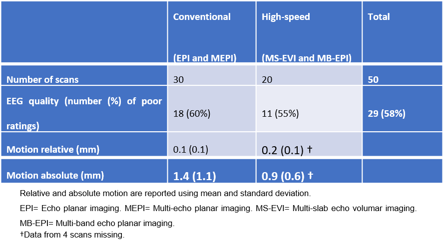
Using concurrent high-density EEG and different high-speed fMRI methods, we investigate safety of RF heating, effect on image SNR and assess EEG data quality. RF related electrode heating during a 30-minute scan did not exceed 1.0o C with any of the pulse sequences. No significant differences in the EEG data quality were found between high-speed fMRI and conventional EPI (p=0.78). Residual ballistocardiographic artifacts resulted in 58% of EEG data being rated as poor quality. This study demonstrates that high-density EEG can be safely implemented in conjunction with high-speed fMRI and that high-speed fMRI does not adversely affect EEG data quality.
Abstract
Introduction
Concurrent EEG and fMRI is increasingly used to characterize the spatial-temporal dynamics of brain activity. Concurrent acquisition of fMRI and EEG has also emerged as a new tool for localizing the epileptogenic zone. However, most studies to date have been limited to conventional echo-planar imaging (EPI). There is considerable interest in integrating recently developed high-speed fMRI methods with high-density EEG to increase temporal resolution and sensitivity for task-based and resting state fMRI, and for detecting interictal spikes in epilepsy. A key concern is RF related heating of the EEG leads and electrodes (1).
In the present study using concurrent high-density EEG and recently developed high-speed fMRI methods, we investigate safety of radiofrequency (RF) related heating, the effect of EEG on cortical signal-to-noise ratio (SNR) in fMRI, and assess EEG data quality.
Methods
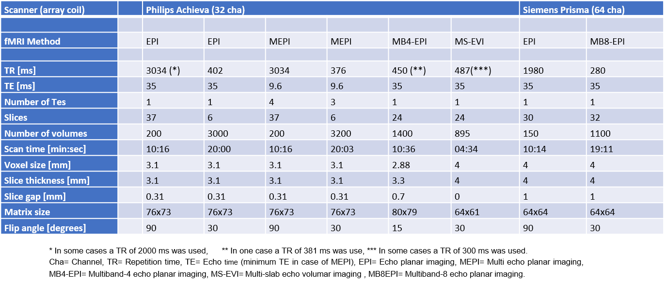
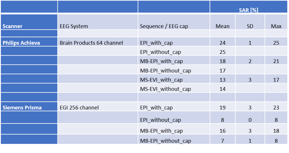
The study compared EPI, multi-echo EPI, multi-band EPI (2,3) and multi-slab echo-volumar imaging (4) pulse sequences, using a Philips Achieva scanner (Philips, Best, The Netherlands) and a Siemens Prisma Fit (Siemens, Erlangen, Germany) scanner equipped with a 64-channel MR Cap (BrainCapMR, Brain Products GmbH, Munich, Germany) and a Geodesic EEG System (Electrical Geodesics, Inc., Eugene, OR) with 256 EEG channels, respectively and array head RF coils. Multi-band EPI data on the Achieva scanner was acquired with 4-fold acceleration (MB4-EPI). On the Siemens Prisma scanner, additional data were acquired using a multi-band EPI sequence with 8-fold acceleration (MB8-EPI). The acquisition parameters for the different fMRI methods are listed in Table 1. Data were collected in 11 healthy controls (3 males, age range 18-70 years) and 13 patients with epilepsy (8 males, age range 21-67 years). Three of the healthy controls were scanned with the 256-channel EEG system, the other subjects were scanned with the 64-channel EEG system. Scalp surface temperature, SNR in occipital cortex and head movement were measured with and without the EEG cap. The degree of artifacts and the ability to identify background activity was assessed by visual analysis by a trained expert in the 64 channel EEG data (7 healthy controls, 13 patients).
Results
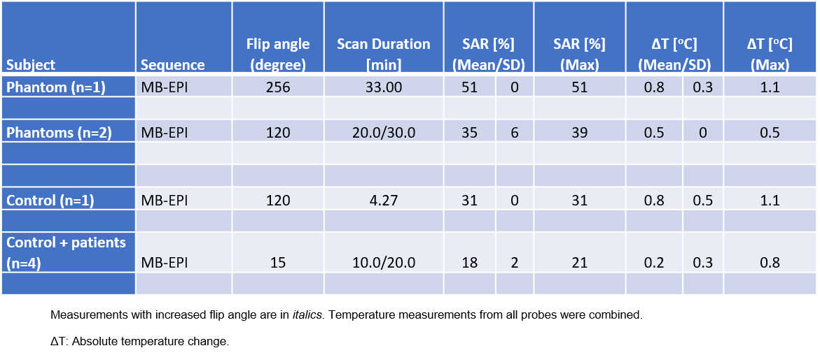
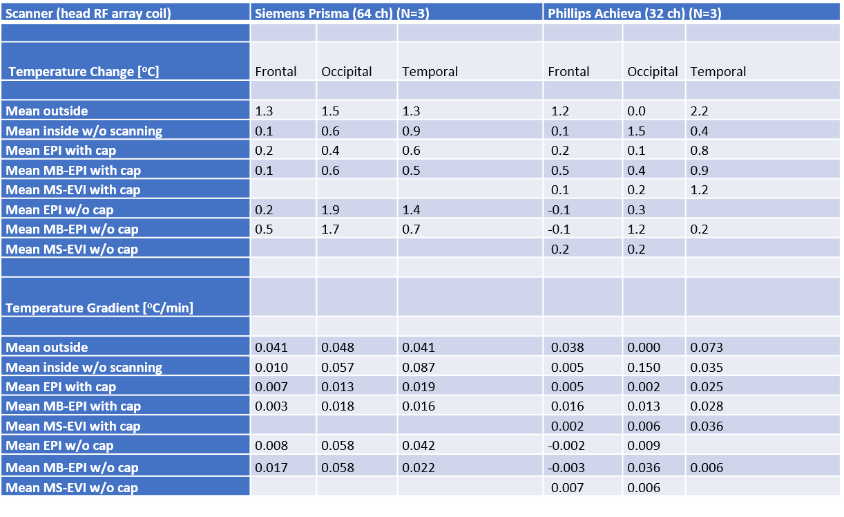
RF induced heating at the surface of the EEG electrodes during a 30-minute scan period with stable temperature prior to scanning did not exceed 1.0o C with either EEG system and any of the pulse sequences used in this study (Tables 2-4). There was no significant decrease in cortical SNR due to the presence of the EEG cap (p > 0.05). No significant differences in the visually analyzed EEG data quality were found between EEG recorded during high-speed fMRI and during conventional EPI (p=0.78). Residual ballistocardiographic artifacts resulted in 58% of EEG data being rated as poor quality (Table 5).
Discussion
The present study demonstrates that concurrent EEG and fMRI can be performed safely across the wide range of fMRI acquisition methods and the body coils used in this study. Although temperature rises of up to 1o C underneath some of the electrodes were measured in a 30-minute scan period, they were on average of such small magnitude that RF induced heating was not a limiting factor for the experimental settings in this study. Temperature drifts of a similar and even larger magnitude were frequently noted even without scanning. In the Prisma scanner a significant increase in SAR was observed when the subjects wear the EEG cap. The maximum SAR values in the present studies, even for scans exploring the safety margins, were well below those reported in previous studies using Fast Spin Echo and Turbo Spin Echo pulse sequences that can reach up to 100 % of the FDA approved SAR limits (5,6). The EEG data quality using the 64 channel EEG system during MR scanning was considerably degraded compared to EEG data measured outside of the scanner. However, EEG data quality was not significantly different between the conventional and the fast sequences. The data quality of the high-density 256 channel EEG array could not be evaluated due to technical difficulties.
Conclusion
This study demonstrates that high-density EEG can be safely implemented in conjunction with high-speed fMRI and that high-speed fMRI does not adversely affect EEG data quality. However, the deterioration of the EEG quality due to residual ballistocardiographic artifacts remains a significant constraint for routine clinical applications of concurrent EEG-fMRI.
Acknowledgements
The Lundbeck Foundation provides salary for MTF during her PhD study. The Lundbeck Foundation provided funding, including salary, allowing SP to be visiting professor in Copenhagen for 6 months.References
1. Carmichael DW, Thornton JS, Rodionov R, Thornton R, McEvoy AW, Ordidge RJ, et al. Feasibility of simultaneous intracranial EEG-fMRI in humans: a safety study. NeuroImage. 2010 Jan 1;49(1):379–90
2. Moeller S, Yacoub E, Olman CA, Auerbach E, Strupp J, Harel N, et al. Multiband multislice GE-EPI at 7 tesla, with 16-fold acceleration using partial parallel imaging with application to high spatial and temporal whole-brain fMRI. Magn Reson Med. 2010 May;63(5):1144–53.
3. Feinberg DA, Moeller S, Smith SM, Auerbach E, Ramanna S, Gunther M, et al. Multiplexed echo planar imaging for sub-second whole brain FMRI and fast diffusion imaging. PloS One. 2010;5(12):e15710.
4. Posse S, Ackley E, Mutihac R, Rick J, Shane M, Murray-Krezan C, et al. Enhancement of temporal resolution and BOLD sensitivity in real-time fMRI using multi-slab echo-volumar imaging. NeuroImage. 2012 May 15;61(1):115–30.
5. Nöth U, Laufs H, Stoermer R, Deichmann R. Simultaneous electroencephalography-functional MRI at 3 T: an analysis of safety risks imposed by performing anatomical reference scans with the EEG equipment in place. J Magn Reson Imaging JMRI. 2012 Mar;35(3):561–71.
6. Lazeyras F, Zimine I, Blanke O, Perrig SH, Seeck M. Functional MRI with simultaneous EEG recording: feasibility and application to motor and visual activation. J Magn Reson Imaging JMRI. 2001 Jun;13(6):943–8.
Figures