1452
Impacts of 3.0 Tesla magnetic resonance imaging noise on hearing function in neonates with hearing protectionHuifang Zhao1, Chao Jin1, Xinyu Li1, Heng Liu1, Xiaoyu Wang1, Xingxing Tao1, Yannan Cheng1, and Jian Yang1
1Department of Diagnostic Radiology, the First Affiliated Hospital of Xi'an Jiaotong University, Xian', China
Synopsis
Loud acoustic noise generated from magnetic resonance (MR) imaging remains the great concern for neonatal exams. This study therefore aims to clarify whether this noise would cause the hearing loss to neonates who underwent MRI exam by auditory brainstem response (ABR). Results indicated that there was no significant difference in all the six ABR indices (waves I, III, V amplitudes and wave I-III, III-V, I-V intervals) between before and after the MRI examinations. Our findings may suggest the rarely temporary impact of MRI noise on ABR in neonates who underwent a 3.0T MRI.
Introduction
Magnetic resonance (MR) imaging of the brain has been an indispensable examination for the neonates in clinics. 1However, the loud acoustic noise generated from MR imaging remains a great concern. Peak sound pressure levels (SPLs) of 3.0T MR imaging can currently reach 130 dBA, presenting high risk of hearing loss for all the participants.2 Efficient hearing protection is thus necessary. Despite using hearing protection, previous studies still found the temporary hearing loss in adults who underwent 3.0T MR imaging.2-6 Differently from adults, neonatal auditory system is being developing. Whether the 3.0T MR imaging noise could cause the hearing loss of neonates should be urgently clarified, especially for a longer-time MR imaging in clinical-research scenario. Therefore, this study aims to investigate the impacts of acoustic exposure to a single 51-minute MR neuroimaging at 3.0T on hearing function of neonates with conventional hearing protection. Auditory brainstem response (ABR) was used to longitudinally monitor the alterations in hearing function, i.e. electrophysiological responses along the auditory pathway before and after MR imaging.Method
This study was approved by the local ethics committee, and parental informed consents were obtained from all participants. From October 2014 to July 2015, 20 neonates with no risk factors for hearing impairment were enrolled in this study from the First Affiliated Hospital of Xi'an Jiaotong University and performed by 3.0T MRI head examinations. The protocols of each sequence were as follows: (1) a three-dimensional fast spoiled gradient-echo T1-weighted sequence (TR/TE, 10.2ms/4.6ms; NEX of 1; isotropic 1×1×1mm3;FOV 18cm); (2) a transverse fast spin-echo T2-weighted sequence (TR/TE, 4200ms/123.90ms; NEX of 1.5; matrix = 256×256; thickness of 4mm; FOV18cm); (3) DTI (30 directions; b value = 600s/mm2; TR/TE, 1000ms/67.4ms; NEX of 1; thickness of 2.5mm; FOV18cm; matrix =128×128/172×172; acquisition time of 7min 42secs);(4) DKI (directions; b value = 1000s/mm2; TR/TE, 11000ms/91.7ms; NEX of 1; thickness of 4mm; FOV18cm; matrix =128×128/172×172; acquisition time of 21min 42secs); (5) T2 FLAIR (TR/TE, 10.2ms/4.6ms; NEX of 1; isotropic 1×1×1mm3; FOV18cm). ABR tests were performed to longitudinally monitor the alterations in hearing function (ABR electrophysiological response indices: waves I, III, V amplitudes and wave I-III, III-V, I-V intervals) along the auditory pathway within 24 hours before and within 20 minutes after the MR imaging. Paired-t test was used to test the difference of ABR indices between the two tests. All statistical analysis were performed by using SPSS 18.0 (SPSS, Chicago, IL, USA); p<0.05 was considered as statistically significant difference.Result
ABR results showed that there was no significant difference in all the six ABR indices, (ABR electrophysiological response indices: waves I, III, V amplitudes and wave I-III, III-V, I-V intervals) between before and after the MRI examinations.Discussion
Our results indicated that no significant difference in ABR electrophysiological indices was found between before and after MRI. This suggested the rarely temporary impact of MRI noise on the neonatal auditory electrophysiological response. In contrast to the temporary hearing loss in adults, such findings on neonates may root in the developmental differences between neonates and healthy adults. Specifically, neonatal cochlea has adult dimensions and developed into a very adult-like configuration; brainstem also reaches a mature state. While, neonates show a lower sensitivity to high-frequency sound. The MRI noise is measured to mainly be 1-2 kHz. It may be such fact that leads to the no significant alteration in ABR electrophysiological indices. Although our results have observed the rarely temporary alteration in neonatal auditory responses, safety issue of MRI remains the great concern, especially for the long-term effects of noise on neonatal hearing function. And more efficient hearing protection and noise-reduction techniques (e.g. ‘silent’ imaging sequence) are therefore in urgent needed.Conclusion
Our findings suggested the rarely temporary impact of MRI noise on auditory electrophysiological responses in neonates who underwent a 3.0T multi-sequence MR imaging.Acknowledgements
This work was supported by the National Key Research and Development Program of China (2016YFC0100300),National Natural Science Foundation of China (No. 81471631, 81771810 and 51706178), the 2011 New Century ExcellentTalent Support Plan of the Ministry of Education, China (NCET-11-0438) the Clinical Research Award of the First Affiliated Hospital of Xi’an Jiaotong University (No.XJTU1AF-CRF-2015-004)References
1.Nora Raschle, Jennifer Zuk, et al. Pediatric neuroimaging in early childhood and infancy:challenges and practical guidelines. Ann. N.Y. Acad. Sci. ISSN 0077-89232.P Radomskij, M A Schmidt,et al.Effect of MRI noise on cochlear function.Lancet 2002; 359: 1486–883.Elizabeth Yenn Lynn Lim ,Ing Ping Tang,et al.3 Tesla magnetic resonance imaging noise in standard head and neck sequence does not cause temporary threshold shift in high frequency.Eur Arch Otorhinolaryngol (2015) 272:3109–31134.Wolfgang Wagner, MD; Irene Staud,et al.Noise in Magnetic Resonance Imaging: No Risk for Sensorineural Function But Increased Amplitude Variability of Otoacoustic Emissions.Laryngoscope, 113:1216–1223, 20035.Brigitte Strizek, Jacques C. Jani, et al.Safety of MR Imaging at 1.5 T in Fetuses: A Retrospective Case-Control Study of Birth Weights and the Effects of Acoustic Noise1.Radiology: Volume 275: Number 2—May 20156.R.E. Venn , A.R. McBrearty ,et al.The effect of magnetic resonance imaging noise on cochlear function in dogs.The Veterinary Journal 202 (2014) 141–1457.Joint Committee on Infant Hearing,Year 2007 Position Statement:Principles and Guidelines for Early Hearing Detection and Intervention Programs,PEDIATRICS Volume 120, Number 4, October 2007Figures
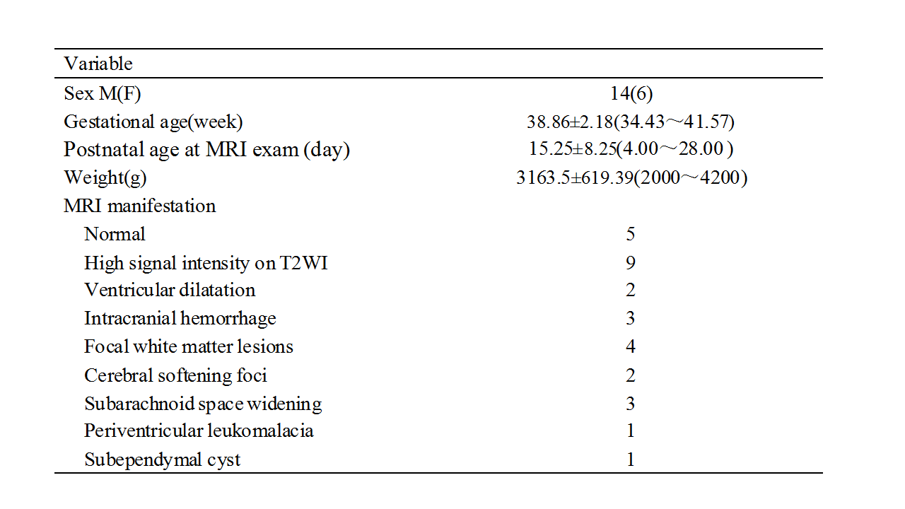
Table 1 Participant demographics
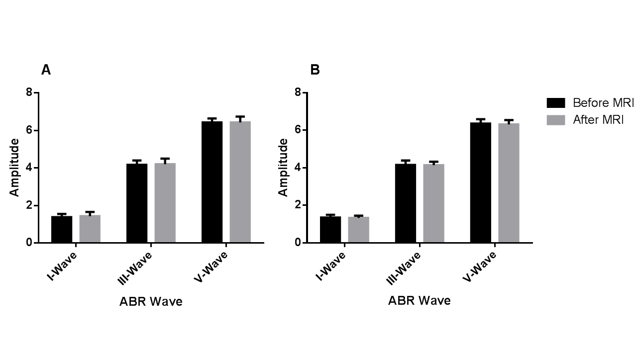
Figure 1 Comparisons of ABR wave amplitude of hearing function between before and after MRI in (A) left ear and (B) right ear.
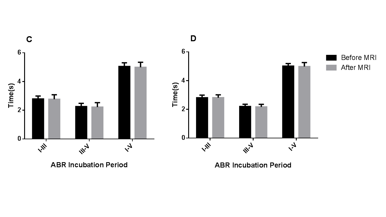
Figure 2 Comparisons of ABR wave interval of hearing function(wave I-III, III-V and I-V intervals) between before and after MRI in (C) left ear and (D) right ear.