1451
Assessment of Peripheral Nerve Stimulation due to MR gradient induced Electric Field around Implantable Device1Abbott, Sylmar, CA, United States
Synopsis
Time varying magnetic gradient fields can induce electric field (E-field) in the human body and may cause peripheral nerve stimulation (PNS) during MR scan. As metallic implant may cause local E-field enhancement, there is speculation that it may increase risk of PNS. In this study, gradient coil modeling is used to investigate induced E-fields around implantable devices. The maximum E-field in the proximity of implants is compared to the whole body maximum E-field of the human body without implant. The result shows that the local enhanced E-field near implants does not exceed the whole body maximum E-field in human body.
Introduction
Previous research1 shows that there is electric field (E-field) enhancement near metallic implantable device when patient with device is exposed to the MR gradient field. This study aims to investigate the induced E-field enhancement due to the presence of implantable device and compare the resultant local E-field magnitude to the whole body maximum E-field induced in a human body without implant. The goal of this study is to assess if a metallic implant can potentially increase the risk of peripheral nerve stimulation (PNS) during MR scan.Methods
Simulations were performed in Sim4Life2. The simulation model contains a y-axis gradient coil with a human model (Duke from the Virtual Family3). The Y-axis gradient field is employed as it is considered the worst-case gradient field for PNS since the y-axis field intersects the largest cross-sectional area of a human body. The IT’IS low frequency tissue conductivity database4 is used for the human body tissues.
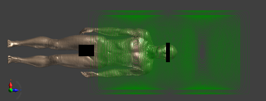
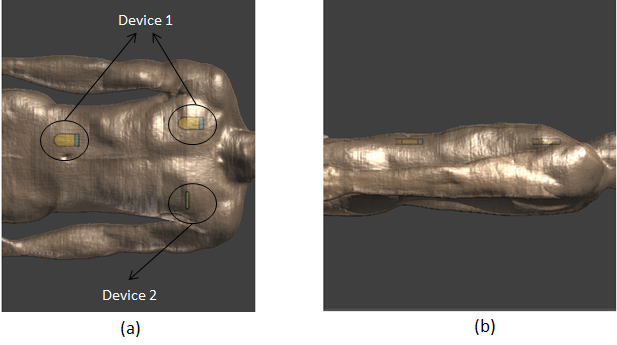
The input current for the gradient coil is fixed for all simulations. The Magneto Static (Biot-Savart) solver was applied for the calculation of gradient fields without implants while the Magneto Static (Vector Potential) solver was applied for the case with implants. The induced E-fields were calculated using the Magneto Quasi-Static solver. The head scan position with the head centered at isocenter is regarded as the 0 cm landmark (shown in Figure 1). The human model was moved from 0 cm to 120 cm landmark along the positive z direction with a 10 cm step size. Two types of implanted devices are modeled (shown in Figure 2). Device 1 consists of a dielectric header and a metal can, and device 2 is a narrow metal can. Two implant sites are considered for device 1, one in the right chest and one in the right abdomen under skin, while device 2 is placed in the left chest under skin. The simulated E-field is averaged along a 5 mm line as recommended in IEEE-C95.1-2005 standard5.
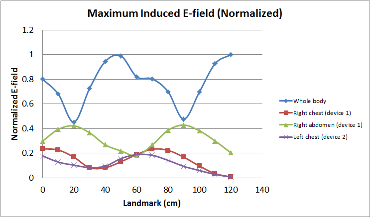
Among all the landmarks, the maximum local E-field enhancement ratio, defined as $$$\frac{E_{with\,implant}}{E_{no\,implant}}$$$, is found to be 1.5 in the right chest, 1.7 in the right abdomen and 2.1 in the left chest. The local maximum E-field near implants in Duke is compared to the whole body maximum induced E-field in Duke without implant. The maximum E-field is considered only in tissue regions where PNS nerves exist. Regions like bone, blood, heart, lung, and central nervous system (CNS) regions are excluded. In previous research6, PNS stimulations are reported in various locations throughout the whole body due to gradient induced E-field. In this study, PNS is assumed to be relevant to the maximum E-field in tissues where PNS nerves exist regardless of in which tissue the maximum E-field was found, i.e., PNS threshold is assumed to be the same over the whole body region.
Results
Figure 3 shows the comparison of whole body maximum induced E-field in Duke (without implant) vs. the local maximum E-field near the implanted devices in the right chest, the right abdomen and the left chest regions. The E-field values in the figure were normalized to the maximum value.Discussion
Although the E-field near implantable device is found to be enhanced compared to the case without implant, the result in Figure 3 indicates that for the investigated scenarios, the maximum E-field near implantable devices is still lower than the whole-body maximum E-field found in the human body without implant. In this study, PNS stimulation is assumed to be triggered by the maximum E-field in tissues where PNS nerves exist. Therefore, as long as the maximum E-field near implantable devices does not exceed the whole body maximum E-field without implant, the implantable device should not be considered as a risk factor of PNS stimulation during MR scan.Conclusion
Numerical simulations were performed for human body model, with and without implants, exposed to gradient fields in different landmarks. For the investigated scenarios, the simulation results reveal that the maximum E-field near implants does not exceed the whole body maximum induced E-field in a human body without implant. Hence, no evidence was found in this study to suggest that a metallic implantable device may increase the risk of PNS during MR scan.Acknowledgements
No acknowledgement found.References
1. X. Chen, J. Edmonson, and M. Steckner, The Potential for Eddy Current Induced Peripheral Nerve Stimulation from an Active Implant Device Canister. ISMRM 24th Annual Meeting and Exhibition
2. https://www.zurichmedtech.com/sim4life/
3. Christ A, et al, The Virtual Family—development of surface-based anatomical models of two adults and two children for dosimetric simulations. Phys Med Biol 55(2): N23-38 (2010)
4. https://www.itis.ethz.ch/virtual-population/tissue-properties/database/low-frequency-conductivity/
5. IEEE Std C95.1-2005: IEEE Standard for Safety Levels with Respect to Human Exposure to Radio Frequency Electromagnetic Fields, 3 kHz to 300 GHz; 2006
6. Liu F, et al, On the Induced Electric Field Gradients in the Human Body for Magnetic Stimulation by Gradient Coils in MRI, IEEE Transactions on Biomedical Engineering, Vol. 50, No. 7, July 2003
Figures