1448
Utility of ZTE for the Characterization of Acute Ankle Fractures1Hospital for Special Surgery, New York, NY, United States, 2Department of Radiology and Imaging - MRI, Hospital for Special Surgery, New York, NY, United States
Synopsis
ZTE MRI provides CT-like tissue contrast, facilitating evaluation of mineralized bone. The utility of ZTE for evaluation of acute ankle fractures was evaluated in a series of 14 patients who underwent preoperative clinical MRI with an additional ZTE sequence, and subsequently underwent surgical fracture fixation. Fractures were characterized in a blinded fashion utilizing ZTE and CT, with subsequent operative confirmation. ZTE provided accurate characterization of fractures relative to both CT and surgery, with excellent inter- and intra-observer reliability.
INTRODUCTION
Magnetic resonance imaging (MRI) provides excellent sensitivity for marrow signal changes in the setting of acute fracture; however, detailed evaluation of mineralized bone is limited on conventional pulse sequences due to limited signal and short T2 values. Because of this lack of signal, CT has traditionally been considered the cross sectional modality of choice for detailed evaluation of fracture morphology. Zero echo time (ZTE) MRI provides CT-like tissue contrast, allowing for improved visualization of mineralized bone relative to conventional MRI sequences. The sequence is not yet in widespread clinical use, but has shown promising results in the shoulder, spine, and cranium1,2,3. The purpose of this study was to evaluate to utility of ZTE for the detection of acute ankle fractures and characterization of fracture morphology, utilizing CT as a gold standard, with operative confirmation.METHODS
In a retrospective series of 14 acute ankle trauma cases, both CT and MRI with ZTE were performed, followed by surgical fracture reduction. Fractures of the posterior, medial, and lateral malleoli detected on ZTE MRI were correlated with the operative report as the reference standard. Raw agreement (%) and correlation (Cohen's κ) were calculated. In addition, eight selected fracture fragments were measured in two dimensions (anterior-posterior and superior-inferior) on corresponding sagittal ZTE and CT images by three independent radiologists. Inter-rater and intra-rater reliability for measurements made using this sequence were calculated (intraclass correlation coefficient, ICC, with 95% confidence intervals).RESULTS
There was substantial to almost perfect agreement between fractures diagnosed on MRI with ZTE and those confirmed at surgery (lateral malleolus, 100% agreement, κ=1; posterior malleolus, 92.9% agreement, κ=0.84, 95%CI: 0.53 to 1.00; medial malleolus, 92.9% agreement, κ=0.85, 95%CI: 0.57 to 1.00). Fragment characterization was accurate and repeatable. Fragment measurements made on ZTE and CT demonstrated excellent agreement between modalities, with ICC between 0.995-0.999 (95% CI 0.985 to 1.000) for each of the 3 readers. Interobserver agreement for fragment measurements made on the ZTE sequence was excellent (ICC 0.995, 95% CI 0.986-0.998).DISCUSSION
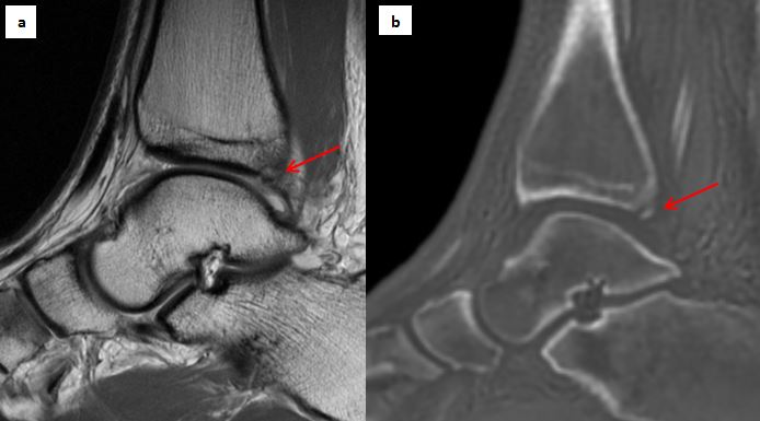
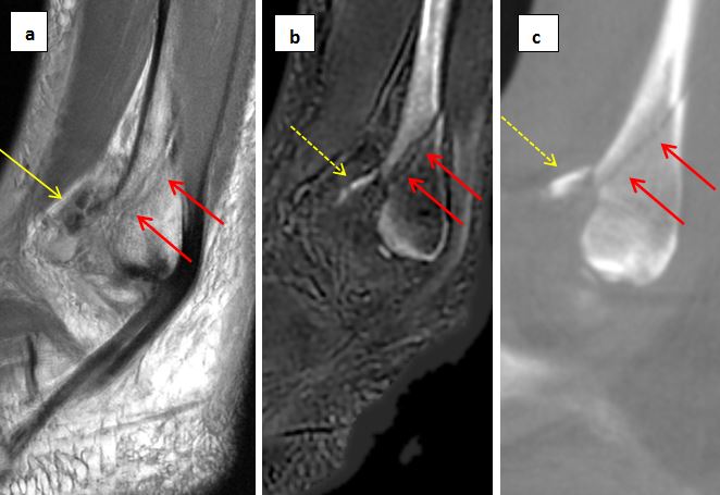
ZTE permits direct visualization of mineralized bone as compared to conventional MRI sequences, allowing accurate characterization of fracture morphology, with improved conspicuity of subtle fracture fragments (Figure 1). Fracture measurements made on ZTE proved repeatable across observers, and correlated well with those made on CT. As conventional MR pulse sequences provide exceptional evaluation of soft tissue and marrow pathology, the addition of the ZTE pulse sequence to conventional imaging algorithms enables complete evaluation of both osseous and soft tissue pathology within a single scanning session, largely obviating the need for CT in many cases, and thereby avoiding additional expense and ionizing radiation, while improving diagnostic efficiency and value.CONCLUSION
The ZTE sequence provided accurate and repeatable characterization of fracture morphology comparable to that of CT in this limited cohort. The addition of this sequence to routine clinical MR protocols allows comprehensive evaluation of both osseous and soft tissue pathology in the setting of acute fracture, potentially obviating the need for an additional CT in many cases.Acknowledgements
HSS has an institutional research agreement in place with GE HealthcareReferences
1. Weiger M, Brunner DO, Dietrich BE, Pruessmann KP. ZTE imaging in humans. Magn Reon Med 2013, 70(2):328-32.
2. Argentieri EC, Koff MF, Breighner RE, Endo Y, Shah PH, Sneag DB. Diagnostic Accuracy of Zero Echo Time MRI for the Evaluation of Cervical Neural Foraminal Stenosis. Spine 2017, epub (PMID: 29095415)
3. Breighner, R., Endo, Y., Konin, G.P., et al. Zero Echo Time Imaging of the Shoulder: Enhanced Detail Using MRI. Radiology (in press).
Figures