1447
Preliminary study of T1rho imaging technique in assessment of early intervertebral disc degeneration in asymptomatic pilots at 3.0T magnetic resonance1Radiology Department, The First Affiliated Hospital of Yangtze University, JingZhou, China, 2Department of CT&MRI, Air Force General Hospital, Beijing, China, 3GE Healthcare, China, Beijing, China
Synopsis
T1rho MRI in the lumbar spine may provide a tool for the diagnosis of early degenerative changes in the disc. In this study, the mean T1rho value of pilots was significantly lower than that of the control group. The degenerative grades of pilots mainly were grade III and IV, but control group were grade I and II. There were significant differences in T1rho values at each age group between pilots and control group. And overload on spine column of pilots may be the important reason in degeneration and accelerate the degeneration process.
Purpose
To evaluate early intervertebral disc degeneration quantified by T1rho and T2-weighted magnetic resonance imaging at 3.0T in asymptomatic pilots and explore the relationship between intervertebral disc degeneration and overload.Introduction
Degenerative disc degeneration is the main cause of low back pain, and also the occupational diseases of pilots. The pathophysiologic mechanism between disc degeneration and overload is not well understood. Studies had shown that T1rho MRI in the lumbar spine may provide a tool for the diagnosis of early degenerative changes in the disc [1-3]. In this study, the mean T1rho value of pilots was significantly lower than that of the control group. The degenerative grades of pilots mainly were grade Ⅲ and Ⅳ, but control group were grade Ⅰ and Ⅱ. There were significant differences in T1rho values at each age group between pilots and control group, but except for <30 years group. we concluded that disc degeneration occurred on pilots was more seriously and the process of degeneration was more quickly. And overload on spine column of pilots may be the important reason in degeneration and accelerate the degeneration process.Method
69 Health volunteers were divided in two groups. pilots group: 34 asymptomatic male pilots in age from 25 to 51(mean 33.47 ± 7.29 years) without history of stopping flight or stopping training. The second, control group: 35 asymptomatic volunteers in age from 24 to 48(mean 31.74 ± 6.99 years). Both sagittal conventional T2- and T1rho-weigted images were acquired at every examination of lumbar disc. T2-weighted imaging were acquired using a fast spin-echo sequence with the following parameters: TR=2556ms, TE=121.3ms, FOV=32×32cm, slice-thickness=4mm, acquisition-matrix=352×320. T1rho-weighted images used 3D spoiled gradient-recalled sequence with parameters as follows: TSL1/TSL2/TSL3/TSL4(time of spin lock) =0/20/40/80ms, Spin Lock Frequency=300Hz, TR=4.2ms, TE=min-full, FOV=260×260mm, NEX=1.0, slice-thickness=5mm, acquisition-matrix=160×160, bandwidth=62.5kHz, PE direction=L/R. The sagittal T1rho maps were reformatted for each disk. Mean T1rho values of the central disk space nucleus pulposus were determined by using post-processing software namely as T2mapping with the Region of Interests that were approximately 49-51mm². The degenerative grade of each lumbar disc was assessed from T2-weighted images according to Pfirrmann degenerative grade. All 69 volunteers were divided into three groups according to age, as <30 years old, 30~40 years old, and≥40 years old groups, respectively.Results
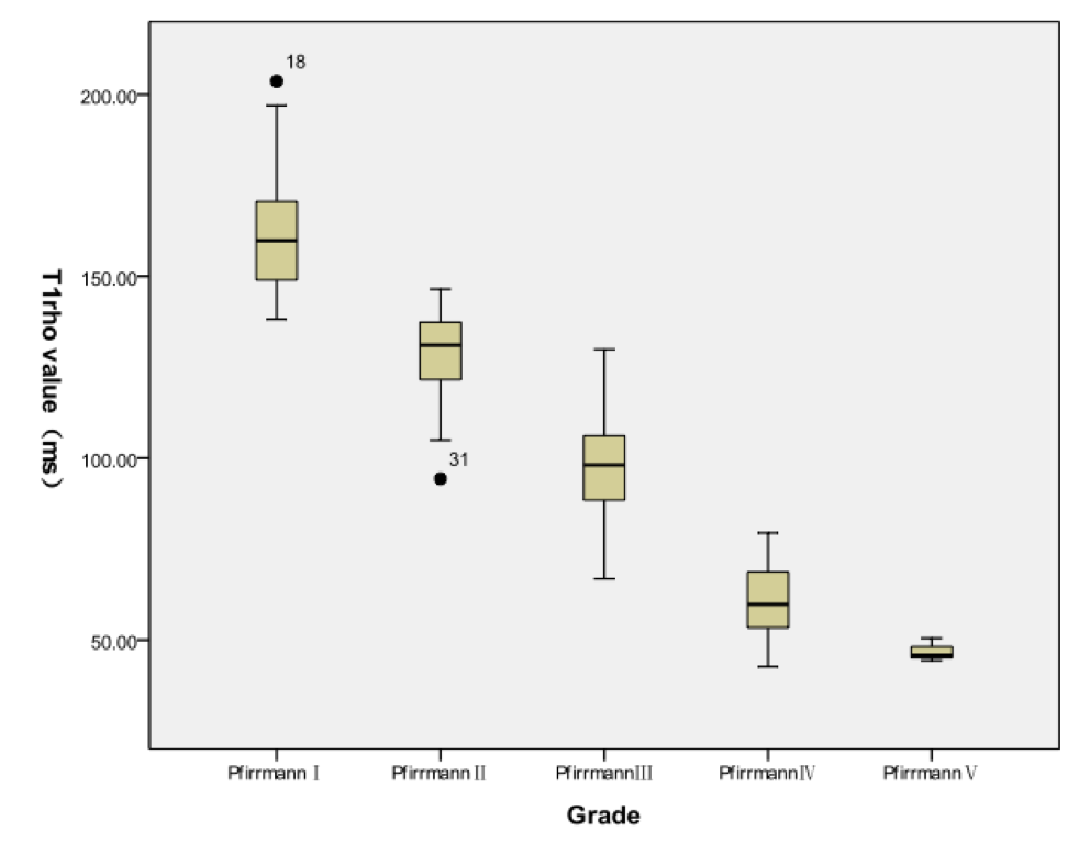
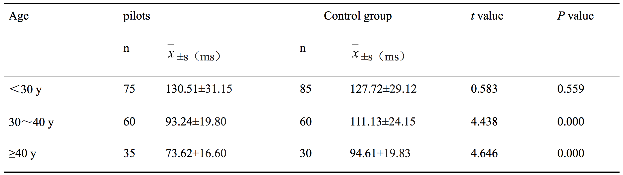
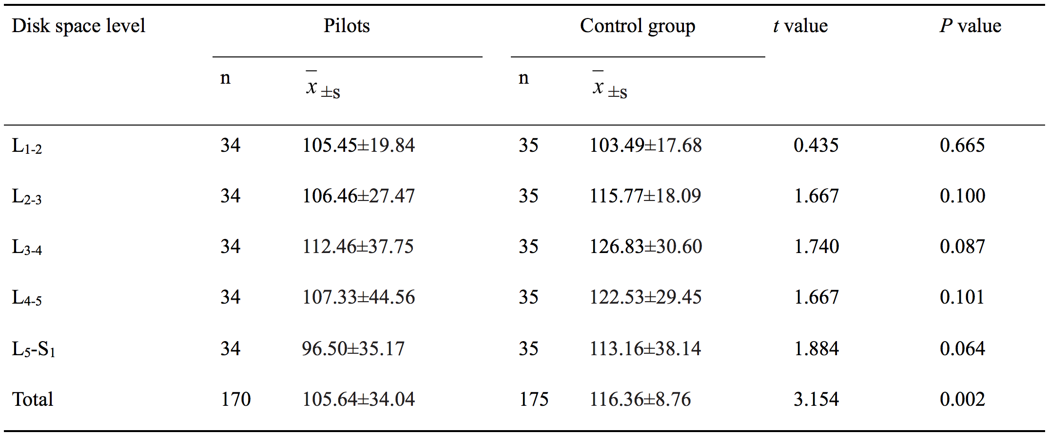
T1rho values significantly negatively correlated with Pfirrmann grade(r=-0.901、-0.921,P<0.01)(Figure.1), The mean T1rho value of pilots was significantly lower than that of the control group (t=3.154,P<0.01).The degenerative grades of pilots mainly were grade Ⅲ and Ⅳ, but control group were grade Ⅰ and Ⅱ(Z=4.04,P<0.01). There were significant differences in T1rho values at each age group between pilots and control group (t=4.438、4.646,P<0.01), but except for <30 years group(P>0.05)(Table.1). Evaluation between pilots and control group showed no difference in T1rho values at each Pfirrmann grade(P>0.05)(Table.2).Discussion and conclusion
Disc degeneration occurred on pilots was more seriously and the process of degeneration was more quickly. Overload on spine column of pilots may be the important reason in degeneration and accelerate the degeneration process. T1rho can be potentially used as a valid clinical tool in quantitative diagnosis of early intervertebral disc degeneration in asymptomatic pilots.Acknowledgements
No acknowledgement found.References
[1]Filippi CG, Duncan CT, Watts R, et al. In vivo quantification of t1ρ in lumbar spine disk spaces at 3 T using parallel transmission MRI. Am J Roentgenol. 2013; 201(1):W110-116.
[2]Zobel BB, Vadalà G, Del Vescovo R, et al. T1ρ magnetic resonance imaging quantification of early lumbar intervertebral disc degeneration in healthy young adults. Spine. 2012; 37(14): 1224-1230.
[3]Blumenkrantz G, Li X, Han ET, et al. A feasibility study of in vivo T 1ρ imaging of the intervertebral disc. Magnetic Resonance Imaging. 2006;24(8): 1001-1007.
Figures