1446
Proton Density Zero Echo Time(ZTE) Imaging for Evaluating the Bone Involvement in the Femoral TumorXin Lou1, Jinfeng Li1, Lin Xu1, Xigang Zhao2, Jianxun Qu2, and Lin Ma1
1Radiology and Imaging, China Army General Hospital, Beijing, China, 2General Electric Healthcare, Beijing, China
Synopsis
MRI can display the compositions of different tissues and adjacent involvements. In the patients of bone tumors, the integrity of cortical bone needed to be assessed for the preoperative planning. This study used proton density ZTE to display the bone involvement in patients of femoral tumors. Substantial agreement was found between CT and ZTE (r=0.98-0.99) and there was not statics significance between the measured diameters from CT and ZTE MRI (p=0.34-0.99). further development of ZTE may obviate the need of CT in evaluating the bone involvement of femoral tumors.
Purpose
When preoperatively assessing the bone involvement in femoral tumor, accurate osseous imaging is necessary for clinical management1. Previously CT was used for the display the cortical bone in bone tumor. Routine MRI fails to direct visualization of Bone because of short tissues relaxation time and low proton density2, 3. This study investigated the possibility of proton density ZTE for the display of the bone involvements in the patients with femoral tumors.Methods
10 patients(7M,3F) with femoral tumors, aged 20.5±4.7(Mean±SD) were enrolled in this study. The local bone involvements in the femora were found in CT scanning. MRI examination were performed in following three days. The Proton Density ZTE pulse sequence was acquired with the following parameters: TR 300-600ms, TE 0.0ms, FA 1 degree, Receiver bandwidth 31.2-62.5kHz, NEX 2, Voxel size 1.2 mm3, FOV 20*20cm. The original images including the CT and ZTE were transfer to the Advanced Workstation 4.6 and were reformatted as axial, coronal and sagittal plane with 2mm slice thickness and 2mm slice space. In the maximum place for the display of bone involvement, the horizontal and vertical diameters of the bone involvement were measured in axial, coronal, and sagittal plane respectively. The reformatted images were displayed with inverted window level. The paired-samples t-test statics were performed for the agreements between the CT and ZTE. Significance was set at p<0.05.Results
In all ten patients with femoral tumors, proton density ZTE imaging clearly displayed the bone involvement showed in figure 1. Perfect agreements of the diameters were found between the CT and ZTE methods (the correlation coefficient was 0.98, 0.98,0.99, 0.99, 0.99, 0.99 respectively). there were not significance difference in the diameters between the two methods (p = 0.98., 0.34, 0.79, 0.49, 0.91, 0.99, respectively).Discussion
The capability of evaluate the bone involvement was necessary for radiological assessment of femoral tumors. Many patients need CT and MRI for preoperative planning4. The results of this study revealed that proton density ZTE imaging could display the femoral cortical bone clearly and acquired nearly same diameters of the bone involvement with CT. The primary limitation of this study was that the measurements required the radiologist to locate anatomy in multi-planar reformatted (MPR) images, potentially introducing additional error in determining the best plane in which to obtain the measure.Conclusion
Proton density ZTE pulse sequence can non-radiatively display the bone involvement in femoral tumor patients. With the continued MRI improvements, proton density ZTE may soon obviate the need the femoral CT and its associated ionizing radiation and additional cost.Acknowledgements
We had an institutional research agreement in place with GE Healthcare.References
1. Delso G, Wiesinger F, Sacolick LI, et al. Clinical evaluation of zero-echo-time MR imaging for the segmentation of the skull. J Nucl Med 2015; 56:417-422. 2. Wiesinger F, Sacolick LI, Menini A, et al. Zero TE MR bone imaging in the head. Magn Reson Med 2015; 75:107-114. 3. Johnson EM, Vyas U, Ghanouni P, Pauly KB, Pauly JM. Improved cortical bone specificity in UTE MR Imaging. Magn Reson Med 2016. 4. Du J, Carl M, Bydder M, Takahashi A, Chung CB, Bydder GM. Qualitative and quantitative ultrashort echo time (UTE) imaging of cortical bone. J Magn Reson 2010;207(2):304-311.Figures
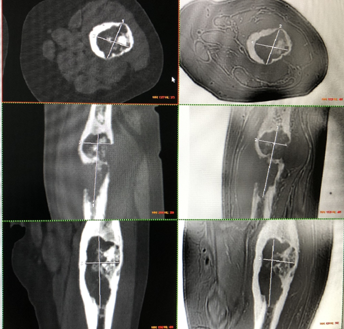
Figure.1 Femoral bone involvement measures
in axial, coronal and sagittal plane, CT (left) and ZTE MRI (right) for the
same patient.