1444
Study of mono-exponential and intravoxel incoherent motion models in differentiation of metastasis from myeloma1Peking University 3rd Hospital, Beijing, China
Synopsis
This study aimed to evaluate the diagnostic performance of diffusion weighted imaging (DWI) to differentiate metastasis from myeloma using the apparent diffusion coefficient (ADC) and parameters derived from the intravoxel incoherent motion (IVIM) theory. 40 patients with metastasis and 12 with myeloma underwent diffusion-weighted magnetic resonance (MR) imaging and dynamic contrast enhanced MRI (DCE-MRI). ADC, diffusion coefficient(D), pseudodiffusion coefficient(D*), and perfusion fraction (f) were calculated.Through our study it is feasible to d ifferentiate metastasis from myeloma by mono-exponential and IVIM models . IVIM-derived D and D* values showed significantly better diagnostic performance than ADC values in differentiating metastasis from myeloma.
INTRODUCTION
At present, metastasis and myeloma are the most common malignant tumors in spine and the imaging findings are similar. It is difficult to distinguish them with the routine imaging methods.However,The treatments of metastasis and myeloma are different, so correct diagnosis is very necessary.
This study aimed to differentiate metastasis from myeloma through the evaluation of the diagnostic performance of diffusion weighted imaging (DWI) by using the apparent diffusion coefficient (ADC) and parameters derived from the intravoxel incoherent motion (IVIM) theory.
METHODS
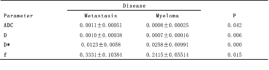
40 patients with metastasis and 12 with myeloma underwent diffusion-weighted magnetic resonance (MR) imaging with 10b(0, 20, 50, 100, 150, 200, 400, 800, 1200, 1500s/mm2)and dynamic contrast enhanced MRI (DCE-MRI). ADC, diffusion coefficient(D), pseudodiffusion coefficient(D*), and perfusion fraction (f) were calculated. Two diseases were compared using independent sample t test or Mann-Whitney U test. Receiver operating characteristic (ROC) analysis of discrimination between metastasis and myeloma.REAULTS
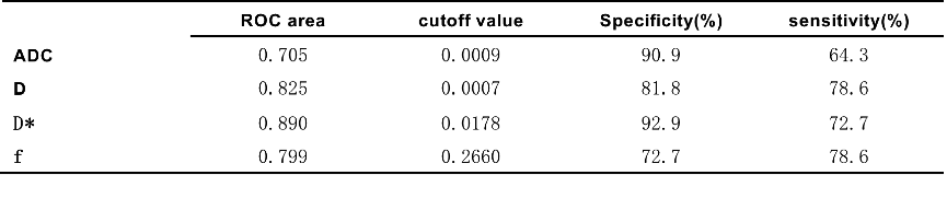
For ADC ,D, and f were significantly lower in myeloma than in metastasis (P<0.05). Parameter of D* was significantly lower in metastasis than in myeloma (P<0.05).However, ROC analysis demonstrated a higher area under the ROC curve value for D than for ADC for differentiating metastasis from myeloma (0.825 vs 0.705; P <0.05).The ROC curve value of D* and f in differentiating the two were 0.890,0.799.DISCUSSION
ADC and D represent diffusion in tissues1. ADC and D were lower in myeloma than in metastasis, which indicated the diffusion effect of myeloma was more significant than in metastasis by diffusion weighted imaging (DWI) of magnetic resonance imaging (MRI).
D* and f react capillary perfusion2.
CONCLUSION
It is feasible to s differentiate metastasis from myeloma by mono-exponential and IVIM models . IVIM-derived D and D* values showed significantly better diagnostic performance than ADC values in differentiating metastasis from myeloma.Acknowledgements
This study is supported in part by the National Natural Science Foundation of China (81701648)and the Beijing Natural Science Foundation (7164309)References
1.Sungmin Woo,Jeong Min Lee,et al.Intravoxel Incoherent MotionDiffusion-weighted MR Imaging of Hepatocellular Carcinoma:Correlation with Enhancement Degreeand Histologic Grade.Radiology.2014;270(3):758-767.
2.A.J. Marchand a, E. Hitti,et al.MRI quantification of diffusion and perfusion in bone marrow byintravoxel incoherent motion (IVIM) and non-negative least square(NNLS) analysis.Magnetic Resonance Imaging.2014;32:1091-1096.
Figures
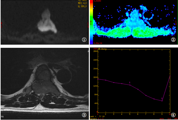
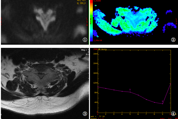
M/51,myeloma.1.b=800s/mm2,DWI imaging 2.pseudo-color map 3.T2-weighted imaging 4.time-signal intensity curve