1437
Automated Seed Points Selection Based Radial-Search Segmentation Method For Sagittal and Coronal View Knee MRI Imaging1Centre of Biomedical Engineering, Indian Institute of Technology Delhi, New Delhi, India, 2BME, ASET, Amity University Haryana, Gurgaon, India, 3Mahajan Imaging Centre, New Delhi, India
Synopsis
Knee disorders are generally marked in tibio-femoral bone junction. Most of available segmentation techniques use time consuming semi-automatic approach as radial search method, in sagittal view only. However, coronal view MRI Knee images are clinically equal important. Proposed approach automates seed points selection process for the radial search method, which work equally good on both sagittal and coronal view for identification of tibio-femoral junction.
Introduction
Segmentation of tibio-femoral bone junction, which includes cartilage, meniscus, ligaments and synovial fluid, is essential for evaluation of knee disorders such as osteoarthritis. Manual segmentation of these image is time consuming. Several segmentation approaches have been proposed where semi-automatic interactive segmentation technique based on edge-detection is one of the widely used because of its high accuracy. These algorithms are designed and work best in sagittal view1,2,3, whereas coronal view is equally important for clinical decision making4.
This semi-automatic radial-search1 method require a selection of single seed point by an expert to initiate the process. This methodology is highly inefficient in segmentation for coronal view in the posterior region of knee where two condyles appear separate. A simple fully automated seed points selection followed by radial search method is implement in this research that works equally good in sagittal and coronal view for segmentation of knee joint.
Methods and Material
In this study, knee MRI data from 4 healthy subjects and 1 patient with meniscal tear were retrospectively analysed. Data-set for four healthy subjects was acquired using Coronal-3D FSPGR (512x512) and for one patient was acquired using Sagittal-3D PD-SPACE (320x320).
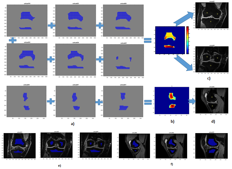
In this study radial-search1 method is used for identification of boundary of bone, which require manual selection of seed point. For segmentation, this approach require two seed-points in sagittal view (one each for tibial and femoral region(fig.1d)), two seed-points for coronal view anterior slices(one each for tibial and femoral region(fig.1c)) and three seed-points for coronal view posterior slices(one each for lateral condyle, medial condyle and tibial region(fig.1c)). We implemented automated seed points selection methodology for radial-search1 for both sagittal and coronal view. These seed points serve as global seed points for all slices of the 3D stack. The methodology works as elaborated below:
Step-1: Images were enhanced using contrast stretching to minimise the intensity of bone area. Images were under sampled by factor of 1/8 for fast computation.
Step-2: Circular kernel (radius 6 voxels) was used, replacing the intensity at each voxel by the sum of values in the kernel space.
Step-3: Voxels with lowest intensities (bottom 1 percentile) were masked (Fig.1a). (marking the voxels belonging to bone region(Fig.1e & 1f).)
Step-4: The mask from step 3 were summed across the z-axis (Fig.1b) followed by a Otsu threshold to have only voxel having bone with highest probability
Step-5: The mask output of step 4 was used to calculate the centroid of object in mask. Specifically, in coronal view segmentation of femur, for anterior images only one centroid was estimated; for posterior images two centroids were estimated for each condyle (fig.1c&d). In sagittal view one centroid would be estimated in femur as both condyles would appear in continuity in the image. For both coronal and sagittal view tibia would require only one centroid.
Radial-search method with manual seed point selection was used for comparison with the proposed methodology. Manual segmentation was performed by experienced Radiologist. Dice Similarity Coefficient (DC) were calculated for manual seed selection and automatic seed selection Radial-search methods against the manual segmented mask.
Results
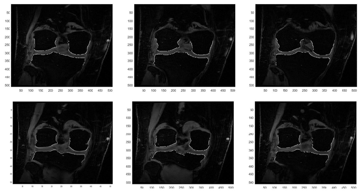
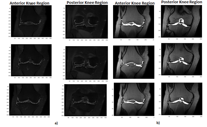
Automatic seed points estimated with the proposed method was found to confine in the center of bone regions in all slices (fig.1c&1d). Table 1 shows the DC for manual and automated seed selection based radial search method. The automated method was found to have high accuracy in both anterior and posterior region of knee joint (DC=~77%) compared in manual method (DC=57-64%). Figure 2 shows identification of bone boundary using automatic seed points for posterior region of knee in coronal view, where two condyles are disconnected. Figure 3 shows the comparison manual and automatic seed selection methods in the posterior region of knee in coronal view, where manual method was found to inefficient in find the bone boundary at intercondylar notch of femur.Discussion
Automated seed point selection method showed a considerable improvement in identification of tibio-femoral bone junction (Table1, Fig.3). The method is shown to be working with high accuracy in both sagittal and coronal. Especially in posterior region in coronal view having disconnected two condyles requiring two manual seed points; however the proposed method automatically identify the seed point if there are two centroid of an object. The automated seed estimated with FSPGR or SPACE sequence can be transformed to other MR sequence images such as PD, T2 (Fig.4) which can help to further segment bone junction in these images.Conclusion
Automated seed point selection based radial search method has been developed for coronal view MR knee imaging, improving the accuracy and time efficiency.Acknowledgements
Authors acknowledge the support of staff at Mahajan Imaging Centre, New Delhi in MRI data acquisition. We would like to thank Dr. Harsh Mahajan and Dr. Vidur Mahajan for their support and insight in the project. The project was funded by IRD, IIT Delhi (project number MI01422).References
- S. Akhtar M.B.B.Ch., et al., An MRI derived articular cartilage visualization framework, Osteoarthritis and Cartilage Vol.-15, 1070-1085, 2007
- Pin Wang, et al., Journal of Medical Imaging and Health Informatic, Vol.-6, 1-9, 2016
- Julio Carballido-Gamio, et. al., Medical image analysis, 12(2):120-135, 2008.
- Braun and Gold, Diagonsis of Osteoarthritis: Imaging, Bone,2012, 51(2), 278-288
Figures