1436
Significant Metabolic Differences Between Benign Lipomatous Lesion and Liposarcoma Identified by High-Resolution 1H and 31P MRS: A Pilot StudySantosh Kumar Bharti1, Brett Shannon2, Adam Levin2, Carol D Morris2, Laura Fayad3, and Zaver M Bhujwalla1,4
1Division of Cancer Imaging Research, Department of Radiology, Johns Hopkins University, School of Medicine, Baltimore, MD, United States, 2Department of Orthopaedic Surgery, Johns Hopkins University, School of Medicine, Baltimore, MD, United States, 3Musculoskeletal Radiology, Department of Radiology, Johns Hopkins University, School of Medicine, Baltimore, MD, United States, 4Department of Oncology, Johns Hopkins University, School of Medicine, Baltimore, MD, United States
Synopsis
Adipocytic tumors present a spectrum of neoplastic disease including benign lipomas and their variants, atypical lipomatous tumors, and malignant liposarcomas. Distinguishing areas of malignant dedifferentiation from benign and atypical lipomatous tumors is a diagnostic challenge due to overlapping magnetic resonance imaging characteristics, and pre-operative diagnostic accuracy is poor. Here we have identified dramatic differences in the metabolic profile of water-soluble and lipid extracts of adipocytic tumors, suggesting that magnetic resonsance spectroscopy may have the potential to improve diagnostic accuracy. Our data may also lead to potential metabolic targets for treatment.
Introduction
Adipocytic tumors present a spectrum of neoplastic disease including benign lipomas, atypical lipomatous tumors (ALTs), and malignant liposarcomas. Liposarcomas are the most common soft tissue sarcoma, accounting for approximately 20% of all adult soft tissue sarcomas [1]. Some liposarcomas are suspected to arise through dedifferentiation of ALTs, a process that is not well understood. Pleomorphic liposarcomas are high-grade, aggressive tumors with high metastatic potential and overall 5-year survival rate of 20-30% [2]. Distinguishing high-grade liposarcomas from benign and atypical lipomatous tumors can be a diagnostic challenge with implications for surgical and clinical management. The rate of misdiagnosis is approximately 30–40% following radiological detection [3], and 7–17% on histological evaluation [4]. A major objective of the present work is to develop metabolic determinants for adipocytic tumor classification and to identify metabolic targets. Our data demonstrate that high-resolution MRS may be used as an additional method for classification and diagnosis of liposarcomas and benign lipomatous tumors.Methods
De-identified human surgical samples were collected from the Department of Orthopaedic Surgery, The Johns Hopkins University School of Medicine Baltimore, MD. One tissue specimen from a benign pleomorphic lipoma and two tissue specimen from a pleomorphic liposarcoma were examined using 1H and 31P MR spectroscopy. Tissue samples were snap frozen and stored at -80°C until 1H MRS analysis. Dual phase solvent extraction was performed on about 400 mg of tumor tissue. The aqueous phase was separated, freeze dried, reconstituted in 600ul D2O PBS for NMR analysis. Organic phase were dried under nitrogen stream and reconstituted in 600ul of deuterated chloroform and methanol (2:1 ration). All MR spectra were acquired on an Avance III 750 MHz (17.6T) Bruker NMR spectrometer equipped with a 5 mm broad band inverse (BBI) probe. Metabolites were quantified using TSP and TMS as internal standard in aqueous and lipid phases respectively. Spectral acquisition, processing and quantification were performed using TOPSPIN 2.1 software.Results and Discussion
Representative 1H and 31P MR spectra of the aqueous phase of tissue extracts obtained from a pleomorphic lipoma and liposarcoma are shown in Figure 1 and Figure 3. Metabolites were quantified and changes were reported in term of percentage considering metabolites level in pleomorphic lipoma as 100% (Figure 2). Depletion in branch chain amino acids (-10%), tyrosine (-15%), phenylalanine (-25%), lactate (-45%), pyruvate (-35-50%), acetate (-5-25%) and elevation in alanine (40%), succinate (35-60%), glycine (400-500%), histidine (15-25%), glucose (100%), glutamine (50%), glutamate (75%), and myo-inositol (300%) in pleomorphic liposarcoma were observed. Variations in the metabolic levels between two specimens obtained from the same tumor indicate metabolic heterogeneity. 31P MRS of the aqueous phase revealed high levels of phosphocholine in the pleomorphic liposarcoma as compared to the lipoma (not detected). 1H and 31P MRS (representative spectra in Figure 4 and Figure 5) analysis of the lipid phase, summarized in Figure 4, identified depletion in lipid (-65%, CH3 peak at 0.9 ppm was quantified) and an increase in phosphatidylethanolamine (75%) and phosphatidylcholine (30%). Visible variations in TAG glycerol backbone (4.2-4.3ppm) and -CH=CH- peaks from polyunsaturated fatty acids at 2.8ppm and 5.3ppm were also observed. We are currently analyzing additional samples from pleomorphic lipoma and liposarcoma as well as other types of sarcomas to expand this study. Our preliminary data support investigating the use of 1H and 31P MRS of liposarcomas for differentiation between subtypes.Acknowledgements
This work was supported by NIH R01 CA193365 and NIH P30CA06973References
- Dei Tos AP: Liposarcomas: diagnostic pitfalls and new insights. Histopathology 2014, 64(1):38-52.
- Downes KA, Goldblum JR et al: Pleomorphic Liposarcoma: A Clinicopathologic Analysis Of 19 Cases. Modern pathology : an official journal of the United States and Canadian Academy of Pathology, Inc 0000, 14(3):179-184.
- Brisson M, Kashima T et al: MRI characteristics of lipoma and atypical lipomatous tumor/well-differentiated liposarcoma: retrospective comparison with histology and MDM2 gene amplification. Skeletal Radiology 2013, 42(5):635-647.
- Hasegawa T, Yamamoto S et al: Validity and reproducibility of histologic diagnosis and grading for adult soft-tissue sarcomas. Human Pathology 2002, 33(1):111-115.
Figures
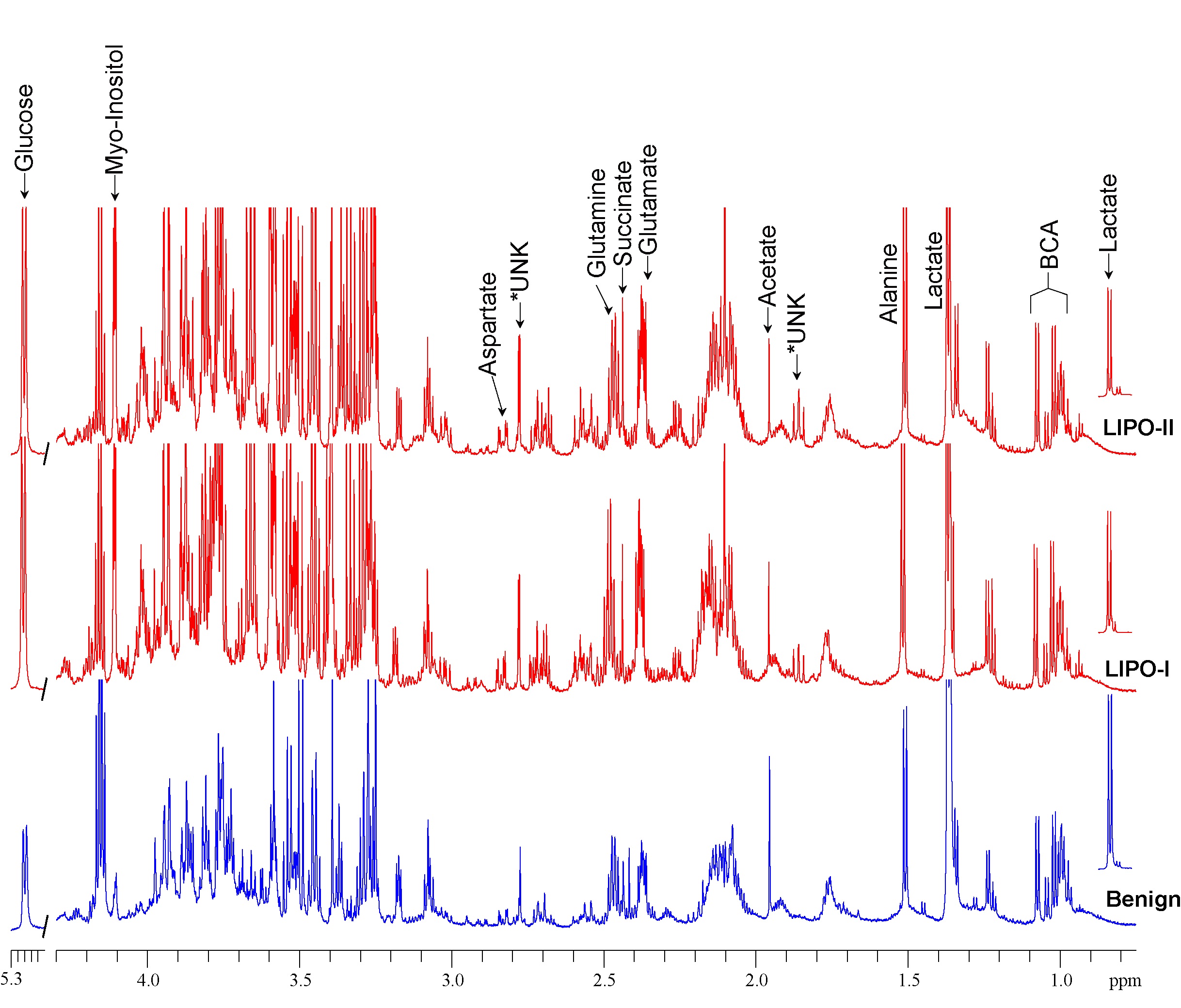
Figure 1: Representative 1H
MR spectra of the aqueous phase of
tissue extracts obtained from pleomorphic lipoma (benign, Blue, lower)
and pleomorphic liposarcoma (LIPO-I, II, Red, upper). To determine the
heterogeneity in the metabolic profile, two different tissue specimens (LIPO-I
& LIPO-II) were analyzed from a malignant liposarcoma. Substantial
metabolic differences were identified between the benign lipoma and the
liposarcoma. These changes include glucose, lactate, glutamine, glutamate,
myo-inositol etc. Some un-identified resonances (1.88ppm, 2.78ppm) were detected
in the liposarcoma that were different compared to benign tissue.
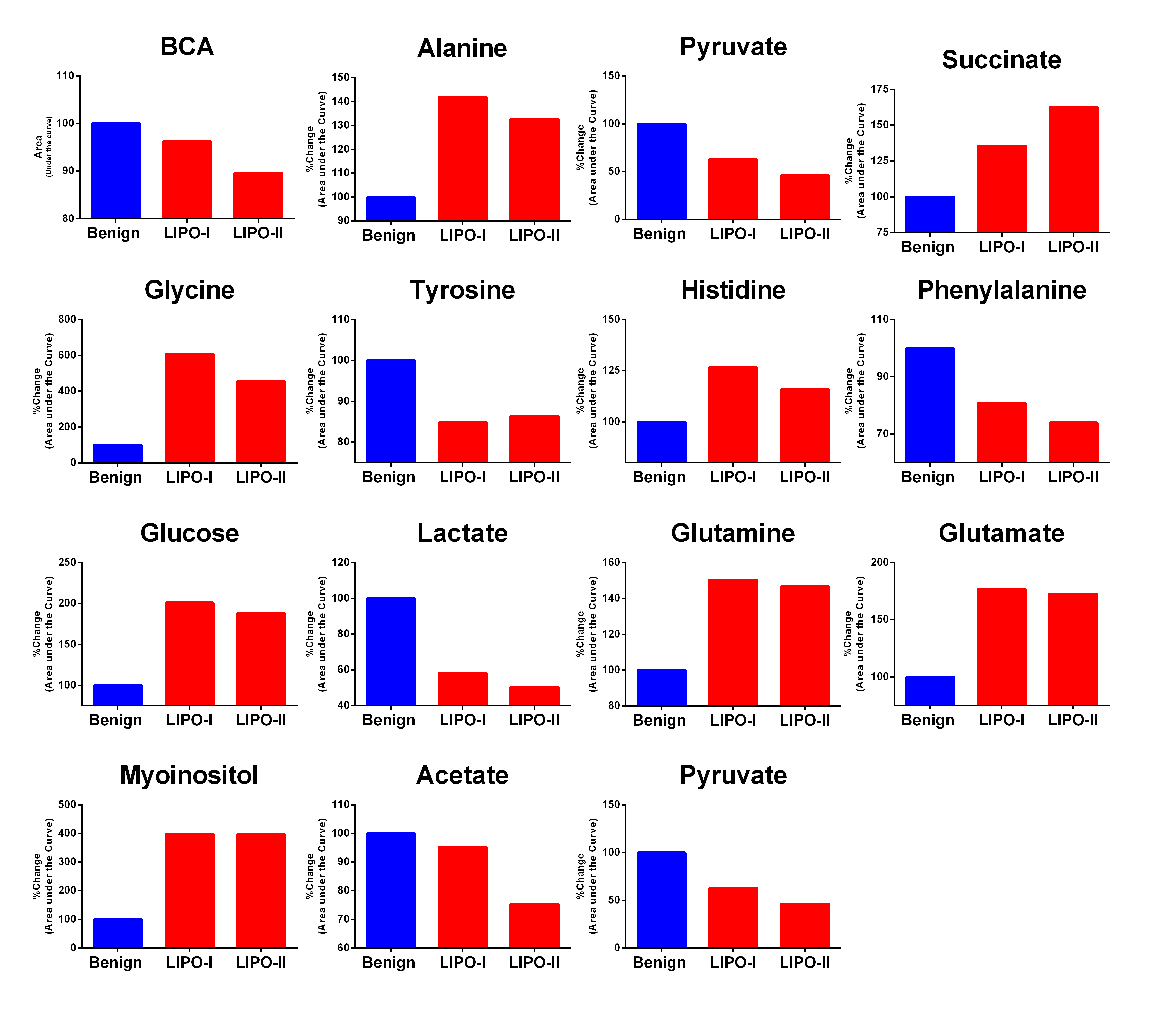
Figure 2:
Quantitative estimation of metabolites in pleomorphic lipoma (benign) and pleomorphic
liposarcoma (malignant) tissue. Spectral area was calculated with respect to
reference and normalized to the tissue weight used for dual phase extraction.
Percentage change as compared to benign (100%) was reported.
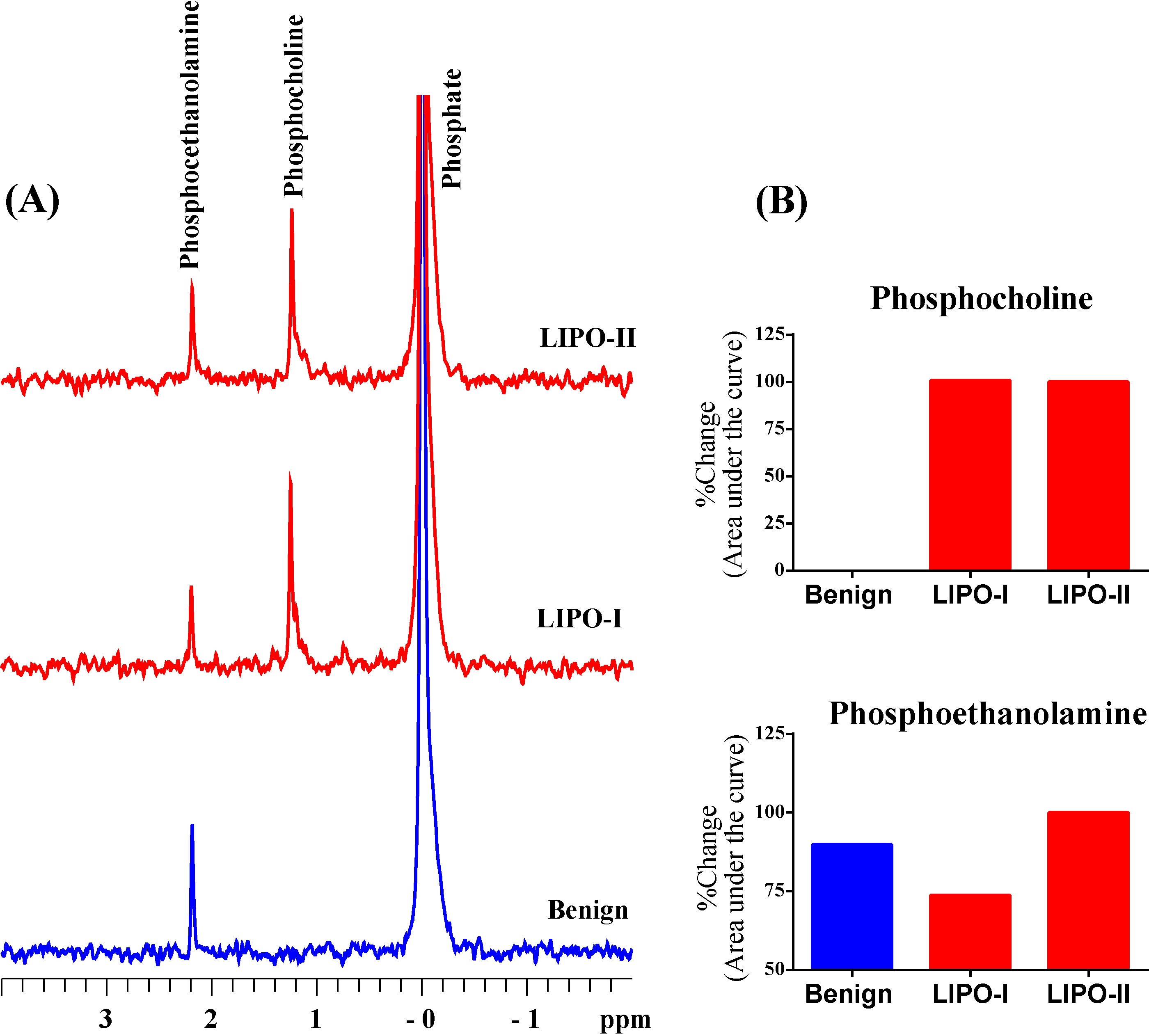
Figure 3: (A) Representative 31P
MR spectra of the aqueous phase of tissue extracts obtained from pleomorphic
lipoma (benign, Blue, lower) and pleomorphic liposarcoma (malignant, Red, upper, LIPO-I, LIPO-II). All
the spectra are plotted with the same vertical scale and were acquired with
identical experimental parameters. (B) Bar plot showing changes in
phosphocholine and phosphoethanolamine. Phosphocholine was not detected in the 31P
MR spectrum of benign tissue.
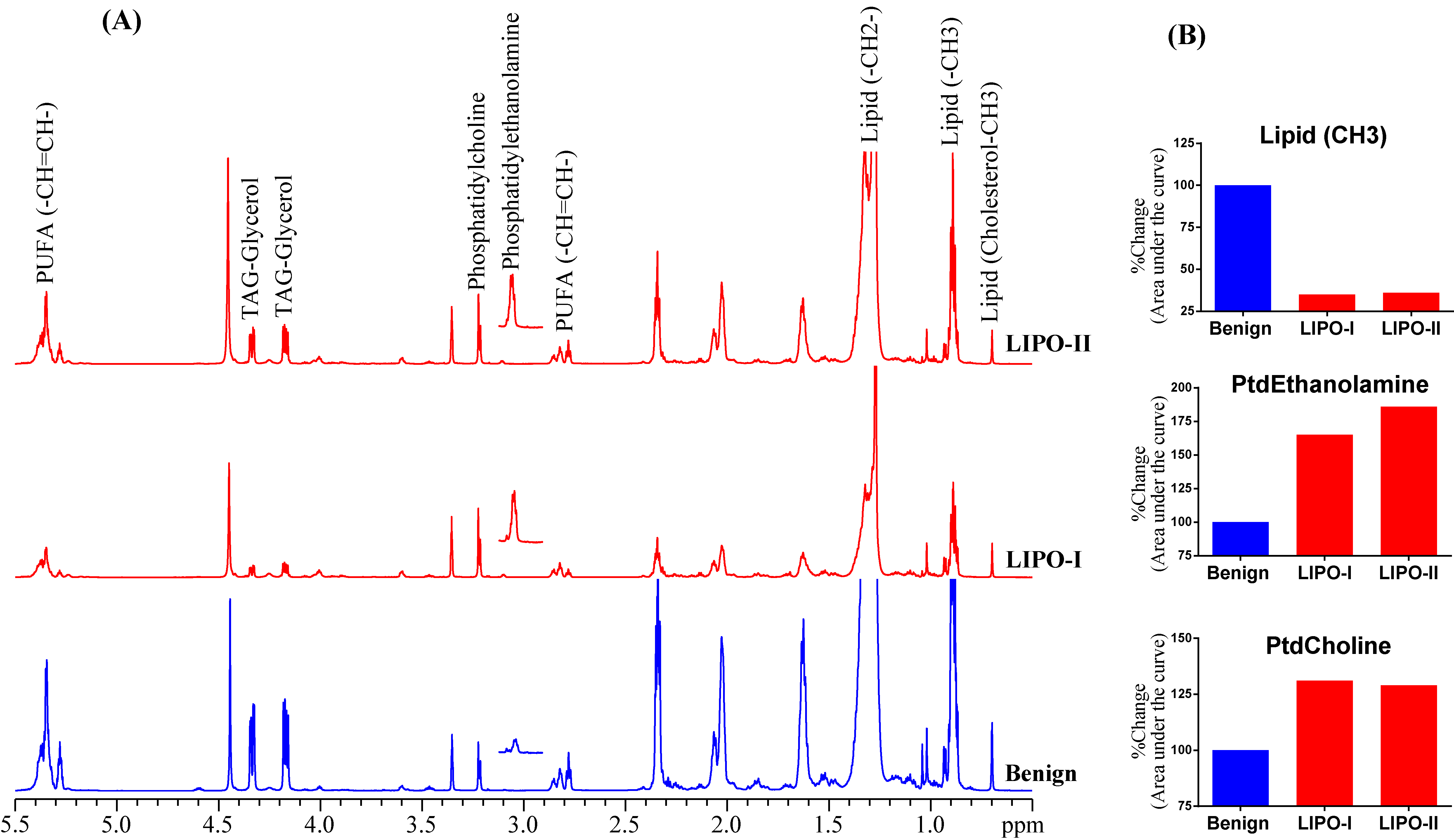
Figure 4: (A)
Representative 1H MR spectra of the lipid phase of tissue extracts obtained
from a pleomorphic lipoma (benign, Blue, lower) and pleomorphic liposarcoma (malignant,
Red, upper). (B) Bar plot showing
changes in lipid, phosphatidylethanolamine (PtdEthanolamine) and phosphatidylcholine
(PtdCholine).
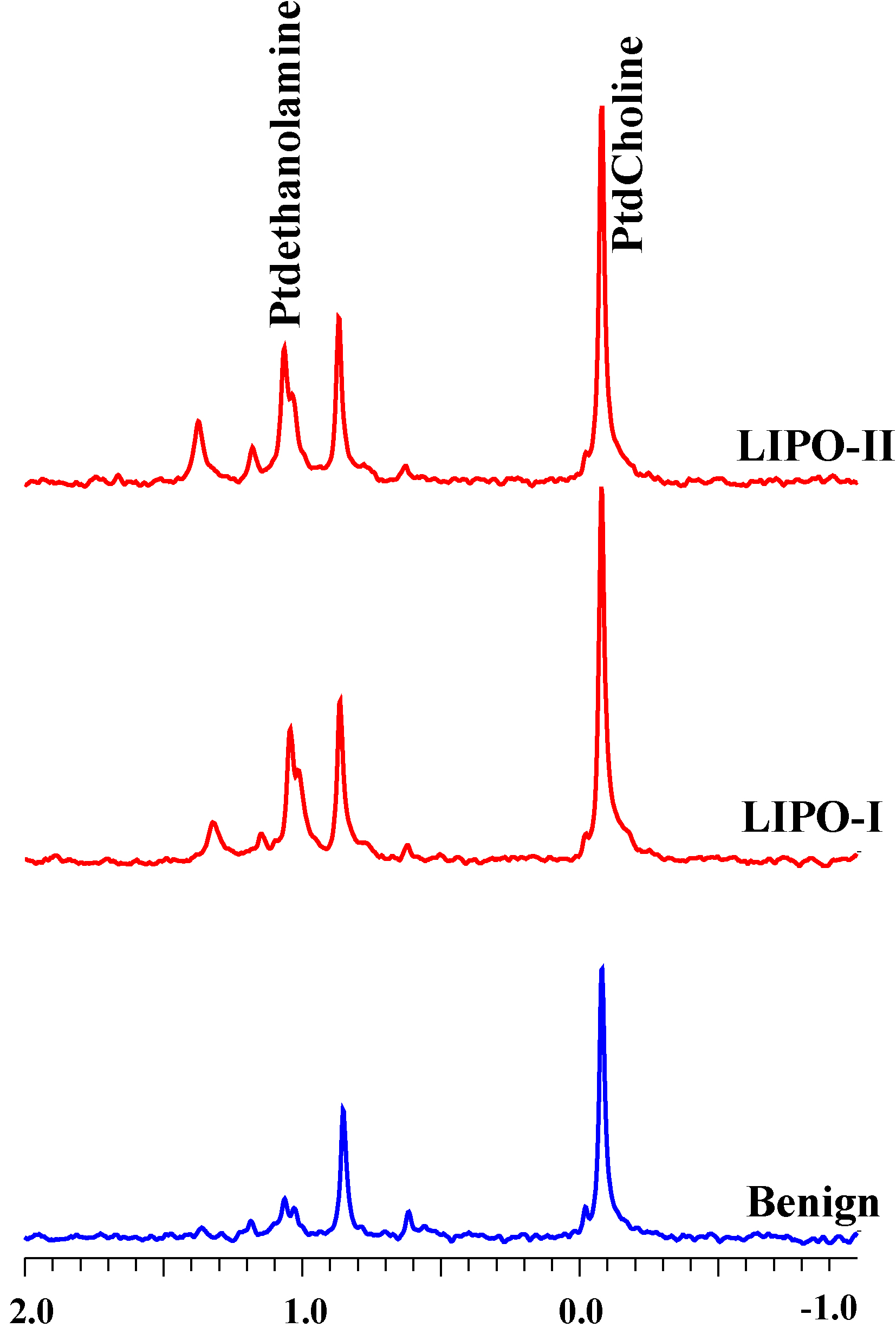
Figure 5:
Representative 31P MR spectra of the lipid phase of tissue extracts obtained
from a pleomorphic lipoma (benign, Blue, lower) and a pleomorphic liposarcoma
tumor (malignant, Red, upper, LIPO-I, LIPO-II). All the spectra are plotted with
the same vertical scale and were acquired with identical parameters.