1434
The role of susceptibility weighted imaging (SWI) in musculoskeletal radiology as an alternative to computed tomography (CT).1Radio-diagnosis, Institute of Nuclear Medicine and Allied Sciences (INMAS), Delhi, India, 2Sir Ganga Ram Hospital, New Delhi, India
Synopsis
SWI has been used for detection of calcification and hemosiderin deposits in diagnosis of the neurological disorders, hemorrhagic disorders and neuroinfectious conditions. Our study tries to answer the question that whether the susceptibility weighted MR imaging can provide alternative to the CT scan and thus decreasing our dependency on the modality which has significant drawback of having radiation dose especially to our young patients. We compared SWi and CT for the characterization of lesion calcification and hemorrhage and we found there was no significant difference in detection rate of these characteristics between two modalities, thus proving SWI as equally sensitive.
Learning objectives
- To highlight the role of SWI in musculoskeletal radiology especially in pediatric population.
- To compare the SWI with CT scan to detect characterizing features e.g. calcification, hemorrhagic foci.
- To go through basics of SWI imaging.
- To present myriad of pictorial assay with SWI imaging including 3D constructed SWI images.
Background
SWI has been used for detection of calcification and hemosiderin deposits in diagnosis of the various neurological disorders including stroke (1), traumatic brain injury (2,3,4), dementia (5,6) hemorrhagic disorders and neuro-infectious conditions (7,8,9), epilepsy (9) , Parkinson's disease (10), We are trying to see the application SWI beyond neuroimaging, extending it in field of musculoskeletal radiology especially in pediatric population as an alternative to CT scan. CT scan is workhorse of the imaging for musculoskeletal system but in pediatric population if MRI shown to be as efficient in detection of calcification it will be an viable alternative to CT especially in follow up scans. While doing literature search we found mostly case reports (11) and very few studies which have done to address this issue (12). It is critical that we find application of MRI in musculoskeletal system to detection calcification to decrease our dependency on CT scan, and inturn decreasing the radiation dose to our patients especially in pediatric population.Methods and procedure details
MRI was performed with 3 Telsa (Skyra Siemen’s), SWI imaging was done by modifying (eg. FOV to suit lesion) the sequence used in brain as SWI sequences dedicated for MSK radiology was not available on our system.
We selected the patients who had already undergone CT scan for characterization of calcification, we performed the routine MR sequences and then incorporated the SWI sequence in it.
We compared the difference in detection of calcification, hemorrhage on CT scan and SWI by 4 radiology trainees and 2 experienced radiologists. We used Chi-square test to know whether there was statistically significant difference in the detection of characterizing features such as calcification or hemorrhage based on modality of susceptibility weighted imaging (SWI) and CT scan.
Results and findings
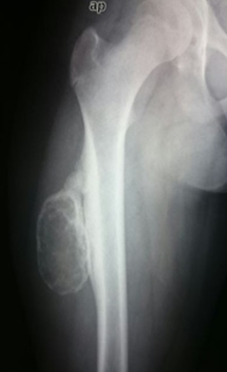
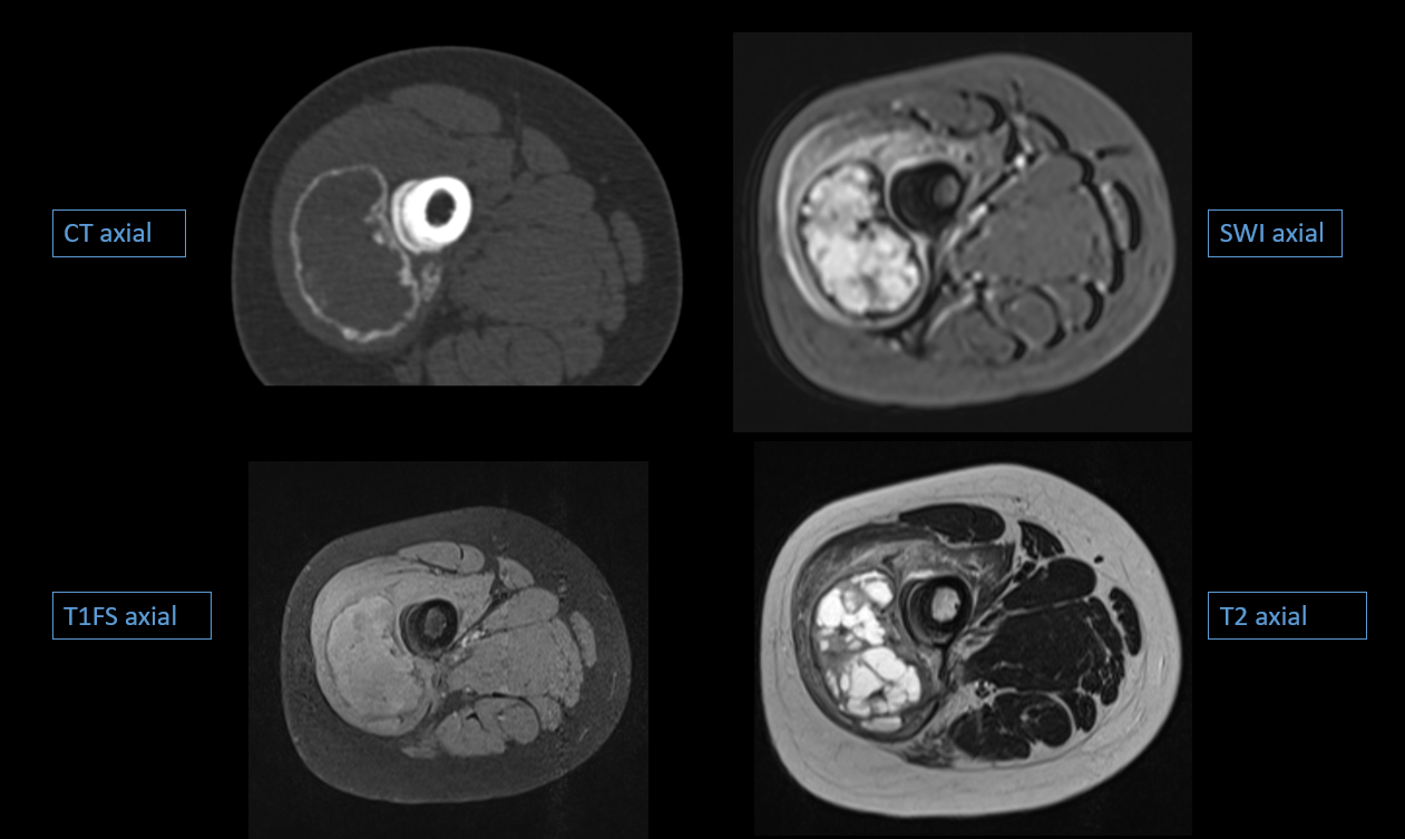
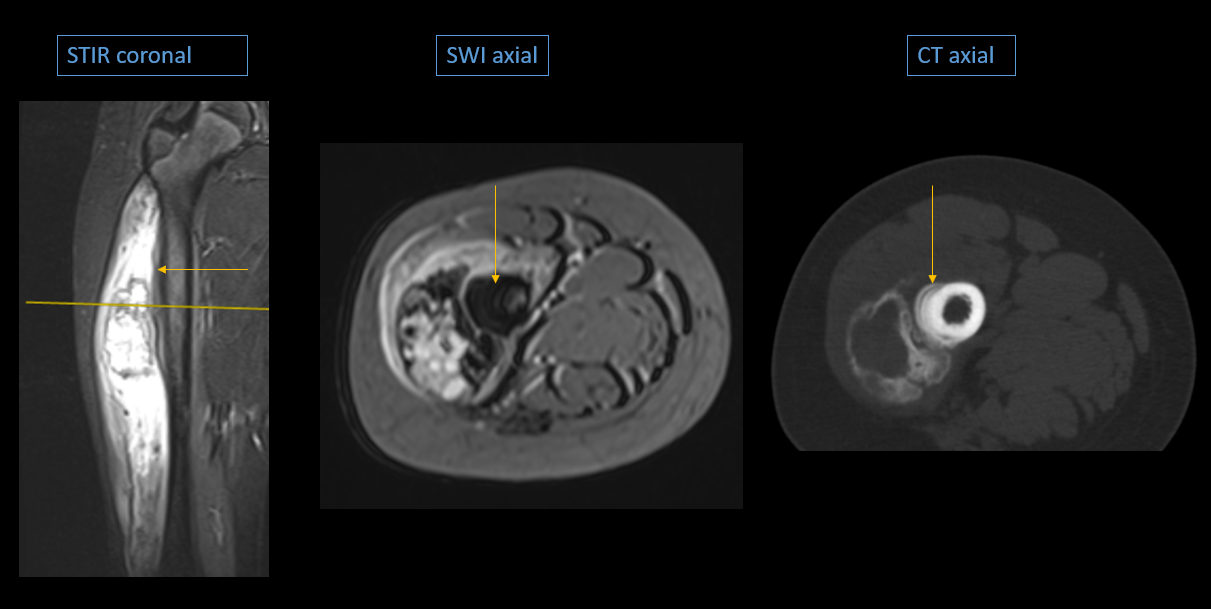
SWI could detect areas of new bone formation in cases of myositis ossificans it was in periphery of lesion (zoning phenomenon) while in cases of osteosarcoma it was in the central of lesion. There was no significant difference in detection of zoning phenomenon on SWI to that of CT scan.
SWI could also identify the areas of hemosiderin deposits due to hemorrhagic foci in tumor which were as blooming on the SWI. There was no significant difference in detection of hemorrhagic foci on SWI to that of CT scan.
SWI also detected periosteal reactions in tumor, myositis ossificans and in chronic osteomyelitis. There was no significant difference in detection of periosteal reactions on SWI to that of CT scan.
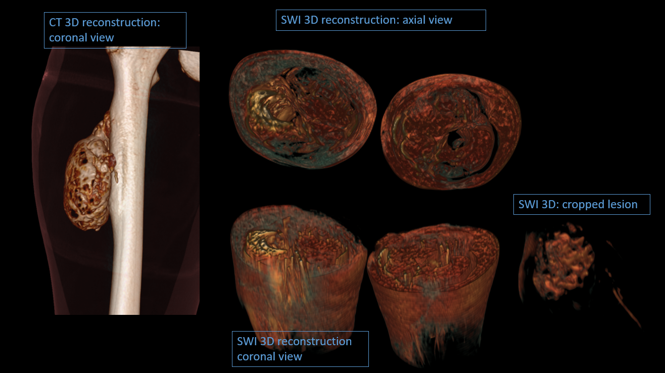
SWI images could also be reconstructed in 3D to help in surgical planning. But the quality of reconstruction is not yet that detailed to be included into the analysis.
Conclusion
The SWI can detect important findings which were thought to be possible only by CT in musculoskeletal radiology. Especially in the pediatric population it would of an immense help as serial follow up imaging is often needed. SWI can be an alternative to CT scan and thus decreasing the radiation dose to young patients.Acknowledgements
No acknowledgement found.References
- Hermier M, Nighoghossian N. Contribution of susceptibility-weighted imaging to acute stroke assessment. Stroke. 2004 Aug 1;35(8):1989-94.
- Roguski M, Morel B, Sweeney M, Talan J, Rideout L, Riesenburger RI, Madan N, Hwang S. Magnetic resonance imaging as an alternative to computed tomography in select patients with traumatic brain injury: a retrospective comparison. Journal of Neurosurgery: Pediatrics. 2015 May;15(5):529-34.
- Ashwal S, Babikian T, Gardner-Nichols J, Freier MC, Tong KA, Holshouser BA. Susceptibility-weighted imaging and proton magnetic resonance spectroscopy in assessment of outcome after pediatric traumatic brain injury. Archives of physical medicine and rehabilitation. 2006 Dec 31;87(12):50-8.
- Huang YL, Tseng YC, Chen DY, Hsu HL, Chen CJ. Susceptibility-Weighted MRI in Mild Traumatic Brain Injury: the Importance of Cerebral Microbleeds. J Neurol Neurophysiol. 2015;6:321.
- Kirsch W, McAuley G, Holshouser B, Petersen F, Ayaz M, Vinters HV, Dickson C, Haacke EM, Britt Iii W, Larsen J, Kim I. Serial susceptibility weighted MRI measures brain iron and microbleeds in dementia. Journal of Alzheimer's Disease. 2009 Jan 1;17(3):599-609.
- Schrag M, McAuley G, Pomakian J, Jiffry A, Tung S, Mueller C, Vinters HV, Haacke EM, Holshouser B, Kido D, Kirsch WM. Correlation of hypointensities in susceptibility-weighted images to tissue histology in dementia patients with cerebral amyloid angiopathy: a postmortem MRI study. Acta neuropathologica. 2010 Mar 1;119(3):291-302.
- Schrag M, Youn T, Schindler J, Greer D, Malhotra A. Susceptibility Weighted Imaging the of Brain in Infective Endocarditis (P1. 003). Neurology. 2015 Apr 6;84(14 Supplement):P1-003.
- Rangarajan K, Das CJ, Kumar A, Gupta AK. MRI in central nervous system infections: A simplified patterned approach. World journal of radiology. 2014 Sep 28;6(9):716.
- Saini J, Kesavadas C, Thomas B, Kapilamoorthy TR, Gupta AK, Radhakrishnan A, Radhakrishnan K. Susceptibility weighted imaging in the diagnostic evaluation of patients with intractable epilepsy. Epilepsia. 2009 Jun 1;50(6):1462-73.
- Gupta D, Saini J, Kesavadas C, Sarma PS, Kishore A. Utility of susceptibility-weighted MRI in differentiating Parkinson’s disease and atypical parkinsonism. Neuroradiology. 2010 Dec 1;52(12):1087-94
- Frouge C, Vanel D, Coffre C, Couanet D, Contesso G, Sarrazin D. The role of magnetic resonance imaging in the evaluation of Ewing sarcoma. Skeletal radiology. 1988 Sep 1;17(6):387-92.
- Özcan E, Sanal HT. The Role of Magnetic Resonance Imaging in the Evaluation of Musculoskeletal System. InMusculoskeletal Research and Basic Science 2016 (pp. 183-195). Springer International Publishing.
Figures