1421
Usefulness of PETRA imaging for frozen shoulder patients1Radiology, Tokyo medical clinic, Tokyo, Japan, 2Radiology, Jyuntendou University Hospital, Tokyo, Japan, 3SIEMENS Healthcare Co., Tokyo, Japan, 4Orthopedics, Tokyo-kita medical center, Tokyo, Japan, 5Orthopedics, Tokyo Women’s Medical University, Tokyo, Japan
Synopsis
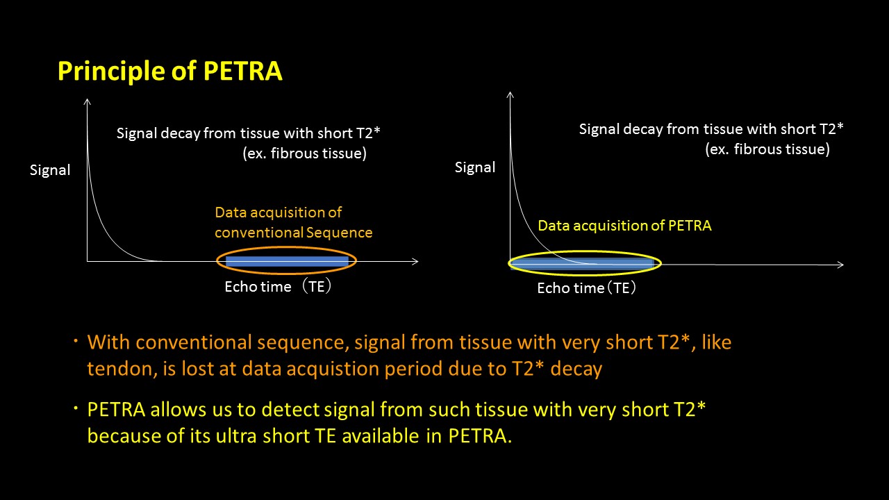
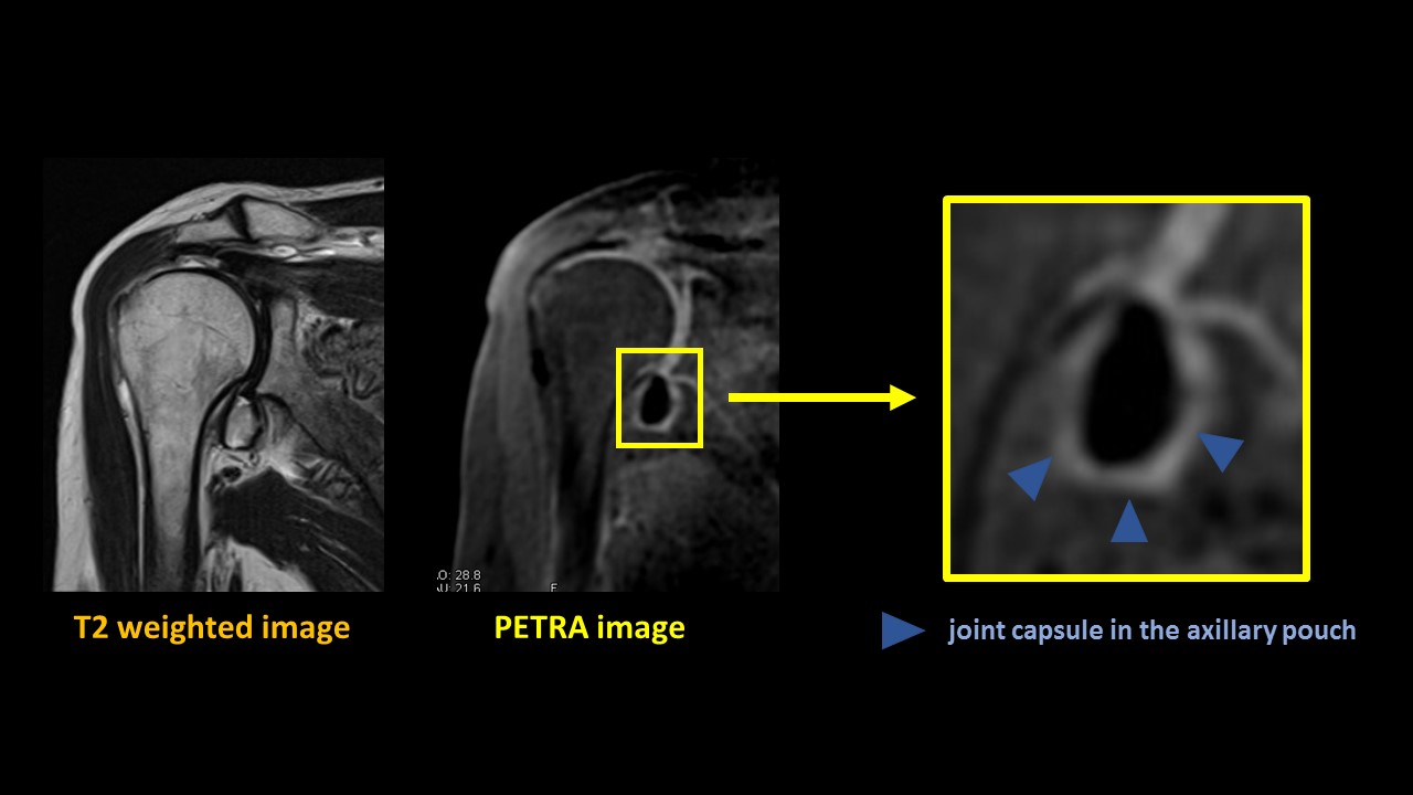
Pointwise encoding time reduction with radial acquisition (PETRA) has made it possible to visualize those tissues which have a short T2* value such as ligaments and tendons as high signal images by using ultra-short echo time (TE). In this study, we evaluated the significant difference of the thickness of the joint capsule in the axillary pouch, depending on the stage or the symptom of patients with frozen shoulder.
Purpose
Using ultra-short echo time (TE), Pointwise encoding time reduction with radial acquisition (PETRA) has the potential to visualize those tissues which have a very short T2* as high signal images while conventional MR imaging visualize them only as low signal images(Figure 1, 2)[1] . The purpose of this study was to evaluate their signal intensities by measuring T2* value of those tissues that traditionally have been visible with the contrast of surrounding tissues and to examine the usefulness of using PETRA for those patients with frozen shoulder.Method
Subjects consisted of 31 patients with shoulder pain. We defined frozen shoulder as follows: range of motion < 110 in forward flexion, < 40 in external rotation and < L3 in internal rotation. These patients were divided into two groups: 19 patients with night pain in their shoulder (5 men and 14 women; mean age, 59 years) were classified to symptomatic group and 12 patients without night pain (5 men and 7 women; mean age, 67 years) were classified to asymptomatic group. A 3T MRI scanner (MAGNETOM Skyra VD13A; Siemens Healthcare) was used to all MR studies. The T2* value in the area where the thickness was measured was calculated with TR 445ms, TE 3.5, 1.9, 5.1, 6.7, 8.4ms. For PETRA imaging, parameters were as follows: TR 4.0 ms, TE 0.1 ms, TI 500 ms, FA 6 deg, base resolution 288, radial views 30000, voxel size 1 x 1 x 1 mm. The thickness of the joint capsule in the axillary pouch was measured with the oblique coronal image by using ZIOSTATION2 (Ver2.4). Two observers measured the thickness and Pearson’s correlation coefficient was calculated. We compared the thickness of the joint capsule in the axillary pouch with patients in the symptomatic group and in the asymptomatic group, and conducted Wilcoxon signed-rank test for both comparisons.Results
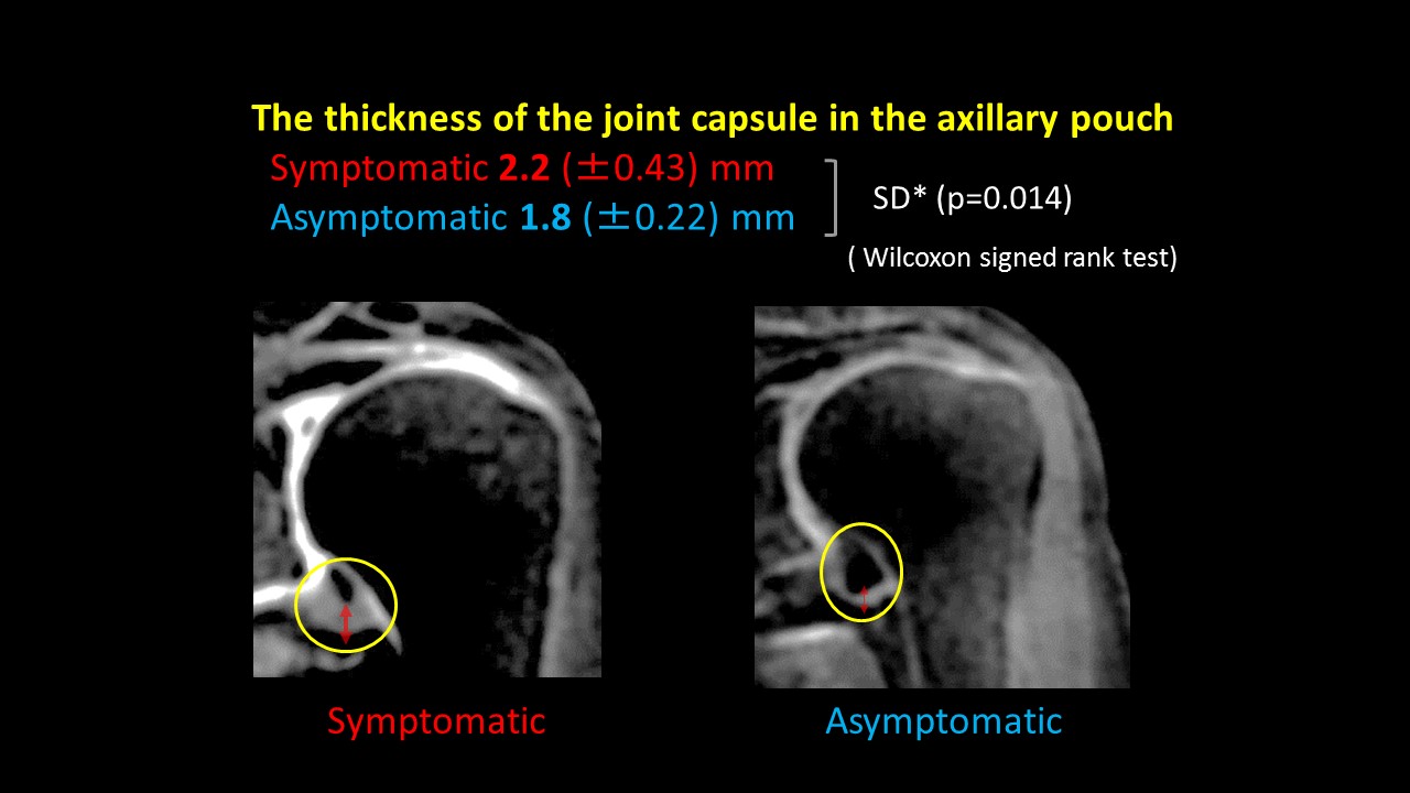
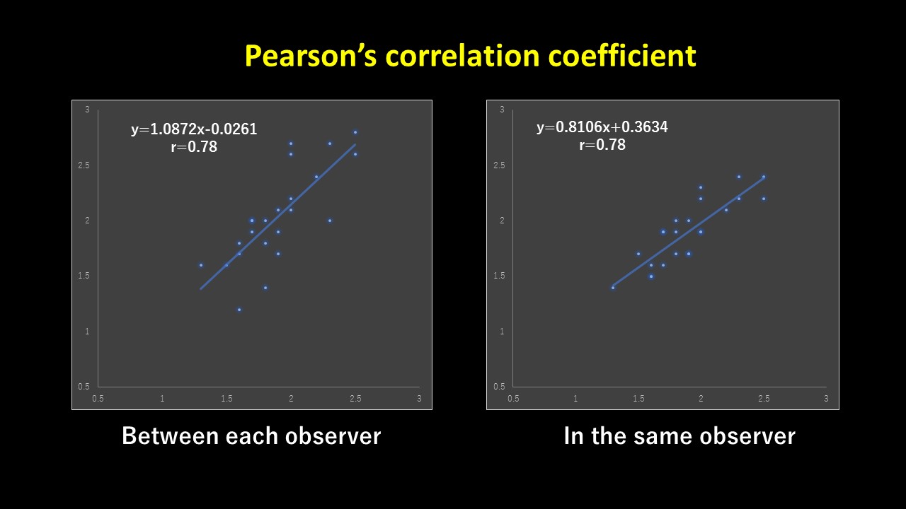
The T2* value of the tissues in the axillary pouch was 9.1 ms. Patients in symptomatic group had more significantly thickened joint capsule in the axillary pouch than in asymptomatic group and their average thickness of the patients in symptomatic group/ in asymptomatic group were 2.2/1.8 mm (p = 0.014 for both comparisons, Wilcoxon signed-rank test; Figure 3). Pearson’s correlation coefficient between each observer/in the same observer were r = 0.78/0.85 (Figure 4).Discussion
The T2* value of the point where the thickness was measured in the axillary pouch was 9.1 ms. According to this result, we found that with the gradient echo sequence, ligaments and tendons were visualized as signal images. Using PETRA imaging based on T1WI made it possible to visualize the contrast more clearly between ligaments or tendons and the joint fluid. Furthermore, we considered that using ultrashort-TE was effective for those studies like this which measure the thickness of joint capsules because it can reduce motion artifacts in the part that is affected by the movement of chest wall when you breathe, such as a shoulder joint. It is said that a symptomatic patient with frozen shoulder has pain during rest and at night which is considered to be caused by the inflammation in the axillary pouch. In the frozen stage, while the pain decreases and fades after the inflammation subside, the shoulder loses range of motion and completes to be frozen. With conventional MR imaging, it has been difficult to visualize the joint capsule of which T2* decay rapidly, however, with PETRA imaging, it is possible to visualize ligaments and tendons as high signal image because its echo time (TE) is 0.1ms, which is very short. In this study, there was difference in the thickness of joint capsule even between patients in symptomatic and asymptomatic group.Conclusion
In this study, we proved the potential of PETRA imaging to visualize the capsular ligament which used to be difficult to be visualized without arthrography. Since the result was related to the stage and the symptom of the patients, it was indicated that using PETRA imaging can be the useful method for those patients who are suffering from shoulder diseases.Acknowledgements
No acknowledgement found.References
[1] D. Grodzki, et al. MRM,2012. 67:510-518
[2] Bernad M, et al. Radiology,2004. 233:486-492
[3] Joon-Yong J, et al. Eur Radiology,2006. 16:791-796
[4] Atsushi T, et al. katakansetsu,2012. 36-3:787-790
[5] Keisuke O, et al. Katakansetsu,1996. 20-1:241-244
[6] Kazuya T, et al. Katakansetsu,1995. 19-2:289-293
Figures