1418
Is the anterolateral ligament affected by the rupture of anterior cruciate ligament? A tentative investigation based on magnetic resonance imaging1The First Affiliated Hospital of Zhengzhou University, Zhengzhou, China, 2Clinical Science, Philips Healthcare, Beijing, China
Synopsis
This study aimed to demonstrate the incidence of injured of ALL following ACL rupture, as well as observe the characteristics of thus injury based on MRI. In the study, we used the high resolution 3D TSE-based sequences including the optimized T1W-VISTA and T1W-VISTA-SPAIR to evaluate the 43 knees of patients who have ligament ruptured through clinical test. Chi-square test was performed to analyze the categorical variables. Binary logistic regression was performed to investigate the main cause. It indicated that ACL injuries has closer association with ACL injuries but less association with LM injuries, and the femoral portions of ALL were easily ruptered
Introduction
Recent studies have characterized the anterolateral ligament (ALL) of the knee injuries with anterior cruciate ligament (ACL) ruptures would cause for anterolateral rotatory instability after ACL reconstruction. However, in China, there was no great importance has been placed on ALL. Once the ACL ruptured, the orthopedist tends to focus on the reconstruction of ACL ignoring the condition of the ALL. The purpose of the study was to assess the incidence of injured of ALL following ACL rupture and observe the characteristics of thus injury based on MRI.Methods
Forty-three patients (28 males, 15 females, aged 27.1±7.4 years old) who have a history of knee injury included.The study was approved by the Ethics Committee of Zhengzhou University First Hospital, Zheng Zhou, China. Each patient underwent clinical Lachmann and a pivot-shift test and all have positive results . All MRI scans were performed on a 3.0T MR scanner (Ingenia, Philips Healthcare, Best, the Netherlands) using a dedicated 8-channel knee coil. The high resolution 3D TSE-based sequences including the T1W-VISTA and T1W-VISTA-SPAIR were optimized for the scanning.The imaging was accessed independently by two consultant musculoskeletal radiologists and they were blinded to all clinical findings. When they had conflicts in the diagnosis, they were asked to give an agreement after discussion. Exclusions criterion were as follows: multi-ligament injuries (any full-thickness injury to posterior cruciate, either collateral ligament), fracture or other inflammation of knees, but findings of meniscus injuries was included . Finally, totally thirty-six patients were included . Injury state was classified into discontinuity of all of the ALL fibers, intra-ligamentous high signal or morphological abnormality. The ALL was regarded as being ruptured in the case that when only parts of the ALL could not be visualized but other parts could. This was according to the fact that for some subjects, the whole ALL could not be visualized even though it was normal. Finally, Chi-square test was performed to analyze the categorical variables. Binary logistic regression was performed to investigate the main cause of ALL damages.Result
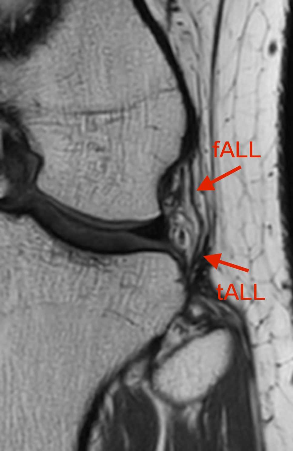
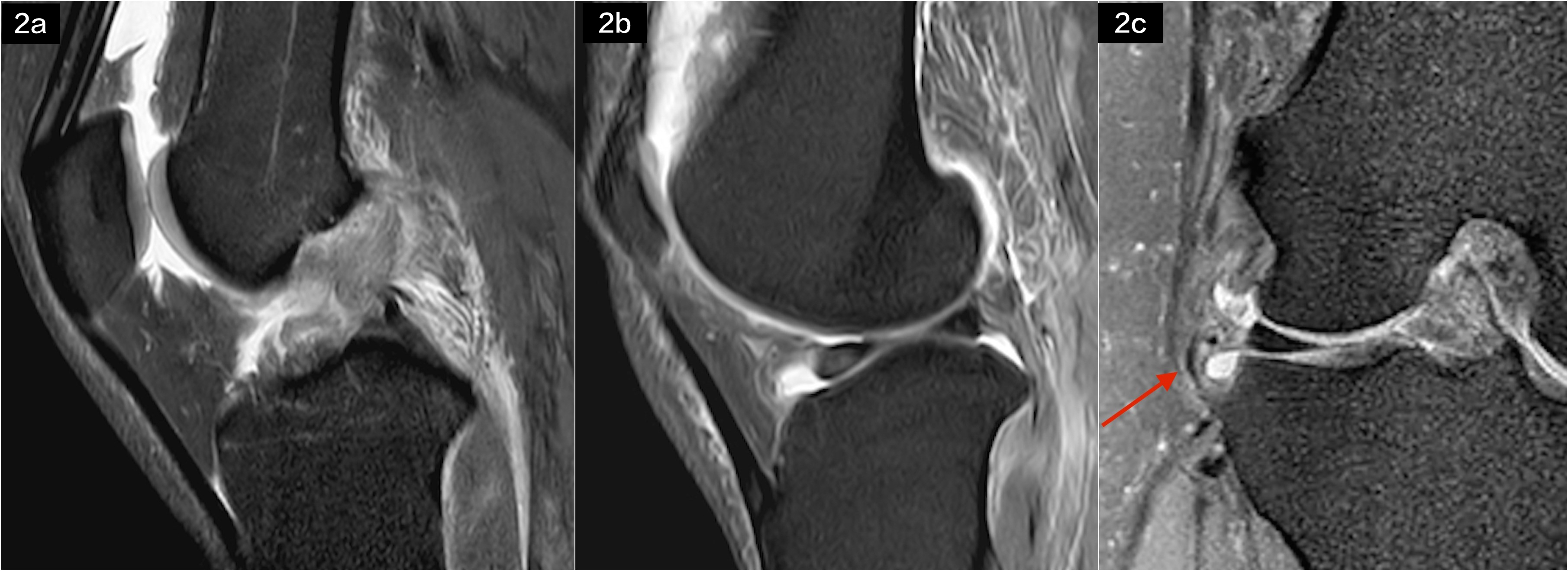
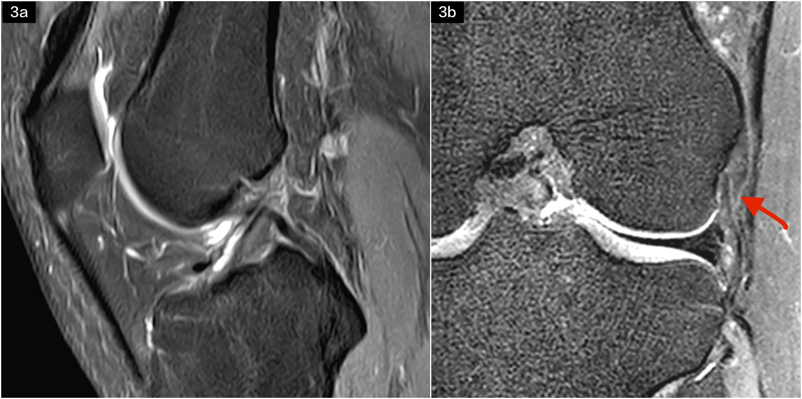
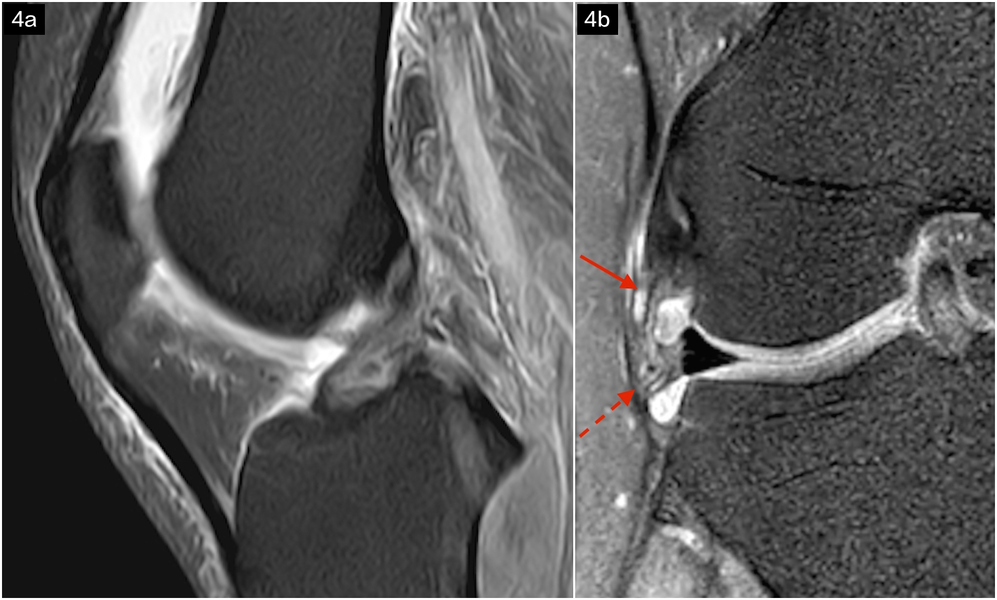
When the ACL and LM both injured, the ALL was the usually injured with a percentage of about 91.7%. When Only the ACL injured, the injured ALL was accounting for 81.8%. When only the LM injured, the injured ALL was only accounting for about 30%. Chi-square results showed that there are significant difference (χ2=9.47,p<0.05) for ALL damages in the three groups. There are significant difference (χ2=9.47,p<0.05) for ALL damage between ACL+ and LM+ groups. And there are significant difference (χ2=7.87,p<0.05) between LM+ and ACL+/LM+ groups. But there is no significant difference (χ2=0.49,p>0.05) between ACL+ and ACL+/LM+ groups. Logistic regression analysis showed the association of ALL damage with ACL injuries and LM injuries with the formula: YALL=0.683×XACL+0.379×XLM (F=10.03, P<0.001).It indicated that ACL injury has closer association with ACL injuries but less association with LM injuries. In all the ALL-injured subjects, the femoral portions of ALL were easily ruptured accounting for 43.8%(14/32) and the whole ALL rupture accounts for 31.3%(10/32). Then the tibial portions rupture only accounts for 6.3% (4/32) . Six cases (18.8%,6/32) with intra-ligamentous edema but with no discontinuity signifying partial thickness injury.Discussion
Our study showed that ALL injuries have closer association with ACL injuries. Because previous studies have demonstrated that both ALL and ACL injuries often occur with pivoting movements. This is due to the knee being put in a precarious position where there is a rotational force applied to the tibia and a valgus moment at the knee as it nears extension, thus causing a pivot shift injury[1.2]. Additionally, we found that the femoral portion of ALL ruptured was involved in most lesions (75.1%), the contusions frequently occurred at the lateral femoral condyle,while the tibial portion was always normal. It is not surprising that the ALL originating at the lateral femoral epicondyle and the transepicondylar axis being the best representation of the knee’s optimal flexion-extension axis[2]. Once the ACL tears, it could result in tension on the meniscus which may contribute to tearing of the LM. And lateral meniscus injury may furtherly destabilise the ACL-deficient knee, thus leading to ALL injury[3]. That’s why when just LM tears, the ALL can keep normal. However, when ACL and LM both ruptures, the ALL always injured. In our previous study, only 20% of the ALL with fibers inserting on the lateral meniscus, which was one of the reason that the association between LM and ALL injured was not so close.Conclusion
This work indicated that there might be association of ACL ruptures with ALL injuries. And T1-VISTA-SPAIR imaging technique would provide the information for clinician to pay attention to the ALL when focus on the ACL reconstructions.Acknowledgements
No acknowledgement found.References
[1]Kosy JD, Schranz PJ, Patel A, et al. The magnetic resonance imaging appearance of the anterolateral ligament of the knee in association with anterior cruciate rupture. Skeletal Radiol. 2017,46(9):1193-1200. [2]Helito CP, Helito PVP, Leão RV, et al. Anterolateral ligament abnormalities are associated with peripheral ligament and osseous injuries in acute ruptures of the anterior cruciate ligament. Knee Surg Sports Traumatol Arthrosc. 2017 ,25(4):1140-1148. [3]Van Dyck P, Clockaerts S, Vanhoenacker FM, et al. Anterolateral ligament abnormalities in patients with acute anterior cruciate ligament rupture are associated with lateral meniscal and osseous injuries. Eur Radiol. 2016,26(10):3383-91.Figures
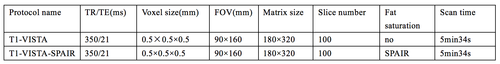
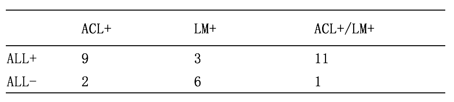
Table2 Information of ALL,ACL and LM injuries
Note: ALL+,ALL injuried;ALL-,ALL not injuried;LM+,lateral meniscus injuried; LM-,lateral meniscus not injuried;ACL+,ACL injuried,ACL-,ACL not injuried