1407
Ex vivo MRS evaluation of severe burn injury in mice shows metabolic changes in skeletal muscle1Pathology, Massachusetts General Hospital, Boston, MA, United States, 2Burns and Plastic Surgery, First Affiliated Hospital of General Hospital of PLA, Beijing, China, 3Burn and Plastic surgery, First Affiliated Hospital of PLA General Hospital, Beijing, China, 4Anesthesia, Critical Care and Pain Medicine, Massachusetts General Hospital, Boston, MA, United States
Synopsis
Patients of severe burn injury often suffer from sepsis, which results in multiple organ failure and prolonged metabolic derangement, leading to higher mortality. Accurate measurements of burn injury-associated metabolic changes may provide the burn clinic with quantitative tools to assess patient status. We tested the efficacy of High-Resolution Magic Angle Spinning (HRMAS) magnetic resonance spectroscopy (MRS) in evaluation of tissue metabolic changes with mouse skeletal diaphragm and gastrocnemius muscles after burn injury. HRMAS measurements indicated that IMTG and plasma FFA levels were increased after severe burn injury, with more pronounced differences detected in diaphragm muscle than in gastrocnemius muscle.
Introduction
Patients of severe burn injury often suffer from sepsis, which results in multiple organ failure and prolonged metabolic derangement, leading to higher mortality1-2. Accurate measurement of burn injury-associated metabolic changes may provide the burn clinic with quantitative tools to assess patient status. Increases of intramuscular fats or intramyocellular triacylglycerols (IMTGs) in skeletal muscle have been proposed as biomarkers of insulin resistance and mitochondrial dysfunction following burn injuries. Here, we tested the efficacy of High-Resolution Magic Angle Spinning (HRMAS) magnetic resonance spectroscopy (MRS) to evaluate tissue metabolic changes inmouse skeletal diaphragm and gastrocnemius muscles after burn injury.Methods
Eight adult male C57BL/6 mice have been studied thus far. After anesthetizing with isofluorane, five animals underwent both dorsal (9 sec) and abdomen (4 sec) full thickness burn with hot water (90oC) to create severe burn injury of approximately 50% of total body surface area. The other three were used as control and underwent anesthesia followed by exposure to 37oC water. Two mice were sacrificed 1 day after burn injury, and the remaining three burn injury mice as well the control animals were sacrificed 3 days after burn injury. Tissues of diaphragm and gastrocnemius muscles were removed and stored at -80oC. HRMAS MRS was used to quantify IMTGs and plasma free fatty acids (FFAs) from ex vivo tissue specimens. Tissue spectra were acquired at 4oC with MAS at 3.6 kHz on a Bruker 600MHz NMR spectrometer. HRMAS spectra were acquired using both a Carr-Purcell-Meiboom Gill (CPMG) pulse sequence with a T2 filter time of 200ms with cw water suppression and a fully relaxed pulse sequence without water suppression. Data were processed and curve fit in Nuts (Livermore, CA) to obtain metabolic intensity of IMTGs.Results
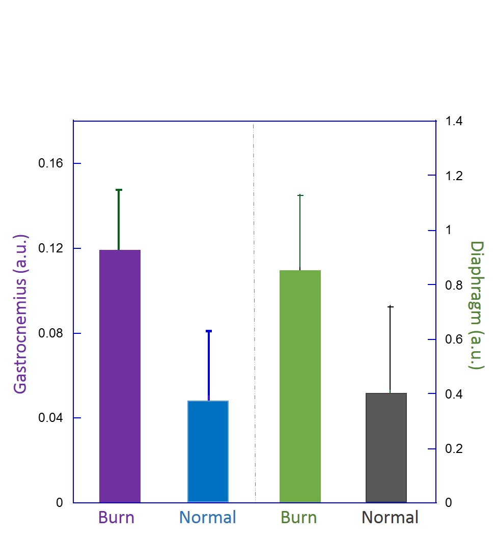
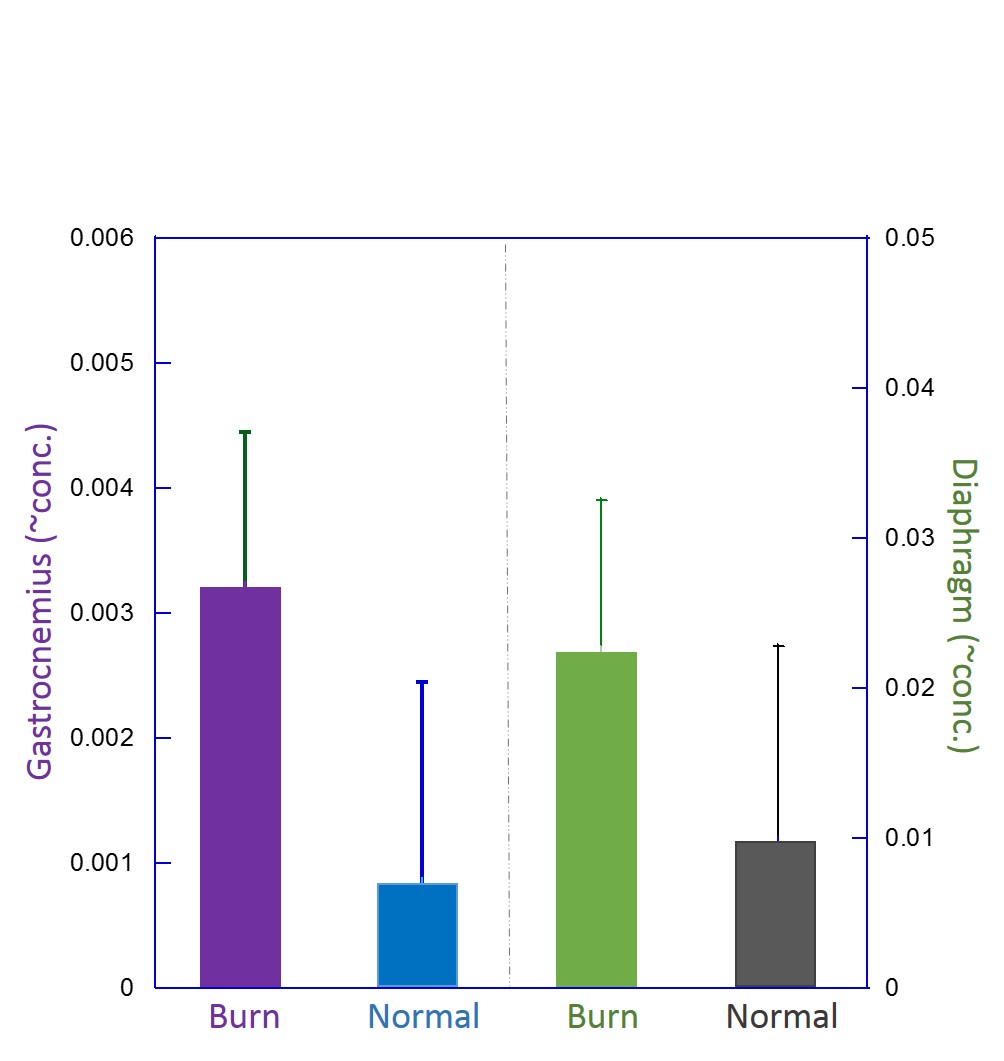
HRMAS measurements indicated that IMTG and plasma FFA levels were increased after severe burn injury. Comparison between control and burn injury mice show increases of IMTGs in skeletal muscle for both the ratio of IMTGs intensity over the intensity of the total metabolic spectral region excluding IMTGs (Figure 1), or as concentration of IMTGs (Figure 2). Moreover, the metabolic difference between burn and normal tissue was more pronounced in the diaphragm than in the gastrocnemius tissue.Discussion and Conclusions
These finding demonstrate that IMTGs and FFAs in unaffected tissues may be useful biomarkers for burn injury and the resulting metabolic issues. Furthermore, they may facilitate the development of novel therapeutic strategies. Although differentiation between burn and normal groups is not yet significant for these small groups, there is a strong trend for the results. Additional analyses and burn injury experiments are underway.Acknowledgements
We would like to acknowledge and thank NIH grant CA115746 and the A. A. Martinos Center for Biomedical Imaging for support.References
[1] Gore DC, Chinkes D, Heggers J, et al. Association of hyperglycemia with increased mortality after severe burn injury. J Trauma. 2001, 51(3): 540-544.
[2] Tuggle DW, Kuhn MA, Jones SK, et al. Hyperglycemia and infections in pediatric trauma patients. Am Surg.2008, 74(3): 195-198.
Figures