1393
Low-field MRI of osteoarthritis in humans: correlations between load-dependent cartilage properties and relaxation parametersErik Roessler1, Carlos Mattea1, Miika Nieminen2, Sakari Karhula2, Simo Saarakkala2, and Siegfried Stapf1
1Ilmenau University of Technology, Ilmenau, Germany, 2University of Oulu, Oulu, Finland
Synopsis
At low magnetic fields, T1 variation within cartilage is a robust parameter that is employed to quantify the layered structure in the tissue and is sensitive to factors such as enzymatic degradation, external load, and diseases such as osteoarthritis. Variable-field relaxometry provides access to the content and local order of glycosaminoglycans and collagen via proton-nitrogen quadrupolar dips. In this study on 20 human cartilage samples, load-dependent low-field and variable-field techniques were combined for the first time to correlate NMR parameters with the severity of osteoarthritis.
INTRODUCTION
While T2 and T1ρ are becoming popular in clinical studies of cartilage, their inherent dependence on sample orientation places a limit to their diagnostic value. T1, on the other hand, is a robust and isotropic parameter, and shows contrast rivalling or exceeding that of T2 when determined at fields below 0.5 T: the range of T1 at low fields across cartilage is much more pronounced than any other tissue in the human body. In addition, the amount of collagen and glycosaminoglycans can directly be determined at fields close to 60 mT due to the signature of 14N nuclei in the 1H relaxation dispersion curve. In this study, low-field and variable-field NMR are combined for the first time with the purpose of quantifying correlations with the degree of osteoarthritic degeneration in humans.METHODS
One-dimensional, depth-dependent scans of bovine and human articular cartilage were carried out with spatial resolutions between 20 and 50 μm on portable, single-sided scanners at magnetic field strengths of 0.27 T and 0.44 T, respectively. The spatial distributions of T2 and T1 were obtained with and without unidirectional compression at 0.6 MPa for the human samples with different degrees of osteoarthritis, covering Mankin grades 0-12. The dispersion of T1 in the 1H Larmor frequency range of 10 kHz to 30 MHz was monitored using a Stelar Fast Field Cycling relaxometer.RESULTS & DISCUSSION
The layered structure of mammalian articular cartilage results in a pronounced T2 variation at all magnetic field strengths1. A similar variation of T1, typically covering a ratio of 3-5 between maximum and minimum values inside the tissue, was identified at a field strength of 0.27 T, while it has been reported as rather small at high magnetic field strengths2. T1 has thus been identified as a suitable parameter to follow changes in cartilage properties by low-field NMR. Average T1, as well as cartilage thickness obtained from T1 measurements of human samples, is found to correlate negatively with Mankin grade. At the same time, a significant correlation was identified for relaxation time reduction before and after uniaxial compression at 0.6 MPa, a typical value for forces appearing in the human knee and hip joint. This finding is of importance since the spatial resolution of 50 μm obtained with the single-sided scanner is about one order of magnitude better than the one in clinical high-field or low-field scanners3, thus allowing a much more reliable definition of thickness change which even includes resolution of the three main cartilage layers. At 1H Larmor frequencies of 2-3 MHz, the so-called quadrupolar dips are superimposed onto a frequency-dependent signature of T1 that can be approximated by power-laws. Varying the composition, water content or structural integrity of cartilage affects both the general frequency dependence of T1 and the shape of the quadrupolar dips, providing a possible diagnostic access to arthropathies such as osteoarthritis (OA)4. In this study, a correlation of the area of the quadrupolar dips with Mankin grade is demonstrated: diseased tissue contains less GAG but more water. The observation is confirmed by artificially altered tissue using trypsin or collagenase5,6.CONCLUSIONS
Low-field MRI and variable-field relaxometry were successfully combined in a study of osteoarthritic human articular cartilage. Spin density and relaxation times were acquired normal to the tissue plane with a spatial resolution of 50 μm or better; in particular, T1 showed a well-pronounced gradient across the tissue, unlike at clinical MRI field strength. The degree of variation of these parameters was followed for samples under load. Correlations of thickness change and T1 change with disease state were observed. In variable-field experiments, the intensity of quadrupolar dips and power-law parameters with disease state could be demonstrated. These results allow for an improved diagnostic interpretation of low-resolution clinical MRI particularly at dedicated extremity scanners.Acknowledgements
No acknowledgement found.References
[1]Xia Y, Magic-angle effect in magnetic resonance imaging of articular cartilage – a review, Invest. Radiol. 2000; 35: 602-621. [2] Rössler E, Mattea C, Mollova A, Stapf S, Low-field one-dimensional and direction-dependent relaxation imaging of bovine articular cartilage, J. Magn. Reson. 2011; 213: 112-118. [3] Rössler E, Mattea C, Stapf S, Feasibility of high-resolution one-dimensional relaxation imaging at low magnetic field using a single-sided NMR scanner applied to articular cartilage, J. Magn. Reson. 2015; 251: 43-51. [4] Broche LM, Ashcroft GP, Lurie DJ, Detection of osteoarthritis in knee and hip joints by fast field-cycling NMR, Magn. Reson. Med. 2012; 68: 358-362. [5] Rössler E, Mattea C, Stapf S, NMRD investigations of enzymatically degraded bovine articular cartilage, Magn. Reson. Med. 2015; 73:2005-2014. [6] Rössler E, Mattea C, Saarakkala S, Lehenkari P, Finnilä M, Rieppo L, Karhula S, Nieminen MT, Stapf S, Correlations of low-field NMR and variable-field NMR parameters with osteoarthritis in human articular cartilage under load, NMR in Biomedicine 2017; 30: e3738/1-14.Figures
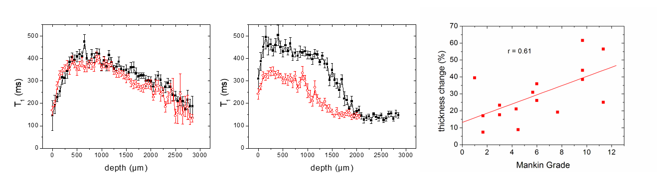
Figure 1: T1 distribution
across cartilage (where 0 denotes the surface) at a pressure of 0.6 MPa (red
curves) compared to 0 MPa (black curves) – left: healthy; middle: severe
osteoarthritis. Right: correlation between relative thickness change and Mankin
grade.