1379
Quantitative evaluation of knee cartilage after anterior cruciate ligament reconstruction using UTE-T2* mapping in a rabbit model1Fudan University affiliated Huashan Hospital, Shanghai, China, 2GE Healthcare, CHINA, Beijing, China
Synopsis
Our study is a prospective longitudinal study conducted to find outcome of anterior cruciate ligament reconstruction in rabbit model. We evaluated degenerative changes of cartilage by UTE-T2* mapping. ACLR knees shows cartilage matrix degeneration at early stage of "ligamentization", though rabbit tibiofemoral cartilage is definitely thin.
Background
Anterior cruciate ligament (ACL) tear is a common and serious knee injury. Although anterior cruciate ligament reconstruction (ACLR) was applied to restore knee function and stability, previous studies have demonstrated that 50-70% of these patients developed radiological changes of osteoarthritis[1]. UTE-T2* quantification has been used widely for detecting degeneration in cartilage retaining intact articular surface. The use of UTE-T2* in rabbit model is less investigated, but may be an effective tool for monitoring cartilage damage[2]. So the objective of this study is to longitudinally evaluate cartilage changes by using UTE-T2* quantification in anterior cruciate ligament (ACL) reconstructed knees in the rabbit model.Mothod
Twelve adult New Zealand White rabbits (3 month old, male/female 6/6) were used in our study. Rabbits were raised at the animal center following the Guidelines for the Care and Use of Laboratory Animals. All animals underwent ACLR surgeries using Achilles tendon on their right knees. Briefly, rabbits underwent general anesthesia by intramuscular injection of 5 mg/kg xylazine and diazepam and intravenous injection of pentobarbital. For each rabbit, the right knee and ankle were shaved and scrubbed by iodine tincture and the animal was transferred to a sterile operation table. All the operations were performed by an experienced surgeon. A 2-3 cm medial curve parapatellar skin incision was made over the right knee. The anterior cruciate ligament was exposed and sectioned. Then the femoral and tibial bone tunnels were made by a 2.5mm diameter drill. The Achilles tendon was obtained from right ankle and then transplanted into right knee as an ACL graft. All rabbits can move freely in their own cages postoperation. MRI scanning was performed at presurgery, 4, 8 and 12 weeks postsurgery by a senior musculoskeletal radiologist. All rabbits were imaged using a 3.0 T MR scanner (Discovery MR 750, GE Medical Systems, Milwaukee, WI, USA) and a rabbit coil. The UTE-T2* mapping was used on all rabbits. Sagittal UTE-T2* mapping parameters used included: TR of 85 ms, echo time (TE) of 0 and 6.8 ms, field view of 10*10 cm2, and slices thickness of 1.0 mm with a 0.0 mm gap. After image acquisition, UTE-T2* values of each regions of interest was measured on two echoes, then calculated by single-index model. Quantitative cartilage UTE-T2* values were measured by sagittal UTE-T2* mapping in four compartments, including medial femoral condyle (FM), medial tibial plateau (TM), lateral femoral condyle (FL), medial tibial plateau (TM) areas. The regions of interest of each slice were assessed in the cartilage layer along the curvature of the joint surface, including the entire thickness of the tibiofemoral cartilage. Two to three consecutive slices were selected for medial and lateral regions of the cartilage, respectively, to cover the entire tibiofemoral cartilage. Additionally, the mean UTE-T2* value of total tibiofemoral cartilage (TFT) was determined by averaging the mean UTE-T2* values of all cartilage compartments. The generalized linear model was used to compare UTE-T2* values of four time points for each cartilage compartment. All statistical analyses were performed with SPSS Statistics version 23.0.0 (IBM, Armonk, NY). P less than 0.05 was considered statistically significant.Results
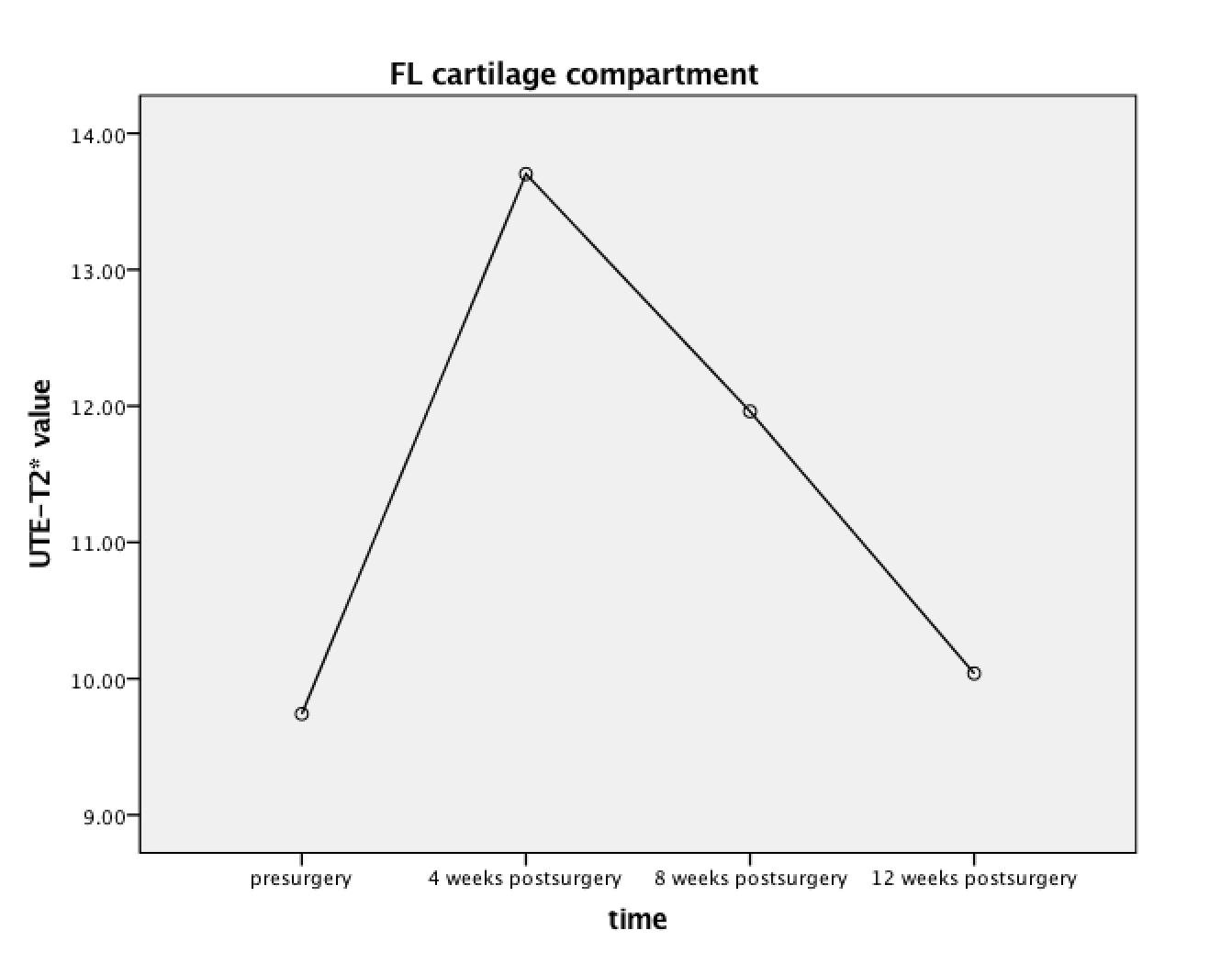
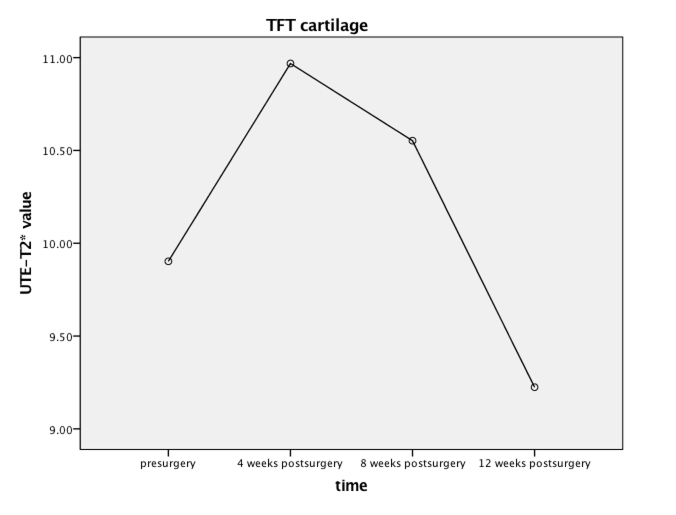
In longitudinal analyses, UTE-T2* values of FL and TFT cartilage compartments elevated at 4 weeks postsurgery, but decreased at 8 weeks postsurgery and decreased to levels similar to those presurgery at 12 weeks, suggestive of healing (P=0.006, P=0.011). No statistically significant changes in UTE-T2* values for FM, TM and TL were observed at four time points (P=0.183, 0.312, 0.125, respectively).Conclusion
This study suggests that UTE-T2* mapping reveals changes in cartilage matrix after ACLR in rabbit model according to postoperative time. FL and TFT cartilage compartments show risk for degeneration at the early stage after ACLR surgery, but getting better at the later stages in ACL grafts “ligamentization”. Longitudinal data in our study provide additional potential for the possibility of high sensitivity of UTE-T2* mapping to occult cartilage injuries in rabbits, though the rabbit tibiofemoral cartilage is definitely thin.Acknowledgements
No acknowledgement found.References
1. Li, X., et al., Cartilage in anterior cruciate ligament-reconstructed knees: MR imaging T1{rho} and T2--initial experience with 1-year follow-up. Radiology, 2011. 258(2): p. 505-14.
2. Fischenich, K.M., et al., A study of acute and chronic tissue changes in surgical and traumatically-induced experimental models of knee joint injury using magnetic resonance imaging and micro-computed tomography. Osteoarthritis Cartilage, 2017. 25(4): p. 561-569.
Figures