1336
Time-domain EPR imaging with slice selection1National Cancer Institute, National Institutes of Health, Bethesda, MD, United States, 2Department of Basic Medical Sciences for Radiation Damages, National Institute of Radiological Sciences, Chiba, Japan, 3Graduate school of Information Sicence and technology, Hokkaido University, Sapporo, Japan
Synopsis
The slice selection imaging has advantages of reducing imaging time and obtaining optimum dynamic range in image for EPR imaging as well as for MRI. However, the slice selection using a selective pulse, which is used in MRI, is difficult to implement in EPR imaging because of ultra-fast relaxation time compared to gradient settling time. Therefore, we used a modulated gradient field to achieve slice selection in pulsed EPR imaging in this study. We demonstrated the slice selection imaging with tubes and a living mouse to show the effect of slice selection in pulsed EPR imaging.
Introduction
In MRI, slice-selection is accomplished using a selective pulse in presence of a slice selective gradient. The spatial encoding and other functional properties of the spins in the selected slice are carried out by the subsequent refocusing pulses and phase or frequency encoding gradients. Such slice selection is difficult in pulsed EPR imaging, due the ~microsecond relaxation times of unpaired electrons which are shorter than gradient settling times. An alternative mode of slice-selection however is feasible by applying a modulated gradient along one of the directions1,2. The selected slice is located at the ‘zero-crossing’ of the modulated gradient. Its thickness depends on the modulation amplitude and frequency. Such slice selection can reduce the imaging time by an order of magnitude since only 2D images are measured, and the slice location can be changed by physically translating the resonator or electrically off-setting the center of the oscillatory gradient. Phantom and in vivo results are shown. Modalities which image a small number of m slices We have incorporated this approach of slice-selection using sinusoidally modulated gradients to generate a set of 2D images of slices that can greatly reduce the measurement time and can thus allow improvement in the SNR and resolution in the selected slices without additional measurement time.Methods
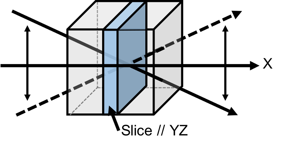
We employ the single point imaging (SPI) scheme, by which two and three dimensional in vivo EPR imaging and relaxation based oximetry have been carried out routinely in our laboratory. In this development, we use the same imaging equipment operating at 300 MHz, with an additional provision of applying a low frequency (100 Hz) sinusoidal field along one of the gradient axes at nominal AC amplitude of about 10 mT/m. The modulation of the gradient along a particular axis introduces inhomogeneity along that axis everywhere except around the midpoint at which the amplitude is zero. A two-dimensional phase encoding in a plane perpendicular to the axis of the modulated gradient retains coherent phase information only from the narrow slice at the center with spin distribution on either side of the slice undergoing total loss of coherence and does not contribute to the detected signal (Fig. 1).
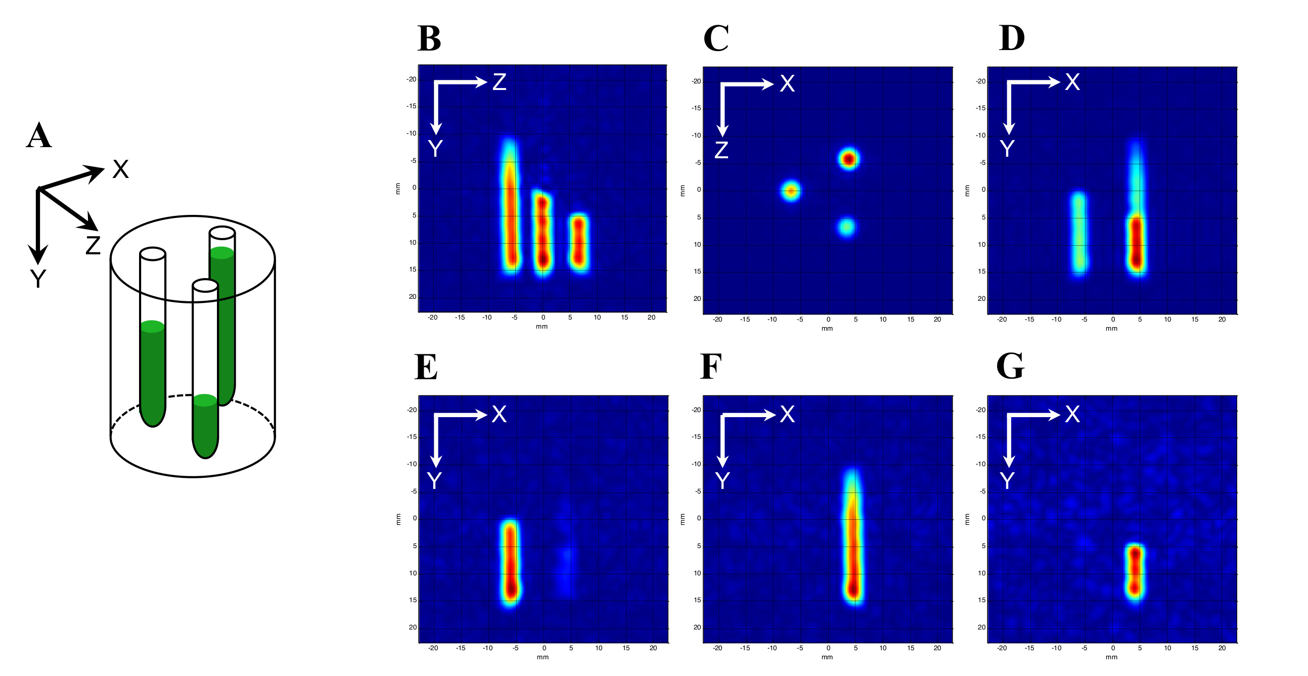
As proof of principle we made a phantom consisting of three tubes(4 mm i.d) filled to different levels with 3 mM Oxo633 (a stable trityl radical with a narrow single ESR absorption) and placed at a spacing interval of 10 mm as shown in Fig. 2A. Two dimensional images were obtained by single point imaging with a maximum gradient of 15 mT/m along the three planes.
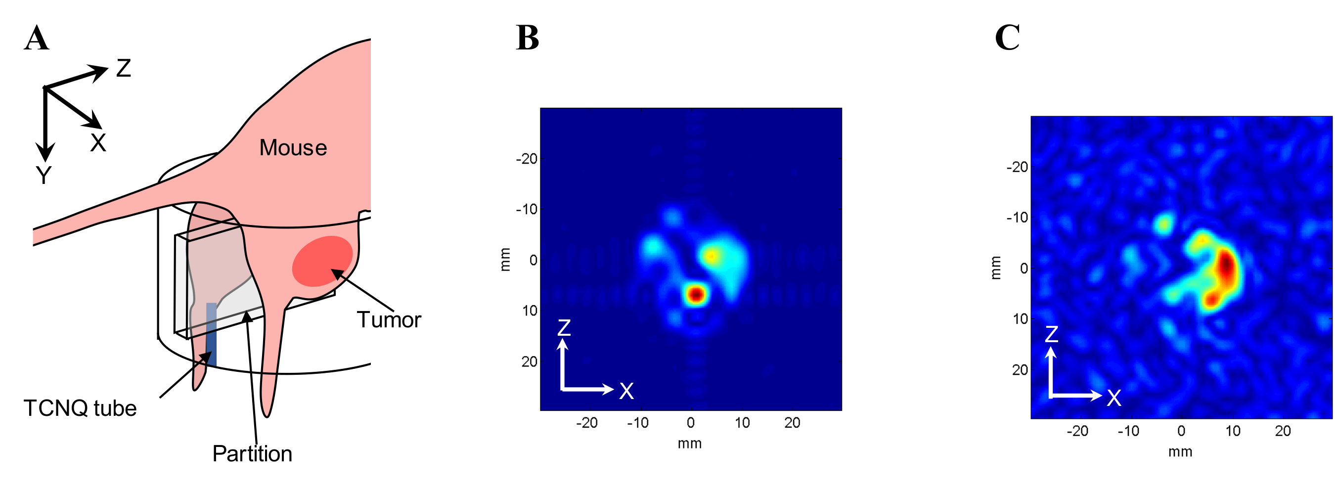
In addition to above phantom experiment, we performed in vivo experiments to investigate how dynamic range was improved using a mouse hind leg. Along with the mouse leg, we placed a TCNQ tube which produces strong signal as shown in Fig. 3A. The mouse was injected 75 mM oxo63 intravenously. Two dimensional images were obtained by single point imaging with a maximum gradient of 8 mT/m.
In order to investigate the minimum slice thickness that can be achieved, we filled a 14 mm glass cuvette (the ones used in optical spectroscopy) with 2 mM Oxo63 and placed the cuvette at the center of the resonator along Y-direction. The EPR spectra were obtained when the Z-gradient was modulated at 100 Hz with a gradual increase in amplitude from 0 to 2 volt.
Results and Discussion
When we carried out the 2D phase encoding in the XY plane with the Z-gradient being modulated at 100 Hz at amplitude of 1.4 volt, we saw only the tube centered at z-coordinate of zero (Fig. 2D). The other two tubes did not produce any signal due to the inhomogeneity imposed by the modulated Z-gradient. By shifting the resonator such that the other tubes were brought to the center sequentially, we could get slices showing exclusive images of each tube. Figure 3B and 3C shows the comparison between images acquired with conventional 2D projection and slice-selection methods. The distribution of Oxo63 acquired by 2D projection imaging was interfered by strong signal of TCNQ, while the slice selection image showed only the distribution of Oxo63. This suggested the optimum dynamic range in signal intensity was achieved by slice selection technique. The minimum slice thickness that could be achieved was around 1.7 mm at and above 1.8 volt.Conclusion
With modulation gradient, we have demonstrated the slice selection in pulsed EPR imaging and succeeded to obtain slice selected images with optimum dynamic range in signal intensity. The method will also enable the study functional dynamics in the images with improved temporal resolution.Acknowledgements
No acknowledgement found.References
- Hinshaw WS. Spin mapping:application of moving gradients to NMR. Phys Lett A. 1974; 48: 87–88.
- Sato-Akaba H, Abe H, Fujii H, Hirata H. Slice-selective images of free radicals in mice with modulated field gradient electron paramagnetic resonance (EPR) imaging. Magn Reson Med. 2008; 59: 885-890.
- Ardenkjaer-Larsen
JH, Laursen I, Leunbach I, Ehnholm G, Wistrand LG, Petersson JS, Golman K. EPR
and DNP properties of certain novel single electron contrast agents intended
for oximetric imaging. J Magn Reson 1998; 133: 1–12.
Figures