1332
A comparison of reference-based methods for removing artifacts in non-water-suppressed 1H MRSI data1New York State Psychiatric Institute, New York, NY, United States, 2Psychiatry, Columbia University, New York, NY, United States, 3Collage of Internet of Things, Hohai University, Changzhou, China, 4Radiology, Columbia University, New York, NY, United States
Synopsis
Sideband artifacts is the major obstacle to 1H MRSI without water suppression. To remove the sideband artefacts, several reference-based methods have been proposed, in which the reference signals are acquired from a water phantom with identical experimental parameters as those of in vivo scan are acquired. The reference-based methods do not suffer scan time penalty and they are compatible with any accelerated sequences such as SENSE-SI. The aim of the present work is to improve and compare the performance of two kinds of reference-based methods, namely, the phase compensation method and the artifact subtraction method.
INTRODUCTION
Proton magnetic resonance spectroscopic imaging (1H MRSI) without water suppression (WS) potentially allows the use of internal water as a standard for metabolite quantification1. The major obstacle to 1H MRSI without WS is the sideband artefacts in the non-water suppressed (NWS) spectrum associated with strong water signal modified by the exciting gradient fields2. To remove these artefacts, several reference-based methods (RBM) have been proposed, in which the reference signals are acquired from a water phantom with identical experimental parameters as those of in vivo scan are acquired3,4. The RBMs do not suffer scan time penalty are compatible with any accelerated sequences. The aim of the present work is to improve and compare the performance of two kinds of RBMs, namely, phase compensation method3 and artifact subtraction method4.METHODS
Data acquisition: We acquired 2D 1H MRSI data, from a water phantom, an MRS phantom, and a healthy volunteer, on a 3T scanner equipped with a 32-channel head coil. The sequence parameters are as follows: Sequence: PROBE-P; FOV = 22x22cm2, slice thickness =10mm; Phase encoding steps=16x16; TR/TE=1500/30ms; SW=2000Hz. The slice was on axial plane and its position was the same for both phantom and human scans in the same scan session but may differ in different sessions. For MRS phantom and human subject, the data were acquired without WS and with WS. For water phantom, only NWS data were acquired.
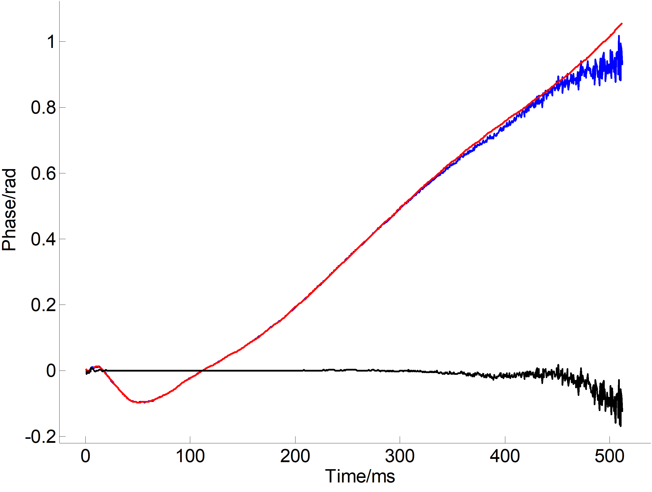
Phase-compensation method (PCM): The time domain NWS signals acquired from a water phantom and an MRS phantom or human subject are given as Sw,m,h(t)=S0w,m,h(t)exp(iφ(t)) where S0 is artifact-free signal and φ is the phase disturbance caused by time-varying magnetic field, which was assumed to be the same under identical experimental conditions4. We obtained φ from Sw and multiplied exp(-iφ(t)) to Sh to obtain the artifact-free signal. We also multiplied exp(-iφ(t)) to Sw to examine if we could get artifact-free water signal or we have residue artifacts.
Artifact subtraction method (ASM): The NWS signal is a combination of artifact-free signal and artifact signal. The latter was supposed to be the same in water phantom and human scans3. However, the differences between them lead to substantial residues. Therefore, we first performed line broadening and lineshape transformation on the water signal to make it match those of in vivo signal, and we then subtract the artifact signal of the water phantom from the NWS of the MRS phantom or human subject. In both PCM and ASM, we used an SVD-based method to model and remove water signal and to do lineshape transformation.1
Comparison: We focused on the artifacts reduction, the robustness, and the pitfalls of the two methods; we also examined approaches to improving the performance of the methods.
RESULTS
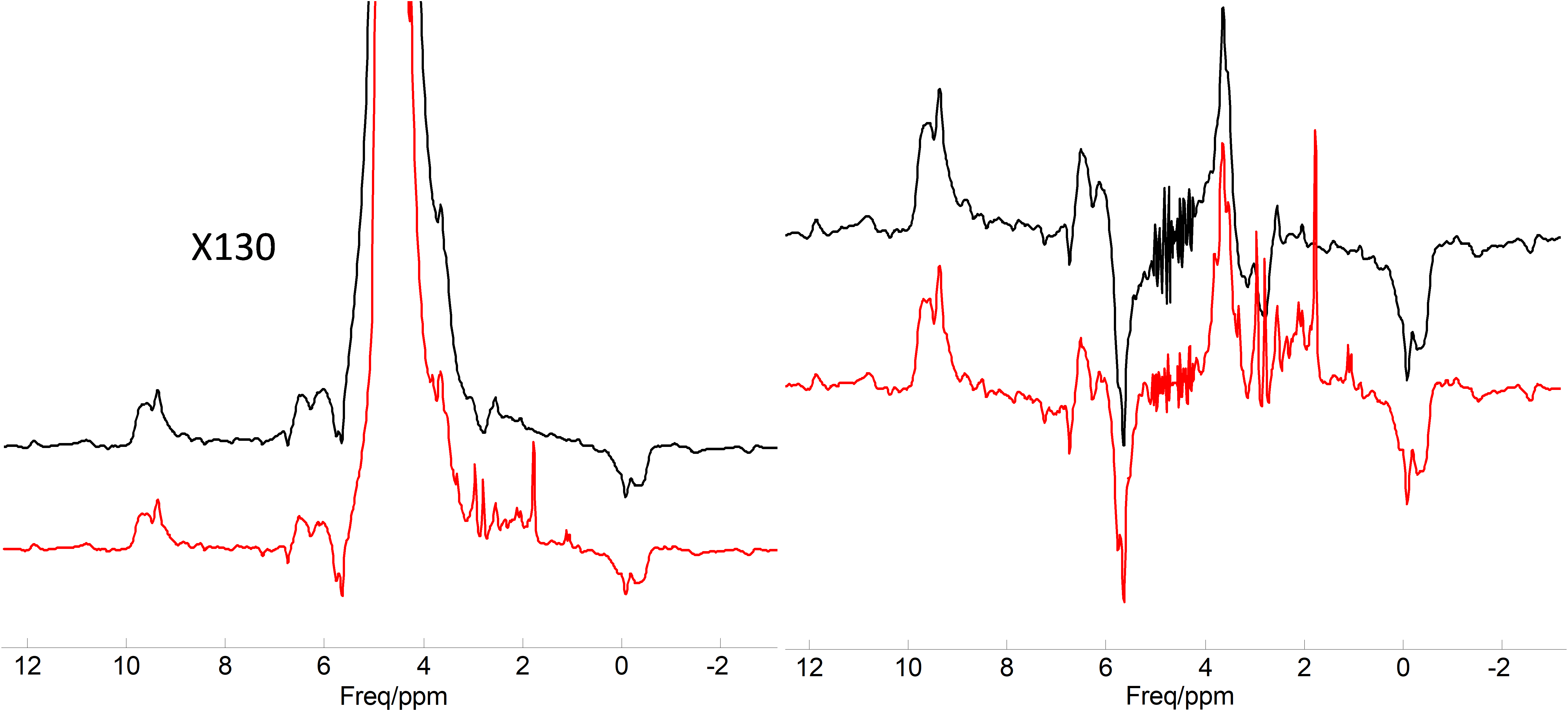
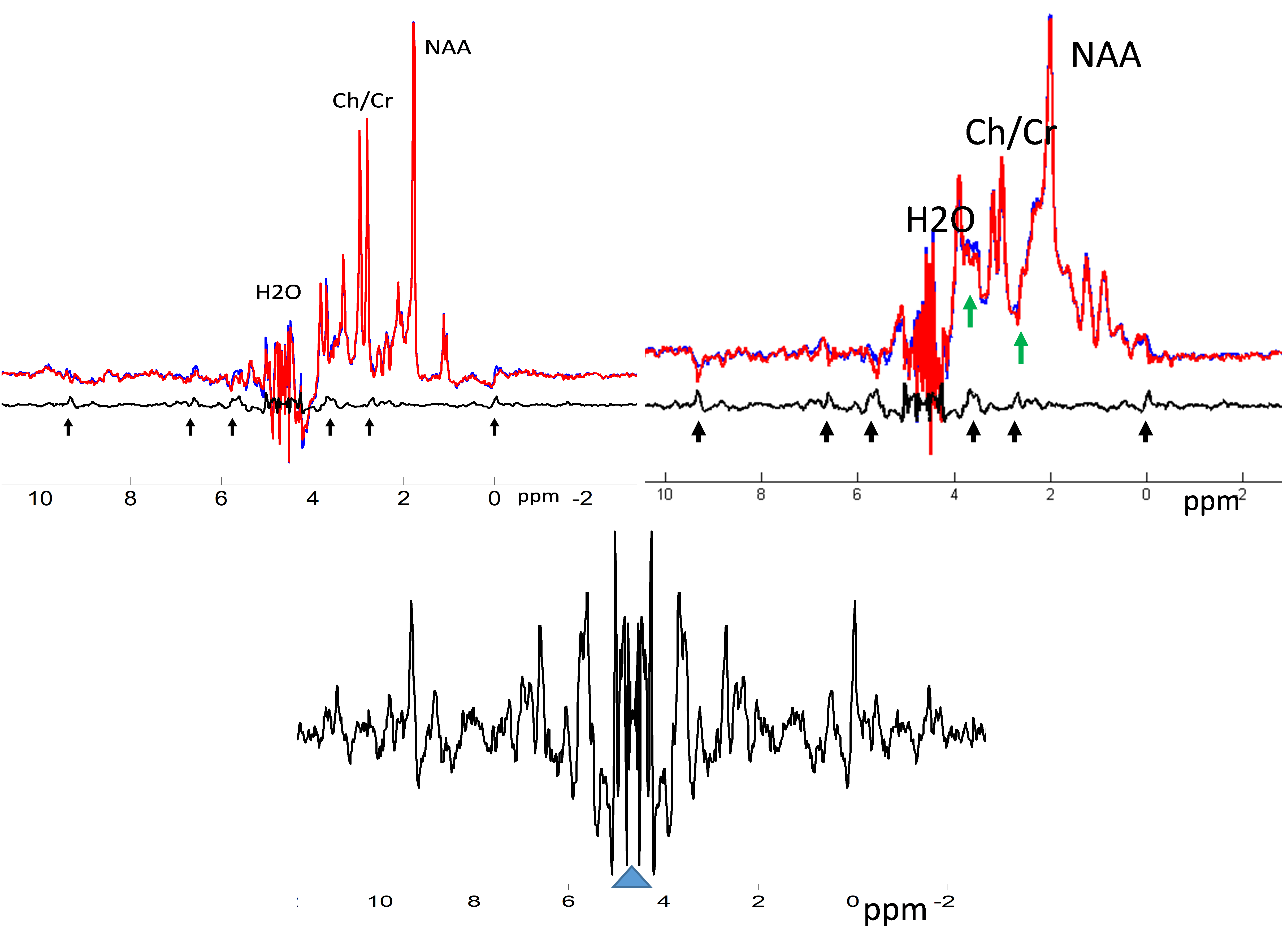
The sideband artifacts in NWS 1H MRS overlay with metabolite peaks and severely distort the baseline (Fig. 1). The phases of the water phantom FID include contributions not only from the varying magnetic fields and eddy current (EC) but also from water frequency offset (Fig. 2). Therefore, multiplying the exp(-iφ(t)) to the NWS signal will not only eliminate the sidebands and EC distortion but also partially compensate lineshape distortion cause by inhomogeneous B0. However, a structured residue signal remains even after phase compensation. We obtained these residue artifacts from reference signal and removed them from the in vivo data (Fig. 3).
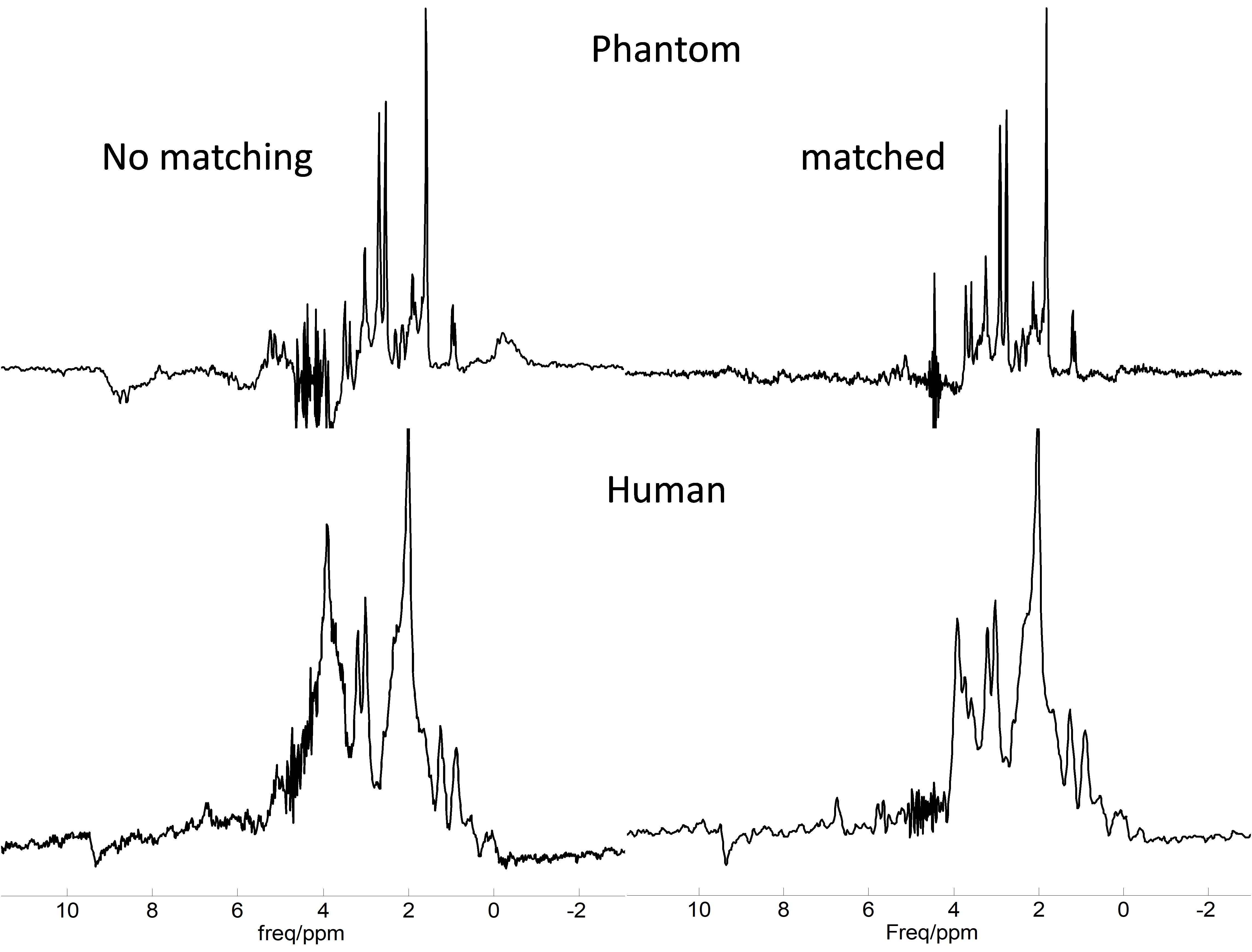
The performance of the ASM depends on matching of linewidth and lineshape between the reference and subject signals. We found that the matching of linewidth plays a major role and can be easily realized. The matching of lineshape relies on accurate water signal modeling and signal deconvolution, which is prone to errors and noise amplification (Fig. 4).
DISCUSSION
The PCM is easy to implement and stable. An important advantage of it is that it can eliminate both sideband and EC artifacts. A major finding of this work is the symmetric residue artifacts seen in the phantom spectrum (Fig. 3). These artifacts can be obtained from the signal of the water phantom and can be removed from the NWS spectra. This may improve the measurement of metabolites around 3.7 ppm and 2.4 ppm (Fig. 4). Fig. 1 shows that good shimming, ie, long FID, of the water phantom is important for the PCM.
The performance of the ASM depends on the matching of the reference and in vivo signals. The matching of lineshape is challenging. The matching of amplitude/linewidth usually suffices when the quality of MRSI spectra is good and is problematic when the linewidth is broad and lineshape distortion is severe.
Acknowledgements
References
1. Dong Z. Proton MRS and MRSI of the brain without water suppression. Prog Nucl Magn Reson Spectrosc. 2015;86-87:65-79.
2. Clayton DB, Elliott MA, Leigh JS, Lenkinski RE. 1H spectroscopy without solvent suppression: characterization of signal modulations at short echo times. Journal of magnetic resonance. 2001;153(2):203-209.
3. Chadzynski GL, Klose U. Chemical shift imaging without water suppression at 3 T. Magn Reson Imaging. 2010;28(5):669-675.
4. Ozdemir MS, Deene YD, Fieremans E, Lemahieu I. Quantitative proton magnetic resonance spectroscopy without water suppression. Journal of Instrument. 2009(4):P06014.
5. Dong Z, Dreher W, Leibfritz D. Experimental method to eliminate frequency modulation sidebands in localized in vivo 1H MR spectra acquired without water suppression. Magn Reson Med. 2004;51(3):602-606.
Figures