1310
Optimization of Radial Echo Planar Spectroscopic Image Reconstruction for Hyperpolarized [1-13C]-Pyruvate Imaging1Imaging Physics, Univ. of Texas-MD Anderson Cancer Center, Houston, TX, United States
Synopsis
Radial echo planar spectroscopic imaging (EPSI) is an efficient method for imaging hyperpolarized (HP) substrates. However, symmetric data sampling between even/odd echo components can lead to ghost artifacts that can interfere with spectral undersampling strategies that enhance SNR. The purpose of this study was to optimize the acquisition and reconstruction of a symmetric radial EPSI sequence for dynamic HP [1-13C]-pyruvate imaging. In this work, we show that the generalized Fourier transform technique preserves spectral bandwidth, reduces ghost and aliasing artifacts, and improves SNR compared to alternative strategies that separately consider even and odd echo subsets.
Purpose
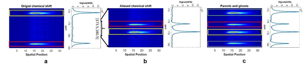
Magnetic resonance imaging of hyperpolarized (HP) agents allows non-invasive assessment of physiological processes that were previously inaccessible.1,2 Imaging HP [1-13C]-pyruvate and its metabolites has been demonstrated as an imaging biomarker of cancer metabolism through quantification of the in vivo conversion of pyruvate to lactate via aerobic glycolysis.3 While this technique provides unprecedented metabolic information, HP signals are short-lived and nonrenewable.4 Therefore, optimized HP image acquisition and reconstruction techniques are critical for the robust measurement of cancer metabolism. Radial echo planar spectroscopic imaging (EPSI) has been demonstrated as a versatile approach to dynamic HP MRI.4 While spectral undersampling in EPSI can improve the signal-to-noise (SNR) and spatial resolution, asymmetric sampling between even and odd echo components in k-t space can result in both Nyquist ghost and aliasing artifacts (Figure 1).5 In this work, we compared several image reconstruction approaches to analyze symmetric EPSI data, in an effort to improve SNR and minimize artifacts.Methods
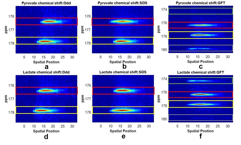
Ghost peaks can be avoided by separate analysis of even and odd echoes from a symmetric EPSI acquisition, with some sacrifice in spectral bandwidth. A generalized Fourier transform (GFT) method6 was implemented to permit reconstruction of the full dataset at full bandwidth and without ghost spectral peaks. Data acquired on a 7T small animal MR scanner (Biospec 70/30 USR, Bruker Biospin Corp., Billerica, MA, USA) was reconstructed using the GFT method, as well as by a separate analysis of the Fourier transform (FT) of the even $$$FE = FT(even)$$$ and odd $$$FO = FT(odd)$$$ echo subsets, and the FT of the even and odd echo sum-of-squares (SOS) combination $$$\sqrt{FE×FE^* + FO×FO^*}$$$. SNR was compared between all image reconstruction techniques, as well as with radial multi-band frequency encoded (MBFE) data with identical image resolution. SNR was defined as the maximum peak value divided by the standard deviation of the signal-free noise. SNR comparisons between these methods was tested using a urea phantom with a radial EPSI pulse sequence (FA = 20°, TR = 9 s, spectral BW = 9 ppm or 20 ppm, NOP = 32, image resolution = 3 mm). A second series of EPSI acquisitions (FA = 20°, TR = 2 s, spectral BW = 7 ppm, NOP = 32, image resolution = 1 mm) were carried out with the scanner frequency adjusted to replicate the chemical shifts of pyruvate and lactate using a thermal [1-13C]-urea phantom, and combined offline to simulate a true multispectral acquisition.Results
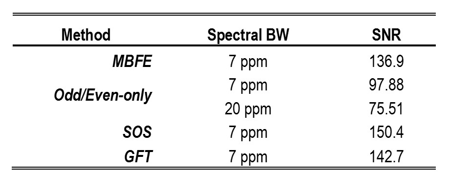
When compared to the odd/even-only components, both GFT and SOS methods improved the SNR by 54% and 46%, respectively (Table 1). EPSI reconstructed with either GFT or SOS methods had a higher SNR than MBFE imaging. A comparison of the results of the reconstruction methods is shown in Figure 2. Reducing the ESPI spectral bandwidth from 20 ppm to 9 ppm improved the SNR by approximately 30% (Table 1). The GFT method supported the full bandwidth of the symmetric EPSI acquisition and successfully suppressed ghost spectral peaks.Discussion
The SNR of EPSI reconstructed by either SOS or GFT methods is superior to even/odd-only echo components, with an SNR of ~1.4 for both methods. The GFT method supports the full spectral bandwidth of the symmetric EPSI acquisition, and reduces ghost and aliasing artifacts that can lead to spectral overlap when spectral undersampling is used to improve SNR.Conclusion
Reconstruction of symmetrically acquired EPSI data using the generalized Fourier transform reduces spectral ghosting and aliasing artifacts, and improves flexibility in protocol optimization that can enhance SNR for dynamic imaging of hyperpolarized substrates.Acknowledgements
This work was supported in part by GE Healthcare, the National Institutes of Health (P30-CA016672) and the Cancer Prevention & Research Institute of Texas (RP140021-P5).References
1. Ardenkjaer-Larsen J, Fridlund B, Gram A, et al. Increase in signal-to-noise ratio of >10,000 times in liquid-state NMR. Proc Natl Acad Sci. 2003;100(18):10158–10163.
2. Najac C, Ronen S. MR Molecular Imaging of Brain Cancer Metabolism Using Hyperpolarized 13C Magnetic Resonance Spectroscopy. Top Magn Reson Imaging. 2016;25(5):187–196.
3. Bankson J, Walker C, Ramirez M, et al. Kinetic modeling and constrained reconstruction of hyperpolarized [1-13C]-pyruvate offers improved metabolic imaging of tumors. Cancer Res. 2015;75(22):4708–4717.
4. Ramirez M, Lee J, Walker C, et al. Radial spectroscopic MRI of hyperpolarized [1-13C] pyruvate at 7 Tesla. Magn Reson Med. 2014;72(4):986–995.
5. Yan H, Braun M. Image reconstruction from Fourier domain data sampled along a zig-zag trajectory. Magn Reson Med. 1991;18(2):405–410.
6. Metzger G, Hu X. Application of interlaced Fourier transform to echo-planar spectroscopic imaging. J Magn Reson. 1997;125(1):166–170.
Figures