1299
Indirect Detection and Spin Amplification of Non-Proton MRS and MRI by Solvent Proton Signals1Chemistry and Biochemistry, UCLA, Los Angeles, CA, United States
Synopsis
A general indirect-detection and spin-amplification scheme has been developed to enhance the sensitivity of heteronuclear MRS and MRI based on dynamic instability of the solvent proton magnetization under collective feedback fields of radiation damping and the distant dipolar field. The heteronuclear solute spins are first detected by the solvent proton spins through various magnetization transfer mechanisms and serve as small “input” signals to perturb the solvent proton magnetization, which is prepared in an unstable state. The weakly detected signal is then amplified through subsequent nonlinear evolution of the solvent proton magnetization to achieve 10x SNR improvement for 13C MRS and MRI.
Introduction
Low sensitivity is particularly problematic in heteronuclear MRS and MRI, where insensitive and/or dilute heteronuclear spins are detected. A general spin amplification scheme was developed to enhance the sensitivity of heteronuclear spins based on dynamic instability of the solvent magnetization under collective feedback fields. The heteronuclear solute spins are first detected by the solvent proton spins through various magnetization transfer mechanisms and serve as small “input” signals to perturb the solvent proton magnetization, which is prepared in an unstable state. The weakly detected signal is then amplified through subsequent nonlinear evolution of the solvent proton magnetization. By manipulating bulk solvent proton spins near the threshold of instability to detect dilute heteronuclear solute spins, sensitivity and signal-to-noise ratios (SNR) of the heteronuclear MR spectroscopy and imaging can be markedly improved to achieve 10x SNR improvement for 13C MRS and MRI.Methods
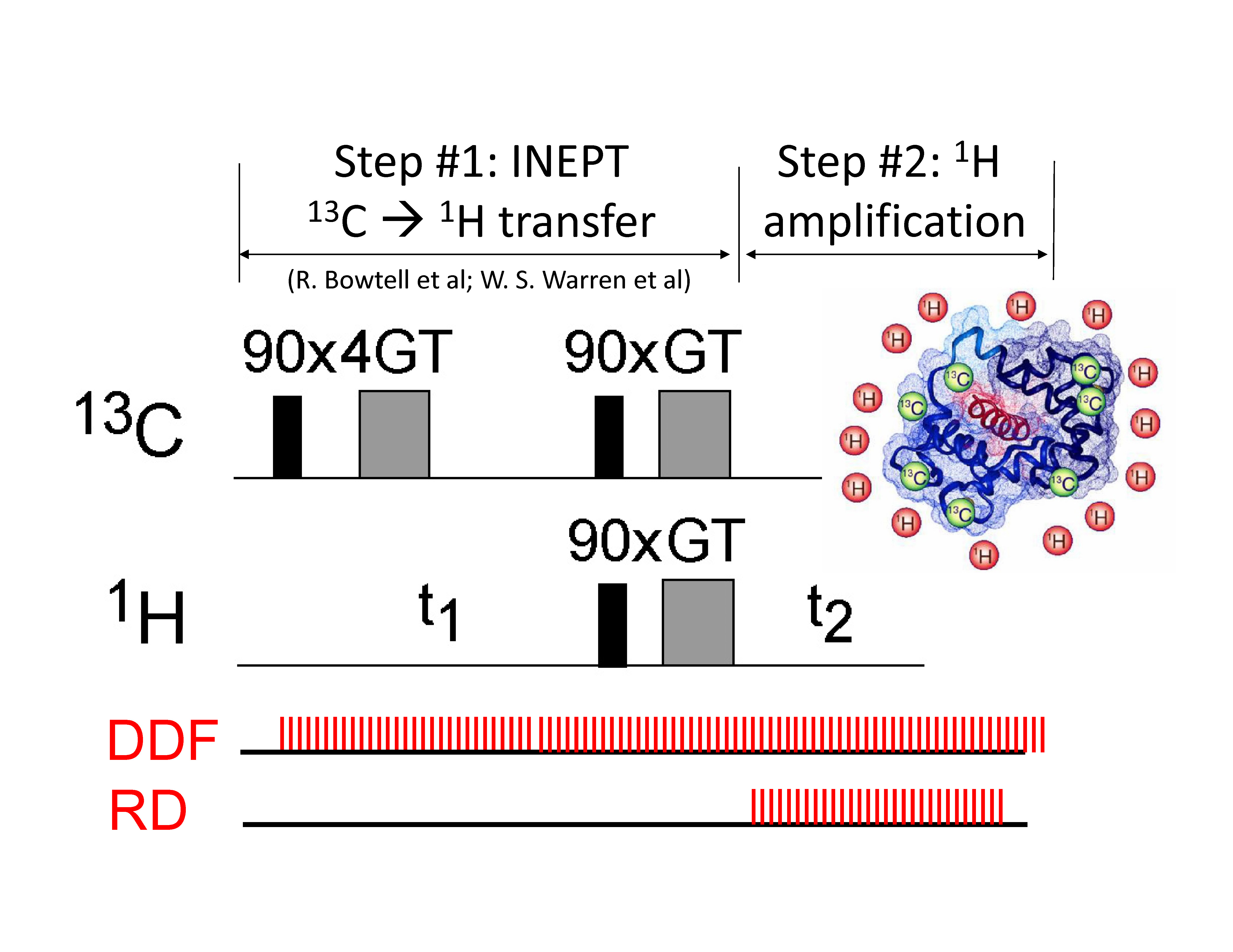
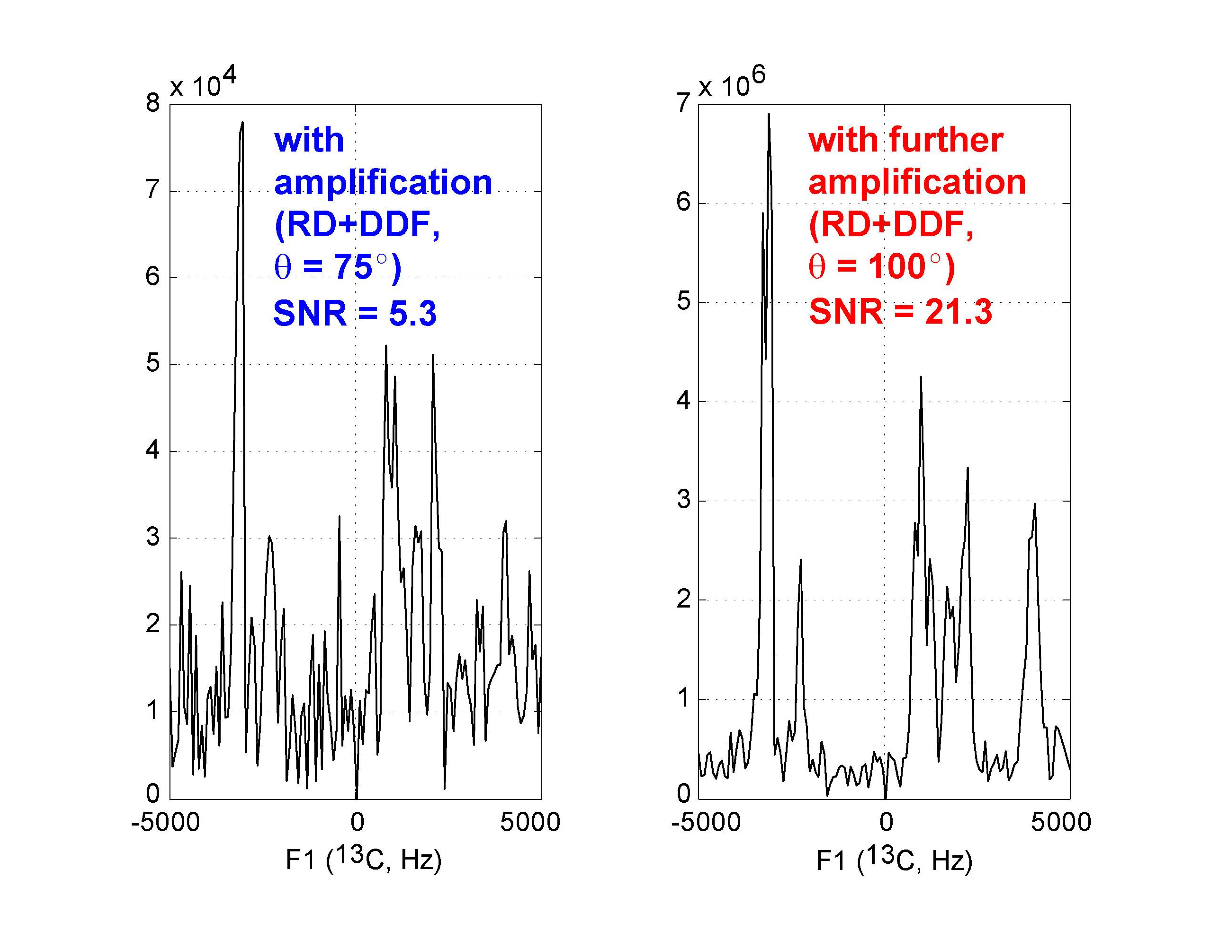
This general spin amplification scheme is shown here to amplify indirectly detected heteronuclear solute signals. Low-gyromagnetic ratio nuclei can be detected through the large 1H solvent magnetization by the distant dipolar field (DDF) [1,2]. As shown in Fig. 1 (pulse sequence at top, a+b), the modulated 1H transverse magnetization precesses under the DDF created by the spatially modulated 13C longitudinal magnetization, generating an echo in the 1H solvent magnetization that carries information about the 13C spins. Recently discovered self-refocusing of dephased solvent magnetization due to the joint action of radiation damping and the DDF [3] is exploited to enhance the indirectly detected echo signal. The extreme sensitivity of the first and largest self-refocused echo's phase and amplitude to the phase and amplitude of the initial triggering magnetization (here, the indirectly detected signal) suggests that the nonlinear spin dynamics can serve as a high-gain spin amplifier to enhance the small initial magnetization transferred to the solvent from the dilute 13C solute spins. The resulting SNR of the amplified indirectly detected echo signal, e.g., 10% 2-13C acetone solution, is improved by ~3-4x (Fig. 2b) compared to without amplification (Fig. 2a, DDF only).Results: C13 MRS
The amplification factor can be further improved by controlling the nonlinear spin evolution under the feedback fields. For example, if the 1H pulse flip angle q>90°, the instability of the inverted net 1H longitudinal magnetization under radiation damping aids in refocusing more 1H transverse magnetization. The resulting SNR is enhanced by an additional 4x, as demonstrated on U-13C glucose (Fig. 3). Moreover, field inhomogeneity or weak continuous gradients may also be exploited to accelerate the self-refocusing process [3] and increase SNR by more than 10x overall (Fig. 2c).Results: C13 MRI
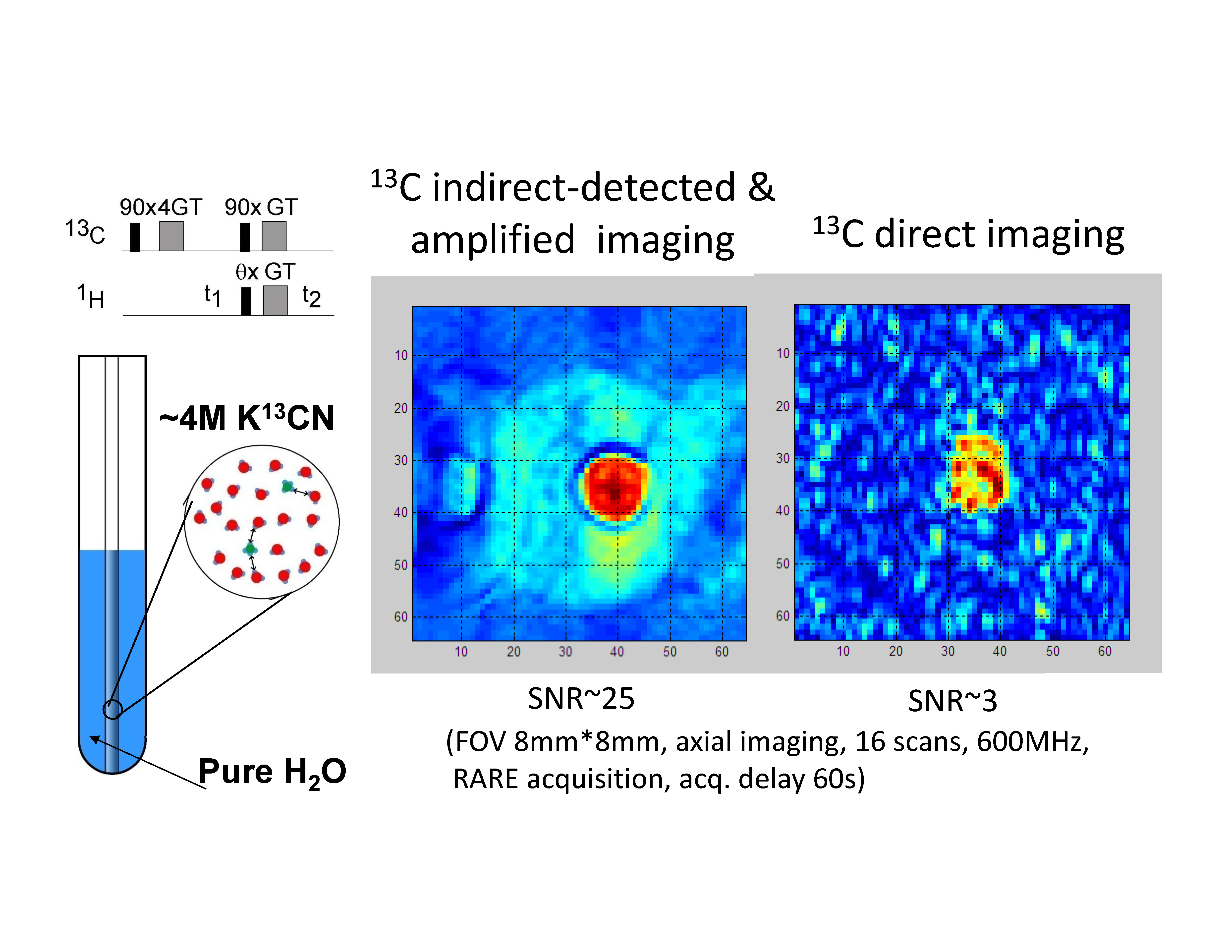
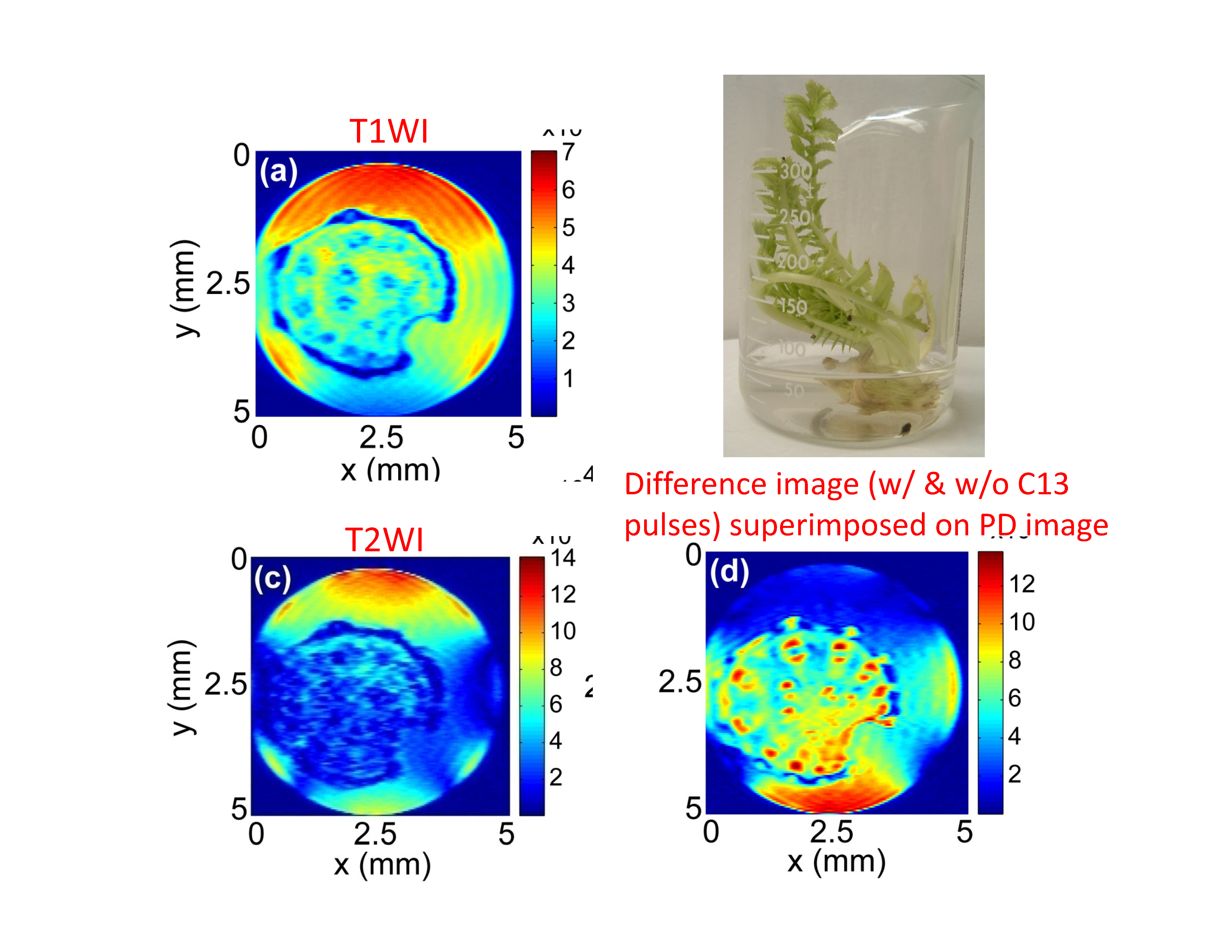
Application of this approach to 13C MRI is shown in Fig. 4 and Fig. 5 for a K13CN phantom sample and in vivo carrot stem, respectively.Conclusion
Sensitivity enhancement by the dynamic instability of solvent proton magnetization represents a new direction for surmounting a long-standing weakness of poor sensitivity in heteronuclear MRS and MRI.Acknowledgements
This work was supported by the Camille and Henry Dreyfus Foundation (TC-05-053), National Science Foundation (DMS-0833863, CHE-1112574, and CHE-1416598), Hirshberg Foundation for Pancreatic Cancer Research, and Taiwan Ministry of Science and Technology (NSC 100-2113-M-002-008, NSC 101-2113-M-002-018, and MOST 103-2923-M-002-006).References
[1] R. Bowtell, J. Magn. Reson. 100, 1 (1992)
[2] Warren et al. J. Chem. Phys. 108, 1313 (1998)
[3] Y.-Y. Lin et al. Science 290, 118 (2000).
Figures