1297
Profiling lipid composition in whole breast tumours using two dimensional (2D) double quantum filtered (DQF) correlation spectroscopy (COSY) and multiple quantum coherence (MQC) magnetic resonance spectroscopy (MRS)1Aberdeen Biomedical Imaging Centre, University of Aberdeen, Aberdeen, United Kingdom, 2Pathology Department, Aberdeen Royal Infirmary, Aberdeen, United Kingdom, 3School of Medicine, University of Aberdeen, Aberdeen, United Kingdom, 4Breast Unit, Aberdeen Royal Infirmary, Aberdeen, United Kingdom
Synopsis
Changes in lipid composition, such as polyunsaturated fatty acids (PUFA), have found to be potential biomarker of breast cancer. It has been shown that PUFA has a role in breast cancer initiation. The relationship in human between lipid composition and breast tumour grading warrants urgent investigation, as a pathway towards improved treatment. Conventional MRS suffers from overlap of nearby lipid and water peaks, and is insufficient for lipid composition measurement. We conducted double quantum filtered (DQF) correlation spectroscopy (COSY) to resolve lipid composition from the whole breast tumour, and multiple quantum coherence (MQC) MRS for further close investigation of PUFA.
Introduction
Lipid composition, the proportion between different types of lipids, plays a significant role in human health, acting as biological signalling agents in adipose tissues such as the female breast. Polyunsaturated fatty acids (PUFA), a category of lipids, was shown to be significantly different in the breast tissues of BRCA1 gene mutation carriers (high risk group) compared to healthy volunteers1, indicating its role in breast cancer initiation. Furthermore, administration of alpha-linolenic acid, a type of PUFA, has been shown to lead to breast tumour regression in mice2. The relationship in human between lipid composition and breast tumour grading warrants urgent investigation, as a pathway towards improved treatment. We hypothesise there is a difference in lipid composition between low and high grade breast cancer. To probe this hypothesis, we conducted double quantum filtered (DQF) correlation spectroscopy (COSY) to resolve lipid composition from the whole tumour, and multiple quantum coherence (MQC) MRS for further close investigation of PUFA.Methods
Thirty female patients with invasive ductal carcinoma (age 39 – 78 years, 15 grade II and 15 grade III), participated in the study. Patients with tumour size less than 1cm, undergoing chemotherapy or hormonal therapy were not eligible. Upon excision, the whole tumour specimens were immediately transported to the imaging centre for scanning. After the scanning, the specimens were transported to Pathology Department for formalin treatment (Figure 1). The study was approved by the regional Research Ethics Service, and written informed consent was obtained from all patients prior to the study.
Magnetic Resonance Spectroscopy
All spectra were collected on a 3T whole body clinical MRI scanner (Achieva TX, Philips Healthcare, Best, Netherlands), using body coil for transmission and a 32-channel receiver coil for high sensitivity detection. Standard T1-weighted anatomical images were acquired with isotropic voxel size of 1mm, TR/TE of 5.2/2.7ms, imaging volume encompassing the whole specimen. The MQC MRS3 was acquired from a single voxel snug-fit to the tumour with TR/TE of 1.25s/130ms, spectral editing frequency at 2.8ppm, 256 averages. The 2D COSY spectrum was acquired from the same location using DQF-COSY sequence4, with TR of 582ms, initial TE of 25ms, a t1 increment of 1ms, 256 increments, 4 averages.
Data Processing
The PUFA to methyl fat (0.9ppm) ratio was calculated from MQC MRS and corresponding reference spectrum using AMARES5 algorithm within the jMRUI software6. All 2D spectral data were quantified in Felix software (v2007, Accelrys Inc., San Diego, USA). Diagonal and cross peak volumes7 were calculated with reference to the methyl fat peak ((0.9,0.9)ppm) to derive the lipid composition, including monounsaturated fatty acids (MUFA) ((2.1,5.3)ppm), PUFA ((2.8,5.3)ppm). The degree of fatty acid unsaturation was calculated by dividing the volume of PUFA peak over MUFA peak8.
Statistical Analysis
All statistical analysis was performed in the SPSS software package (Release 23.0, SPSS Inc., Chicago, USA). T-tests were performed on patient characteristics and lipid composition to assess the group difference between the two grades. Pearson’s correlation tests were performed between PUFA and lipid composition against tumour size, age and body mass index. The statistical results with p value <0.05 were classified as significant.
Results
The patient characteristics are summarised in Table 1, and there was no significant difference in all the parameters between groups. There was no significant difference in PUFA, lipid composition cross peaks and degree of fatty acid unsaturation between grades (Table 2, Figure 2). There was no significant correlation between PUFA and lipid composition against tumour size, age or body mass index of the patients (Table 2, Figure 3). The DQF-COSY spectrum from a typical breast tumour is shown in Figure 2(e).Discussion
We did not observe any significant difference in PUFA, lipid composition and degree of fatty acid unsaturation between grade II and grade III breast cancer. PUFA or lipid composition was neither closely linked to tumour size, age or body mass index of the patients. Therefore, our results do not support the hypothesis of a decrease in unsaturated fatty acids during tumour development, nor support the notion of the association between PUFA or lipid composition and demographic data. However, we showed that MQC MRS minimises the contamination of water on PUFA, while the use of DQF-COSY expands the lipid peaks onto a map, allowing lipid composition to be profiled in a single scan.Conclusion
Our results show that lipid composition is not the major factor associated with breast tumour grading, indicating limited contribution from lipid changes during tumour progression. This study paves the way for further clinical applications of the MRS methods to study the roles of lipid composition in breast cancer.Acknowledgements
The authors would like to thank Mr. Nicholas Senn for conducting data auditing, Dr Matthew Clemence for clinical scientist support, Dr. Tim Smith for biologist support, Ms Bolanle Brikinns for patient recruitment support, Ms Dawn Younie for logistic support, Prof Andrew M Blamire for advice on MRS. This project was funded by Friends of ANCHOR, and Sai Man Cheung is jointly supported by Elphinstone scholarship, Roland Sutton Academic Trust and John Mallard scholarship.References
1. Ramadan, S, Arm, J, Silcock, J, et al. Lipid and Metabolite Deregulation in the Breast Tissue of Women Carrying BRCA1 and BRCA2 Genetic Mutations. Radiology. 2015; 275(3): 675 – 682.
2. Chen J, Power KA, Mann J, et al. Flaxseed alone or in combination with tamoxifen inhibits MCF-7 breast tumor growth in ovariectomized athymic mice with high circulating levels of estrogen. Exp Biol Med (Maywood) 2007; 232(8):1071-1080.
3. He, Q, Shkarin, P, Hooley, RJ, et al. In vivo MR spectroscopic imaging of polyunsaturated fatty acids (PUFA) in healthy and cancerous breast tissues by selective multiple-quantum coherence transfer (Sel-MQC): A preliminary study. Magn Reson Med. 2007; 58(6): 1079 – 1085.
4. Prescot AP, Dzik-Jurasz ASK, Leach MO, et al. Localized COSY and DQF-COSY1H-MRS sequences for investigating human tibial bone marrow in vivo and initial application to patients with acute leukemia. J Magn Reson Imag. 2005; 22(4):541–8.
5. Vanhamme L, van den Boogaart A. Improved method for accurate and efficient quantification of MRS data with use of prior knowledge. J Magn Reson. 1997;129(1):35–43.
6. Naressi A, Couturier C, Castang I, et al. Java-based graphical user interface for MRUI, a software package for quantitation of in vivo/medical magnetic resonance spectroscopy signals. Comput Biol Med. 2001; 31(4):269–86.
7. Thomas MA, Lipnick S, Velan SS, et al. Investigation of breast cancer using two-dimensional MRS. NMR Biomed. 2009; 22(1):77–91.
8. Singer S, Sivaraja M, Souza K, et al. 1H-NMR detectable fatty acyl chain unsaturation in excised leiomyosarcoma correlate with grade and mitotic activity. J Clin Invest. 1996; 98(2):244–50.
Figures
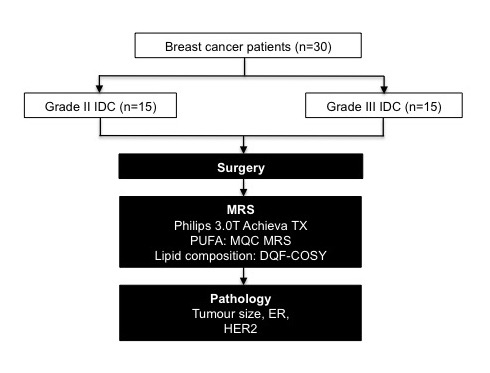
Figure 1. Study design
The study design adopting two-group cross sectional arrangement is shown in a flow chart. Thirty patients with invasive ductal carcinoma (15 grade II and 15 grade III) participated in the study. After wide local excision or mastectomy, the freshly excised tumours were scanned on a 3T clinical MRI scanner to derive PUFA and lipid composition of the whole tumour using multiple quantum coherence (MQC) MRS and double quantum filtered-correlation spectroscopy (DQF-COSY) respectively. Subsequently, histopathological analysis was carried out to derive tumour size, estrogen receptor (ER) and human epidermal growth factor receptor 2 (HER2) status.
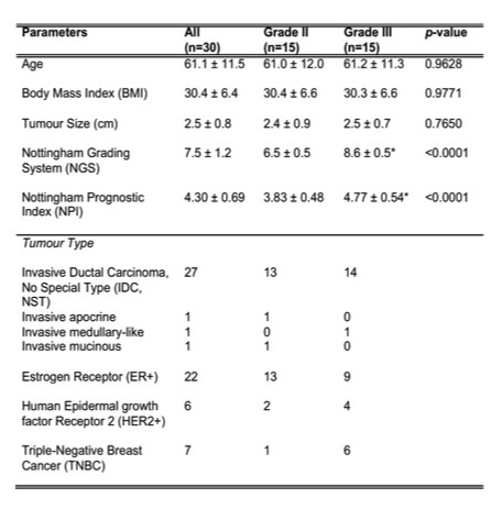
Table 1. Patient demography
Patient demographics and routine histopathological findings of excised breast tumours are shown for each group and the entire cohort. Quantitative data were expressed as mean and standard deviation, while qualitative data expressed as number of positive cases. Significant findings are marked by ‘*’.
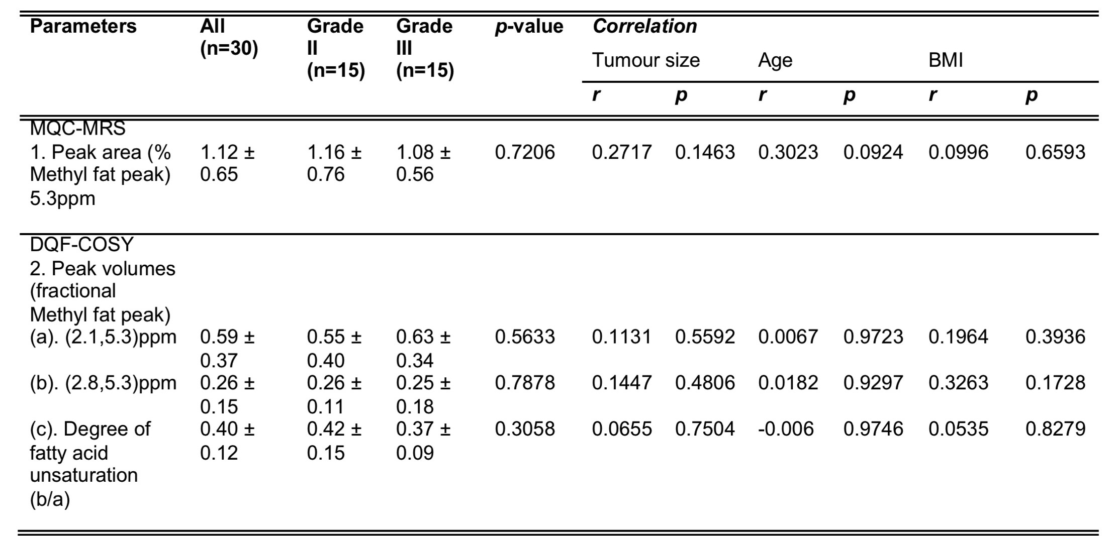
Table 2. Lipid composition and correlation with demographic indicators
PUFA from multiple quantum coherence (MQC) MRS and lipid composition from double quantum filtered-correlation spectroscopy (DQF-COSY) are shown for groups and the entire cohort. Correlation scores of PUFA and lipid composition against demographic indicators are also shown. There are no significant findings. BMI stands for body mass index.
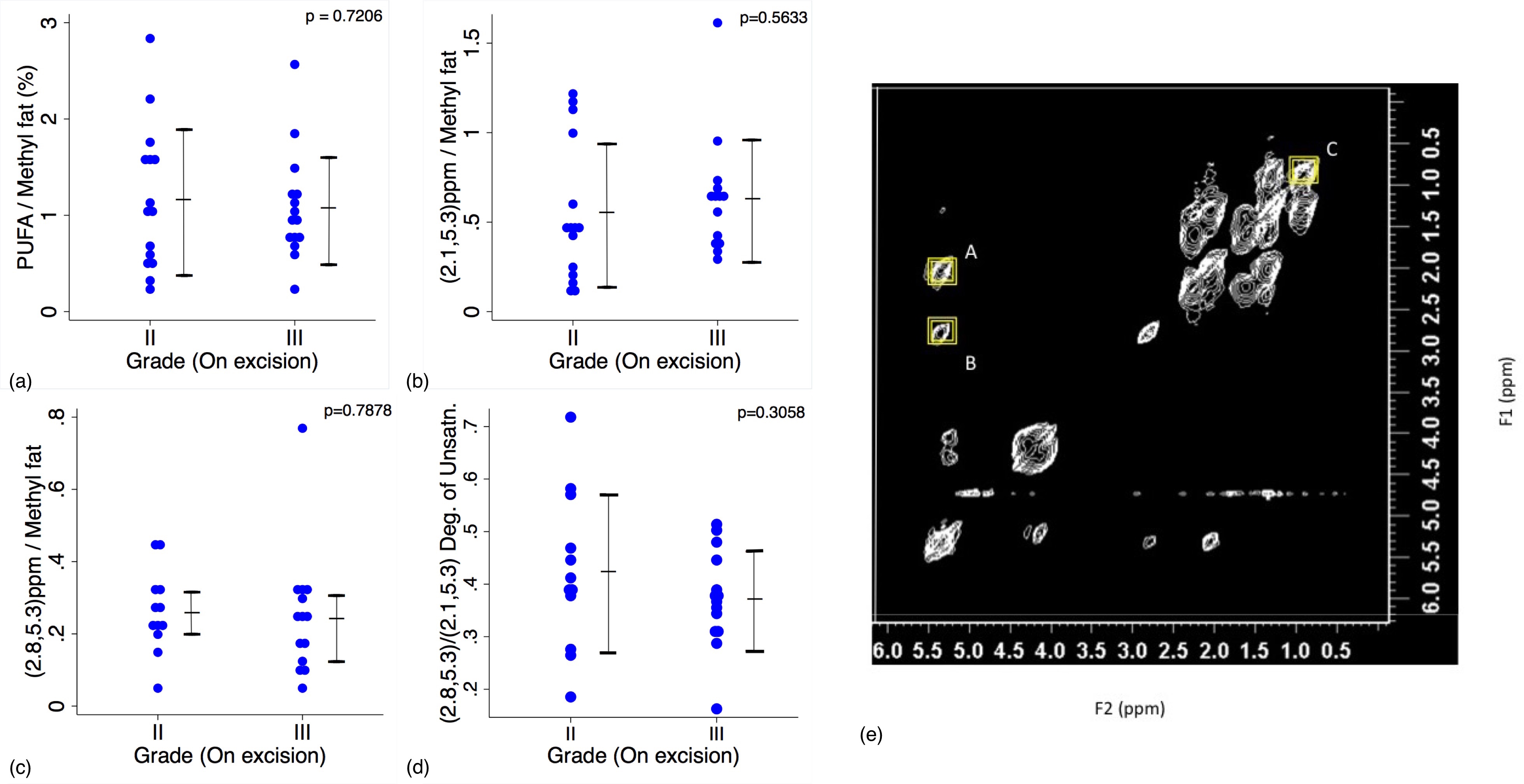
Figure 2. Group difference results and double quantum filtered-correlation spectroscopy (DQF-COSY) spectrum
The group difference in (a) PUFA (5.3)ppm in MQC MRS, (b) MUFA (2.1, 5.3)ppm, (c) PUFA (2.8, 5.3)ppm, (d) Degree of fatty acid unsaturation (PUFA/MUFA) in DQF-DOSY as shown in dot plots. The t-tests were performed between the groups and p value shown for each plot. No significant difference was observed across the comparisons. (e). A DQF-COSY spectrum obtained from a breast tumour specimen. Lipid composition are derived from (A) MUFA (2.1, 5.3)ppm, (B) PUFA (2.8, 5.3)ppm with reference to (C) Methyl fat (0.9,0.9)ppm peak.
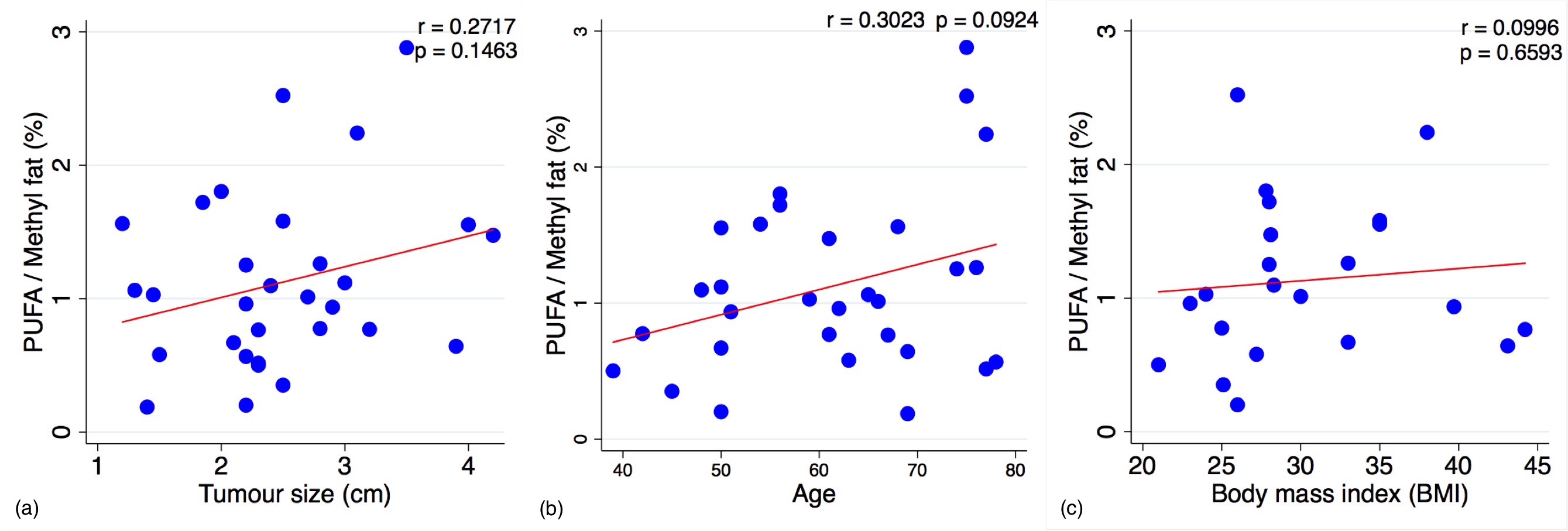
Figure 3. Correlation results
The polyunsaturated fatty acids (PUFA) from MQC MRS was correlated against (a) tumour size, (b) age and (c) body mass index within the entire cohort, and are shown as scatter plots. The regression line is shown, with the corresponding Pearson’s r score and p value displayed. PUFA is independent of tumour size, age or body mass index of the patients.