1285
Macromolecule-suppressed GABA acquisition at 7T with commonly available Gaussian editing pulses.1Imaging Institute, Cleveland Clnic, Cleveland, OH, United States
Synopsis
Co-editing of macromolecule(MM) resonances is a major problem in J-difference based editing (e.g. MEGA-PRESS) at 3T and lower field strengths. Symmetrical pulsing centered at the 1.7 ppm MM resonance alleviates this problem but results in loss of desired GABA signal, in addition to loss of unwanted MM signal, due to high bandwidth of frequency-selective editing pulses. Larger separation of editing pulses at 7T reduces the problem, but large chemical shift displacement errors, especially at low B1, make MEGA-PRESS non-viable at 7T. Using a low-power MEGA-LASER sequence, we measured macromolecule minimized GABA at 7T with editing pulses having bandwidths available in most scanners.
Introduction
Co-edited macromolecule (MM) signals during J-difference editing of GABA (as in MEGA-PRESS) is a major problem in GABA MRS. While symmetric frequency-selective 1.5 and 1.9 ppm pulses (centered at 1.7 ppm MM resonance) mitigates the problem by cancelling MM signal,1 it requires narrow bandwidth (BW) editing pulses, as broader pulses result in cancellation of GABA signal at 3T2 and lower field strengths. MEGA-SPECIAL3 reduces this problem but is highly sensitive to subject/scanner stability due to subtraction used in localization in addition to editing. The problem is largely minimized at 7T due to increased separation of pulses. However, strong chemical shift displacement error (CSDE) at 7T makes PRESS localization almost impossible since very high RF amplitude is needed to generate broadband refocusing pulses to minimize CSDE, especially at low B1, making MEGA-PRESS inviable at 7T.4, 5 Localization by adiabatic refocusing (LASER) method has overcome this difficulty. MEGA-sLASER (semi-LASER) sequence has been used at 7T,4 in which 2 pairs of adiabatic hyperbolic secant (HS) pulses are used, while LASER requires 3 pairs of HS pulses. While sLASER enables scanning with lower specific absorption rate and TE, it comes with the penalty of reduced insensitivity to B1 inhomogeneity, which is very important at high field like 7T, due to the non-adiabatic excitation pulse. Using a low power MEGA-LASER sequence on Siemens 7T scanner we have performed GABA editing without MM contamination with commonly available broad BW symmetric pulsing.Methods
Three volunteers were scanned at Siemens 3T Prisma using 20-channel (16 head / 4 neck) head coil, and 7T Magnetom with SC72 gradient (Siemens Medical Solutions, Erlangen) using a 32-channel head coil (Nova Medical). A 3×3×3 cm3 voxel in the occipital cortex was scanned for each subject at both field strengths. Water suppressed (WS) and unsuppressed (NWS) MEGA-PRESS scans were performed (TR/TE = 2000ms/68ms) with (i) 45 and (ii) 60 Hz BW editing pulses at 1.9 and 7.5 ppm (centered at water resonance) and at 1.9 and 1.5 ppm (centered at MM resonance). At 7T WS and NWS MEGA-LASER scans were performed (TR/TE =5500ms/75ms) with (i) 103 and (ii) 145 Hz BW editing pulses at 1.5 and 1.9 ppm. MEGA-LASER with 1.9 and 7.5 ppm editing pulses was also run on a single subject to obtain GABA+MM signal for comparison. In addition, a MEGA-PRESS scan was also run (TR/TE =5500ms/75ms) at 7T with 103 HZ BW editing pulse. LASER component consisted of GOIA-W(16,4)6 refocusing pulse, GOIA duration: 5 ms, BW = 20 kHz, B1-modulation: 16, G-modulation: 4, VERSE factor: 10, B1 adiabatic threshold: 570 Hz. Because of low power the B1 max was increased by 10% to compensate more for B1 inhomogeneity. Data analysis with MRUI software7 consisted of phase correction, subtracting the sum of OFF spectra from the sum of ON spectra to obtain the edited spectrum, and apodization with a Gaussian filter. Ratio of area under 3 ppm and water peaks from WS and NWS spectra respectively were determined to obtain GABA/GABA+MM levels in arbitrary unit.Results and Discussion
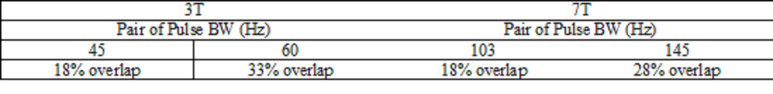
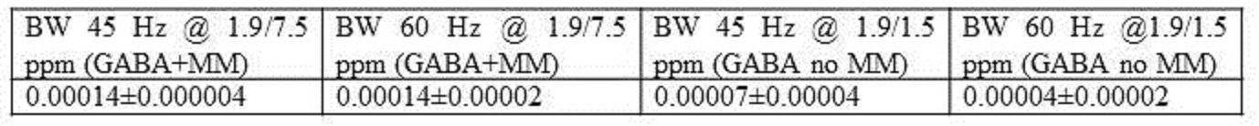
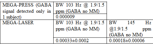
The calculated overlap of different pairs of Gaussian pulses, centered at 1.5 and 1.9 ppm, used in this study are shown in Table 1. The minimum achievable BW in most scanners is ~60 Hz,8 corresponding to ~33% overlap of the 1.5 and 1.9 ppm Gaussian pulses at 3T. At 3T GABA+MM levels are not affected by the editing pulse BW (Table 2) as there is no effect of the 7.5 ppm pulse on 1.9 ppm GABA resonance. For 1.9/1.5 ppm pulsing with 45Hz BW, GABA level is 50% lower than that of GABA+MM, while with 60 Hz BW the spectral overlap causes ~43% reduction in GABA signal than with 45 Hz pulses. At 7T, while GABA signal was barely detectable with MEGA-PRESS (Table 3), it was detected with MEGA-LASER (larger overlap with 145 Hz pulses resulted in 45% GABA signal reduction than with 103 Hz pulses). This data show that MM free GABA could be obtained with no requirement of narrow BW editing pulses at 7T. While MEGA-PRESS is not viable option at 7T,4, 5 MEGA-LASER is effective in obtaining MM free GABA spectra as can be seen from Fig. 1. Fig. 2 shows GABA+MM and GABA spectra from a single subject with MEGA-LASER. The area under the 3 ppm peak in the MM-suppressed scheme is ~70% of that in the MM-unsuppressed scheme, showing that ~30% of the GABA+MM peak is from MM.Conclusion
MEGA-LASER can be used to measure MM free GABA at 7T with Gaussian pulses available in most scanners.Acknowledgements
We thank Dr. Ovidiu Andronesi of Massachusetts General Hospital for providing us with MEGA-LASER sequences used at 7T. We thank Drs. Sineyob Ahn and Mark Brown of Siemens Healthineers for MEGA-PRESS sequence. FASTESTMAP sequence used for shimming was developed by Edward J. Auerbach and Malgorzata Marjanska and was provided by the University of Minnesota under a C2P agreement.References
1. Henry PG, Dautry C, Hantraye P, Bloch G. Brain GABA editing without macromolecule contamination. Magn Reson Med. 2001;45(3):517-520.
2. Edden RA, Puts NA, Barker PB. Macromolecule-suppressed GABA-edited magnetic resonance spectroscopy at 3T. Magn Reson Med. 2012;68(3):657-661.
3. Near J, Simpson R, Cowen P, Jezzard P. Efficient gamma-aminobutyric acid editing at 3T without macromolecule contamination: MEGA-SPECIAL. NMR Biomed. 2011;24(10):1277-1285.
4. Andreychenko A, Boer VO, Arteaga de Castro CS, Luijten PR, Klomp DW. Efficient spectral editing at 7 T: GABA detection with MEGA-sLASER. Magn Reson Med. 2012;68(4):1018-1025.
5. Bhattacharyya P, Lowe M, Andronesi OC. GABA editing with reduced sensitivity to B1 inhomogeneity and improved detectability at 7T using MEGA-LASER. Proc. Intl. Soc. Mag. Reson. Med. . 2017;25:3012.
6. Andronesi OC, Ramadan S, Ratai EM, Jennings D, Mountford CE, Sorensen AG. Spectroscopic imaging with improved gradient modulated constant adiabaticity pulses on high-field clinical scanners. J Magn Reson. 2010;203(2):283-293.
7. http://www.mrui.uab.es/mrui/.
8. Mikkelsen M, Barker PB, Bhattacharyya PK, Brix MK, Buur PF, Cecil KM, Chan KL, Chen DY, Craven AR, Cuypers K, Dacko M, Duncan NW, Dydak U, Edmondson DA, Ende G, Ersland L, Gao F, Greenhouse I, Harris AD, He N, Heba S, Hoggard N, Hsu TW, Jansen JFA, Kangarlu A, Lange T, Lebel RM, Li Y, Lin CE, Liou JK, Lirng JF, Liu F, Ma R, Maes C, Moreno-Ortega M, Murray SO, Noah S, Noeske R, Noseworthy MD, Oeltzschner G, Prisciandaro JJ, Puts NAJ, Roberts TPL, Sack M, Sailasuta N, Saleh MG, Schallmo MP, Simard N, Swinnen SP, Tegenthoff M, Truong P, Wang G, Wilkinson ID, Wittsack HJ, Xu H, Yan F, Zhang C, Zipunnikov V, Zollner HJ, Edden RAE. Big GABA: Edited MR spectroscopy at 24 research sites. Neuroimage. 2017;159:32-45.
Figures