1278
What is the optimal ROI size for single voxel MRS in global brain pathology?1Depts. Radiology and Biomedical Research, University of Bern, Bern, Switzerland
Synopsis
The purpose of this study was to investigate optimal voxel size (VS) as a compromise between increasing SNR and decreasing linewidth under the side-constraint of minimal artifact levels and to investigate potential benefits from considering signals from single coil elements separately. Eight different VS were evaluated; hinting at optimal VS of 60 cm³ and indicating that lineshape information from unsuppressed water should be included in the fitting process. Differences in single coil elements show substantial impacts on spectral quality, indicating that individual processing and exclusion of certain channels is superior to the standard procedure of an indiscriminate weighted sum.
Introduction
The optimal choice of acquisition parameters is vital in clinical MRS. In non-focal disease, the location in the brain and the size of the volume-of-interest (VOI) can often be chosen to maximize SNR as well as quality and stability of the acquired spectra and derived metabolite data. Optimal acquisition parameters are reflected in minimal Cramer-Rao Lower Bounds (CRLB), which are investigated in this work as a function of VOI size (VS) and fitting conditions. For very large VS, the spectral resolution deteriorates and outer-volume artifacts (spurious lipids) may arise[1-3]. It is thus unclear which VS will minimize the CRLB as a compromise between increasing SNR and decreasing linewidth under the additional condition of minimal artifacts. Furthermore, single channel coils have been replaced by multichannel receive arrays, which offer different spatial views on the overall signal and cannot only be used for speeding up chemical shift imaging scans (e.g. based on SENSE techniques[4]) but also – as suggested in this contribution – for improved spatial signal averaging or simultaneous evaluation of individual coil element spectra. In addition, this lends ideally to the use of lineshape information for modeling spectra with different spectral quality.Methods
Six human volunteers (supraventricular VOI), measured with a 3T MR Scanner (Prisma, Siemens), 64 channel receive head coil (44 used), semi-Laser localization sequence[5], accurate and misset 2nd order shim to simulate limited field homogeneity; Eight different VS (8 to 99 cm3 with 32 acquisitions each. Data processing by jMRUI[6] and model fitting using FiTAID[7,8]. In vitro verification in studies on a braino phantom[9].Results and Discussion
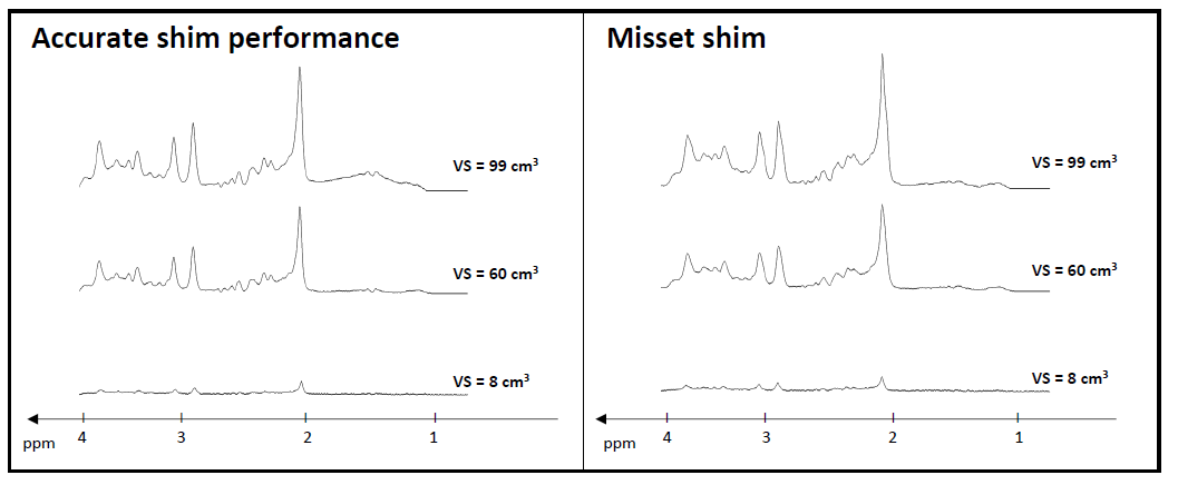
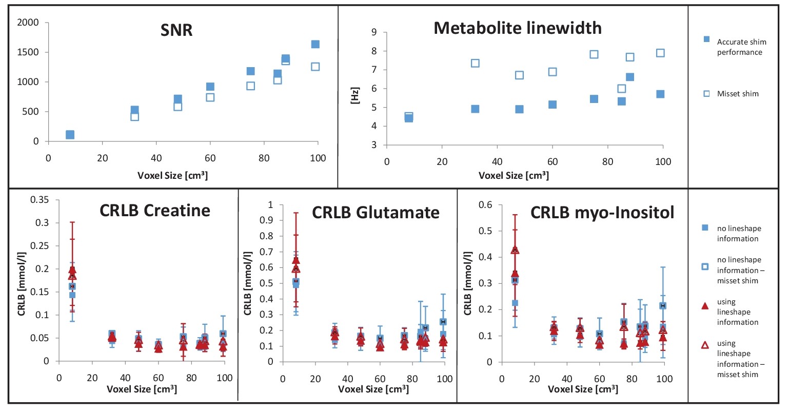
Spectral quality and SNR achieved for different VS is illustrated in Fig 1 by representative spectra for optimized and misset shimming conditions. Large VS clearly comes with degraded resolution. This is evident from Fig 2, where VS dependence of SNR and linewidth are plotted for a single case. Fig 2 also contains cohort data for some CRLBs obtained for estimated areas as function of VS. They consistently decrease with VS to a minimum and increase (with large interindividual spread) for larger VS, hinting at an optimal VS of ~60 cm3, which is larger than most of what has been used in the literature so far, but a bit less than what had been suggested in a preliminary study[9]. The increase of CRLB beyond 60 cm3 and the interindividual spread can be mitigated by inclusion of lineshape information into the fitting model.
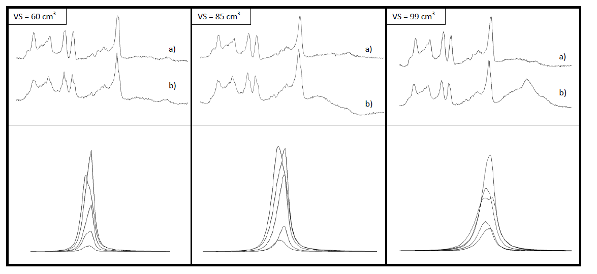
Fig 3 illustrates that the spectral quality from different coil elements can be vastly varying, especially for larger VS. This is also reflected in the peak shapes obtained from unsuppressed water, indicating that simultaneous fitting of separate spectra with inclusion of lineshape information may be superior to weighted data combination before fitting the summed spectrum. Alternatively, signals from single coil elements with large artifacts (outer-volume lipids or bad linewidth) can be omitted from the coil average, and in case of frequency shifts across the ROI, it may be beneficial to align the spectra before averaging.
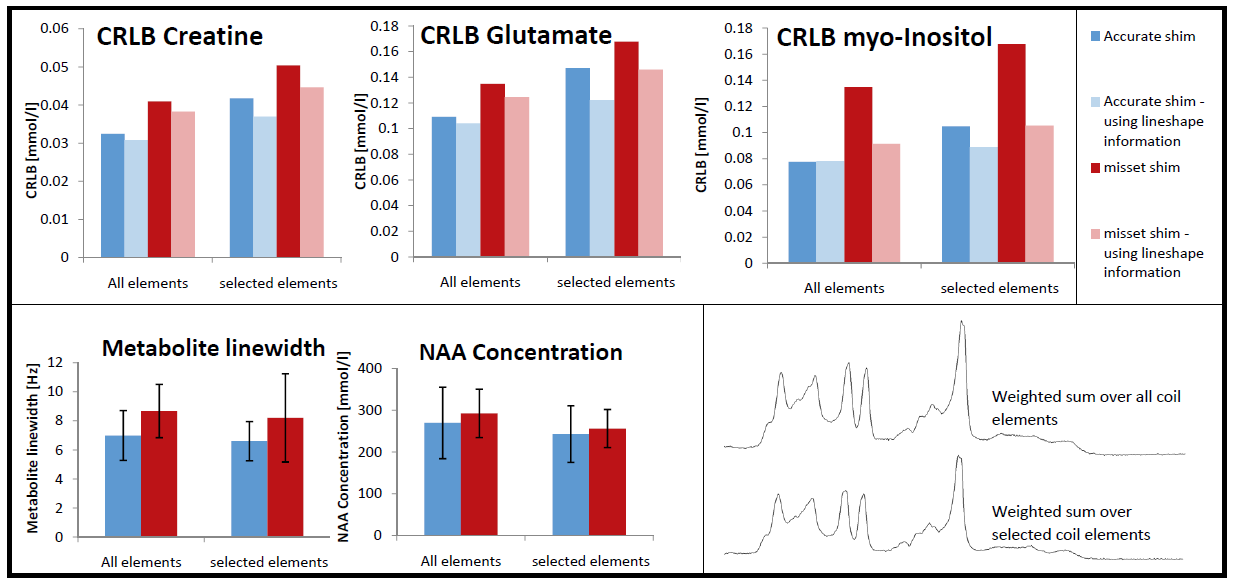
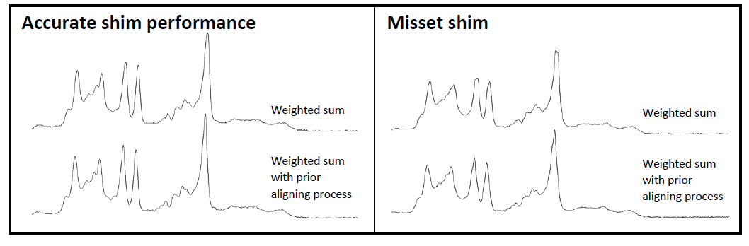
The former suggestion is illustrated in Fig 4 for the VS of 75cm3 where some coil elements show large artifacts that were excluded from the weighted sum, which then features less artifacts at the cost of 10-38% reduction in signal intensity and an increase of CRLB of ~25%. The benefit in signal quality and reliability from reduced artifacts is harder to quantify. The effect of increasing CRLB could be reduced to ~17% by using averaged lineshapes when fitting the averaged spectrum. The benefit of aligning spectra not only to reduce temporal experimental instabilities (e.g. patient motion), but also to align spectra from different coil elements within each acquisition is documented in Fig 5 where resulting coil-averaged spectra with and without alignment are shown suggesting that especially for non-ideal shim conditions linewidths and artifact levels can be reduced.
Conclusion
Increasing VS with 2nd-order fieldmap shimming yielded decreasing CRLB up to ~60 cm³. In addition, the combination of multi-channel data should not only be based on SNR and coil-coupling, but also on artifact levels and lineshapes of subspectra from single coil elements. Elimination of subspectra with bad quality or frequency alignment of subspectra may increase overall spectral quality. Inclusion of lineshape information promises to even extend the useful range of VOI sizes beyond 60 cm3. Simultaneous fitting of subspectra enforcing lineshape and coil sensitivity information may be superior to signal combination with subsequent fitting but will have to be tested with appropriate consideration of SNR in the fitting process for each of the coil elements.Acknowledgements
Supported by the Swiss National Science Foundation (#320030_156952, #320030_175984)References
1. Fleysher et al., Magn Reson Imaging (2009), 27: 222-232
2. Bartha, NMR in Biomedicine (2007) 20: 512-521
3. Juchem and de Graaf, Analytical Biochemistry (2016) doi: 10.1016/j.ab.2016.06.003
4. Dydak et al., Magn Reson Med (2001) 46:713-722
5. Marjanska and Auerbach, Center for Magnetic Resonance Research, University of Minnesota, USA; https://www.cmrr.umn.edu/spectro/
6. Stefan et al., Measurement Science & Technology (2009) 20: 104035
7. Chong et al., Magn Reson Mater Phy (2011) 24: 147-164
8. Adalid et al., Magn Reson Mater Phy (2017) doi: 10.1007/s10334-017-0618-z
9. Hoefemann et al., Magn Reson Mater Phy (2017) 30 (Suppl 1): S343–S499 #503
Figures