1274
JSASSI: A B1 Insensitive Technique for J-Resolved 2D Magnetic Resonance Spectroscopy at 7T1Radiology, Icahn School of Medicine At Mount Sinai, New York, NY, United States, 2Biomedical Engineering, City College of New York, New York, NY, United States
Synopsis
Magnetic resonance spectroscopy (MRS) can be used to investigate metabolite concentration changes correlated to neurological and psychiatric diseases. Improved spectral resolution and metabolite quantification in these disorders would add to our understanding of neurodegenerative diseases. JSASSI is a novel technique for localized two-dimensional (2D) MRS, based in part on the JPRESS spectroscopic sequence while implementing pulses from the SASSI sequence. An incrementing Δt1 time delay is introduced for resolving J-coupled metabolites from overlapping resonances. JSASSI was applied in phantoms and in vivo. Metabolite peaks for NAA, Glx, Cr and others were clearly identified using JSASSI. Unambiguous detection and resolution of J-coupled metabolites could facilitate reliable quantification of metabolites such as GABA, with potential applications in characterization and treatment monitoring in psychiatric disorders.
Introduction
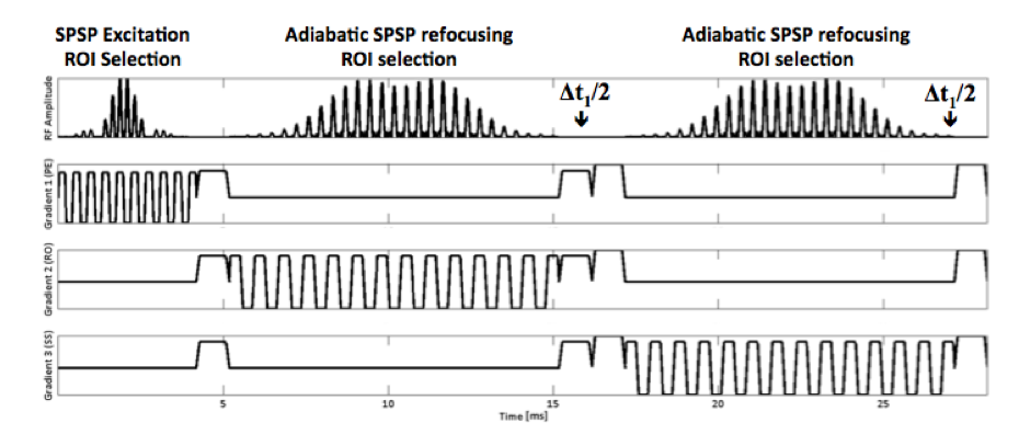
Magnetic resonance spectroscopy (MRS) can be used to investigate metabolite concentration changes correlated to neurological and psychiatric diseases1-2. Improved spectral resolution and metabolite quantification in these disorders would add to our understanding of neurodegenerative diseases. JSASSI is a novel technique for localized two-dimensional (2D) MRS, based in part on the JPRESS spectroscopic sequence3 while implementing pulses from the SASSI4. Like JPRESS, JSASSI has volume selection performed using three slice-selective radiofrequency (RF) pulses: 90°-180°-180° and an incrementing Δt1 time delay can be utilized for resolving J-coupled metabolites from overlapping resonances. SASSI pulses were selected because they are spectrally selective, less susceptible to chemical shift localization errors, and have less B1 inhomogeneity compared to conventional 180° pulses, resulting in better signal to noise ratio (SNR) and spatial coverage4. These pulses are also specific absorption rate (SAR) intensive compared to other B1 insensitive RF pulse schemes such as LASER5.Methods
An incrementing time delay was introduced before and after the final 180° RF refocusing pulse to produce a 90°-180°-(Δt1/2)-180°-(Δt1/2). Incrementing the time delay allows for indirect sampling of a second spectral dimension and thereby resolves metabolites exhibiting J-coupling from overlapping resonances. We used 7-Tesla (7T) MRI with a 32-channel Nova Medical head coil to demonstrate JSASSI in two phantoms and one healthy volunteer.
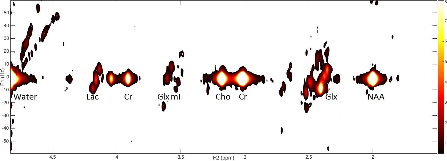
The JSASSI sequence was implemented in a gamma-Aminobutyric acid (GABA) phantom containing GABA and choline as a well as a Braino phantom containing multiple metabolites including N-acetyl-aspartate (NAA), creatine (Cr), choline (Cho), myo-inositol (mI), lactate (Lac), GABA citrate, and glutamate/glutamine (Glx). The sequence parameters were: TE = 55 ms, TR = 1500 ms, Δt1 = 0.4 ms in GABA phantom; 2.5 ms in Braino phantom, voxel size 2.2x2.2x2 cm3 [9.68 ml], F1 Bandwidth = 400 Hz, F2 Bandwidth = 2000 Hz, Measurements = 128, Averages = 1, Scan time = 3 minutes 12 seconds.
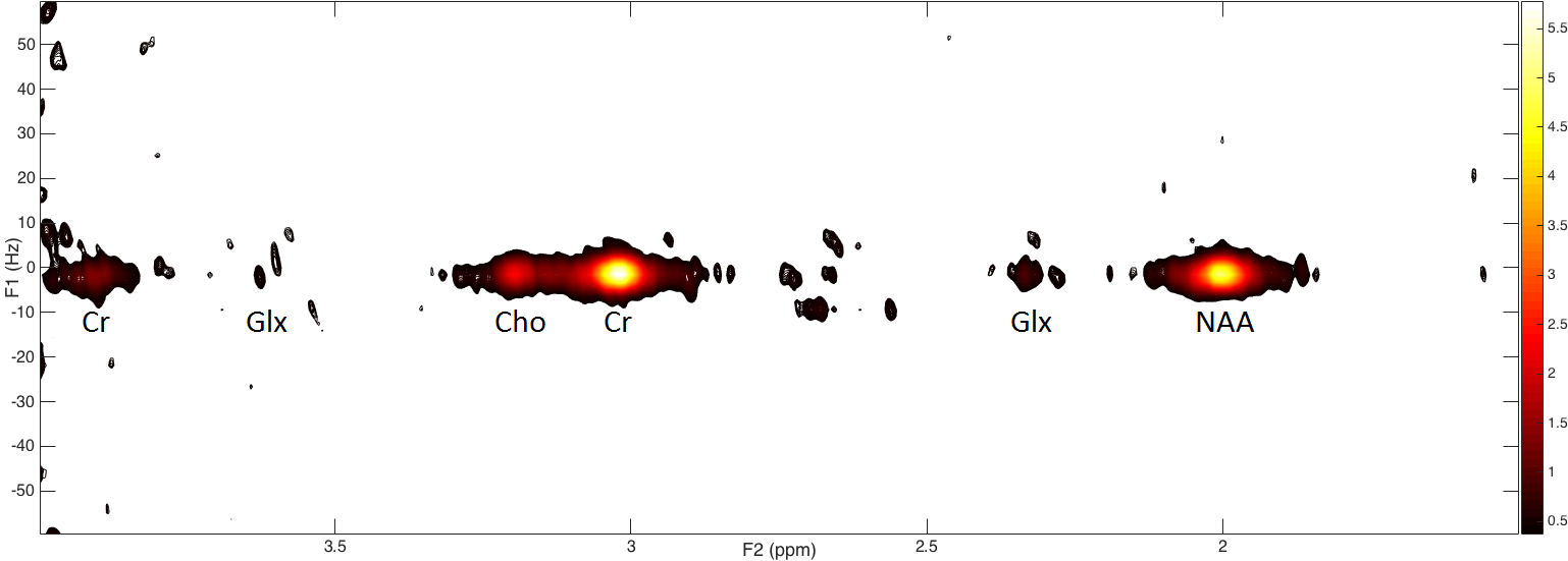
A healthy volunteer was scanned to demonstrate an in vivo application of JSASSI with the following parameters: TE = 55 ms, TR = 1500 ms, Δt1 = 2.5 ms, voxel size 3x3x3 cm3 [27 ml], F1 Bandwidth = 400 Hz, F2 Bandwidth = 2000 Hz, Measurements = 128, Averages = 2, Scan time = 6 minutes 24 seconds.
A 2D CSI implementation of JSASSI in the Braino phantom was performed, with the following parameters: TE = 55 ms, TR = 1500 ms, Δt1 = 2.5 ms, voxel size 2x2x2 cm3 [8 ml], F1 Bandwidth = 400 Hz, F2 Bandwidth = 2000 Hz, Measurements = 128, Averages = 1, Scan time = 51 minutes.
Results
We were able to identify all of the peaks in the spectra through their resonances in the JSASSI spectra. Observed J-coupling in the lactate spectrum agreed with literature values of 7 Hz J-coupling constant. In the in vivo implementation of JSASSI, we were able to observe NAA, Glx, Cr, and Cho peaks.. No water or lipid suppression pre-pulses or saturation bands were applied. Observed spectra showed minimal presence of water or lipids, confirming previous observations of spectral specificity in the SASSI pulses4.Discussion
This pilot study demonstrates the successful implementation of JSASSI in phantoms and in vivo. Future implementations of the sequence will employ acceleration strategies including echo-planar spatial acquisition and sparse sampling of the second frequency dimension to improve clinical feasibility of the sequence. Unambiguous detection and resolution of J-coupled metabolites could facilitate reliable quantification of metabolites such as GABA, with potential applications in disease characterization and treatment monitoring in psychiatric disorders.Acknowledgements
NARSAD Young Investigator Grant
NIH R01 MH109544
Icahn School of Medicine Capital Campaign
Translational and Molecular Imaging Institute
References
1. Pouwels PJ, Frahm J. Regional metabolite concentrations in human brain as determined by quantitative localized proton MRS. Magn Reson Med 1998;39(1):53-60.
2. Wijtenburg SA, Yang S, Fischer BA, Rowland LM. In vivo assessment of neurotransmitters and modulators with magnetic resonance spectroscopy: Application to schizophrenia. Neurosci Biobehav Rev 2015;51:276-295.
3. Thomas, M.A., N. Hattori, M. Umeda, T. Sawada, and S. Naruse, Evaluation of two‐dimensional L‐COSY and JPRESS using a 3 T MRI scanner: from phantoms to human brain in vivo. NMR in Biomedicine, 2003. 16(5): p. 245-251.
4. Feldman, R.E. and P. Balchandani, A semiadiabatic spectral‐spatial spectroscopic imaging (SASSI) sequence for improved high‐field MR spectroscopic imaging. Magnetic resonance in medicine, 2016. 76(4): p. 1071-1082.
5. Lin, M., A. Kumar, and S. Yang, Two‐dimensional J‐resolved LASER and semi‐LASER spectroscopy of human brain. Magnetic resonance in medicine, 2014. 71(3): p. 911-920. 1.
Figures