1265
Comparison of long TE 1H-MRS to gas chromatography-mass spectrometry for analysis of adipose tissue fat composition1German Diabetes Center Düsseldorf, Düsseldorf, Germany
Synopsis
Long TE 1H-MRS of fat results in an improved baseline and more narrow peaks but may be impacted by J-coupling effects. Here we compare long TE 1H-MRS of adipose tissue fat composition to gas chromatography-mass spectrometry (GC-MS) of adipose tissue biopsies. There was a close correlation between the two methods for both unsaturation =CH/CH2 (R = 0.719, P < 0.00001) and saturated chain length CH2/CH3 (R = 0.782, P < 0.00001). MRS overestimated unsaturation and underestimated saturated chain length. Long TE 1H-MRS allows assessment of adipose tissue fat composition, however correction factors are needed for comparison to other methodologies.
Introduction
Interest in using non-invasive magnetic resonance spectroscopy to analyse fat composition in humans in vivo has gained attention in recent years 1,2,3,4,5 . Long TE 1H-MRS of fat results in an improved baseline and more narrow peaks and can even be used to resolve the omega-3 peak in vivo 1,2,3. There are however concerns as to how much the analysis is impacted by T2 and J-coupling effects, which has led some to favour short TE 1H-MRS for analysis of fat 4,5. However, to date only two studies have validated in vivo MRS of fat composition using analysis of biopsies 1,6 and only one of those for 1H-MRS 1. Here we present further validation of using long TE 1H-MRS of adipose tissue fat composition by using gas chromatography-mass spectrometry (GC-MS) analysis of biopsies.Methods
Thirty-eight (38) volunteers were recruited for the study. The volunteers arrived 7 AM in the morning for MRS measurements and were biopsied immediately after. MRS was performed on a 3.0 Tesla MRI research scanner (Achieva, Philips, Best, The Netherlands). The volume of interest (VOI) was placed in the deep subcutaneous adipose tissue (DSAT) at the height of the umbilicus. The biopsy sample was also obtained from the DSAT at the same height under ultrasound guidance. The spectra were acquired using PRESS with 200ms/4000ms/16 (TE/TR/NSA) and a set 12x12x12mm VOI. All spectra were individually phase and frequency corrected before averaging. Spectra were analysed for unsaturation (=CH/CH2) and saturated chain length (CH2/CH3) using jMRUI. Lipid extraction and analysis from DSAT biopsies was performed according to previously described methods 7 with FAs analyzed as fatty acid (FA) methyl esters using GC-MS. The following FA were analysed from the biopsies; 12:0, 14:0, 16.0, 16:1n-7, 18:0, 18:1n-9 and 18:2n-6. The biopsy derived FA were expressed as percentage of total FAs wherefrom the unsaturation and saturated chain length was calculated, which corresponds to the (=CH/CH2) and (CH2/CH3) indices obtained by MRS. The small letter ‘c’ is used to indicated number of carbons in the saturated chain length.Results
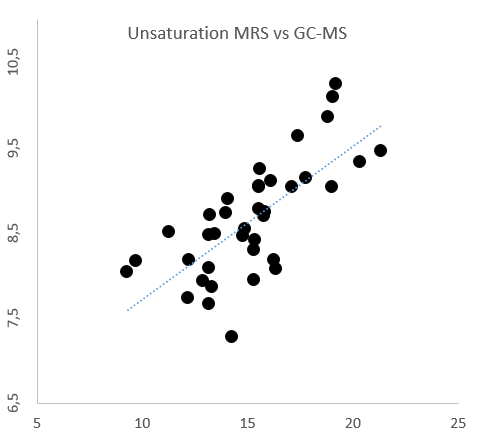
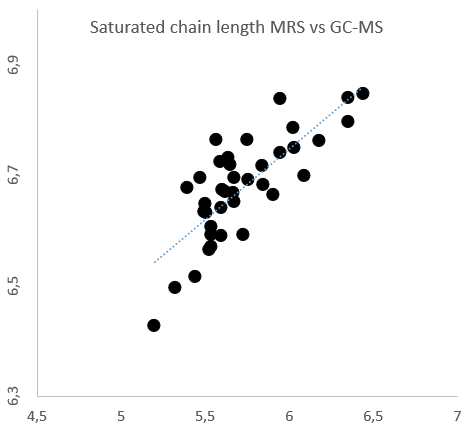
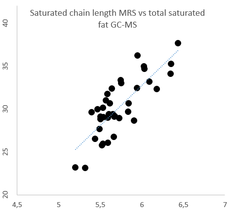
Comparison of the unsaturation (=CH/CH2) and saturated chain length (CH2/CH3) between the two methods are shown in figures 1 and 2. There was a close correlation between the two methods for both unsaturation =CH/CH2 (R = 0.719, P < 0.00001) and saturated chain length CH2/CH3 (R = 0.782, P < 0.00001). MRS overestimated unsaturation (15.2% ± 2.7%) vs GC-MS (8.7% ± 0.7%) and underestimated saturated chain length (5.7c ± 0.3c) vs GC-MS (6.7c ± 0.1c). The MRS derived saturated chain length also correlated closely with the GC-MS derived total saturated fat content (R = 0.804, P < 0.00001), see figure 3.Discussion
Long TE 1H-MRS indices of adipose tissue fat composition provides good agreement with GC-MS analysis. However, the long TE 1H-MRS derived unsaturation and saturated chain length are overestimated and underestimated, respectively. J-coupling effects do play a role, but also the limitations of the GC-MS analysis will result in inaccuracy. Long chain polyunsaturated fats are not always observed by GC-MS, which leads to an underestimation of the true unsaturation and overestimation of the true saturated chain length. The long TE 1H-MRS derived saturated chain length seems to be a good indicator of total saturated fat content.Conclusions
Long TE 1H-MRS allows assessment of adipose tissue fat composition, however correction factors are needed for comparison to other methodologies.Acknowledgements
We thank the participants
for their invaluable contributions. The German Diabetes Center is funded by the
German Federal Ministry of Health (Berlin, Germany) and the Ministry of
Innovation, Science and Research of the State of North Rhine Westphalia (Düsseldorf,
Germany). This study was supported in part by grants from the German Federal
Ministry of Education and Research (BMBF) to the German Center for Diabetes
Research (DZD e.V.), from the Helmholtz Alliance Imaging and Curing
Environmental Metabolic Diseases (ICEMED) and the Schmutzler-Stiftung.
References
1. Lundbom J, Hakkarainen A, Fielding B, et al. Characterizing human adipose tissue lipids by long echo time 1H-MRS in vivo at 1.5 Tesla: validation by gas chromatography. NMR Biomed. 2010 Jun;23(5):466-72.
2. Lundbom J, Hakkarainen A, Lundbom N, et al. Deep subcutaneous adipose tissue is more saturated than superficial subcutaneous adipose tissue. Int J Obes (Lond). 2013 Apr;37(4):620-2.
3. Lundbom J, Hakkarainen A, Söderlund S, et al. Long-TE 1H MRS suggests that liver fat is more saturated than subcutaneous and visceral fat. NMR Biomed. 2011 Apr;24(3):238-45.
4. Machann J, Stefan N, Wagner R, et al. Intra- and interindividual variability of fatty acid unsaturation in six different human adipose tissue compartments assessed by 1H-MRS in vivo at 3 T. NMR Biomed. 2017 Sep;30(9).
5. Hamilton G, Schlein AN, Middleton MS, et al. In vivo triglyceride composition of abdominal adipose tissue measured by 1H-MRS at 3T. J Magn Reson Imaging. 2017 May;45(5):1455-1463.
6. Thomas EL, Cunnane SC, Bell JD. Critical assessment of in vivo 13C-NMR spectroscopy and gas-liquid chromatography in the study of adipose tissue composition. NMR Biomed. 1998; 11: 290–296.
7. Hutchins PM, Barkley RM, Murphy RC. Separation of cellular nonpolar neutral lipids by normal-phase chromatography and analysis by electrospray ionization mass spectrometry. J Lipid Res. 2008 Apr;49(4):804-13.
Figures