1210
In-Plane Superresolution with Phaseless Subpixel Encoding1Institute for Biomedical Engineering, University of Zurich and ETH Zurich, Zurich, Switzerland
Synopsis
A method to obtain a high-resolution 2D image from a series of subpixel-encoded low-resolution 2D scans is presented. The phaseless character of this encoding allows unknown signal phase fluctuations to be easily discarded and makes the method a promising alternative to k-space segmenting in motion-sensitive experiments such as diffusion MRI. Based on an analogy with the structured illumination method used in superresolution optics, important improvements have been introduced that reduce artefacts caused by k-space truncation and magnetic field inhomogeneity. The utility of the method is demonstrated by a 3-fold resolution enhancement of diffusion-weighted EPI of human head.
Introduction
The method of super-resolution (SR) MRI based on microscopic tagging (1) can be regarded as an analogue of the harmonic excitation (or structured illumination) used in SR optics (2,3) to surpass the diffraction limit by shifting the normally inaccessible spatial frequencies into the aperture of the imaging system. The phaseless character of the tag-based encoding (4) makes this method a promising tool for MRI applications where the signal phase is instable, e.g., diffusion tensor imaging. We explore this optical analogy to improve the reconstruction of phaseless SR-MRI by suppressing artefacts resulting from k-space truncation (Gibbs phenomenon) and from tagging pattern distortions caused by magnetic field inhomogeneity.Methods
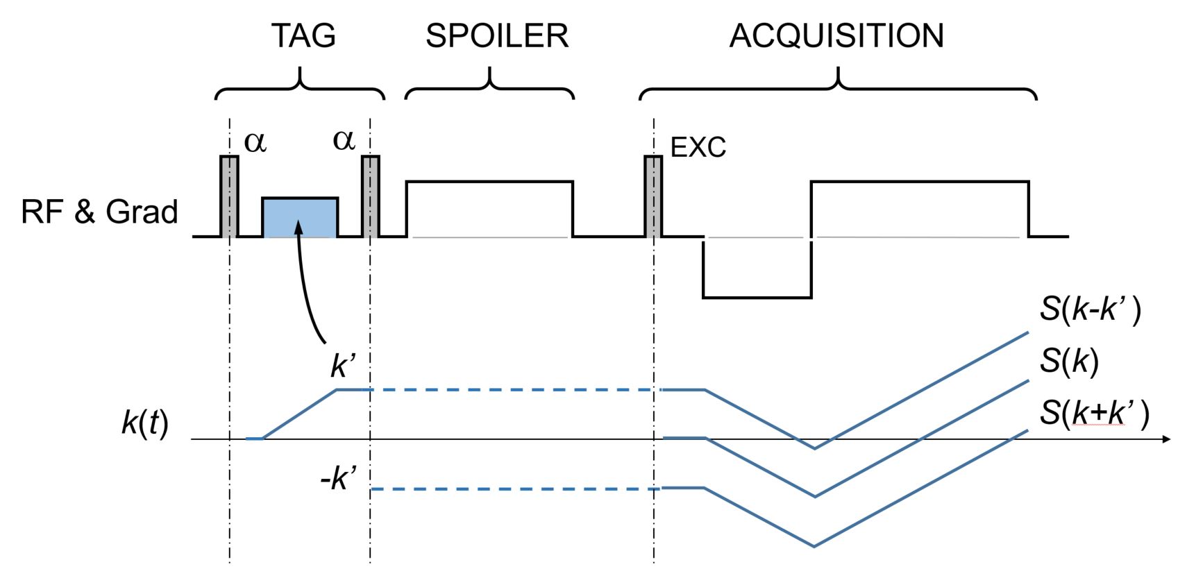
A single-shot imaging sequence such as EPI is preceded by a tagging module consisting of two RF pulses separated by a gradient pulse of integral $$$ k’ $$$ (Fig.1). Low resolution single shot images are reconstructed using a k-space window $$$ W(k) $$$ to limit the Gibbs ringing, and transformed back to k-space following the magnitude calculation to remove the random phase. The contributions from the k-space bands shifted by $$$ \pm k’ $$$, introduced by the tagging, are separated from the central band based on a phase cycle of the RF pulses (which shifts the pattern in space), shifted to their proper positions, Fourier transformed and summed up. The result is equal to the FT of the true k-space data multiplied by three shifted copies of the low-resolution k-space window:
$$ S_{\text{final}}(k) = S_{\text{true}}(k) \times \left[ W(k+k’) + W(k) + W(k-k’) \right] $$
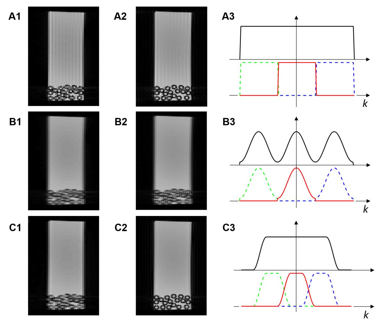
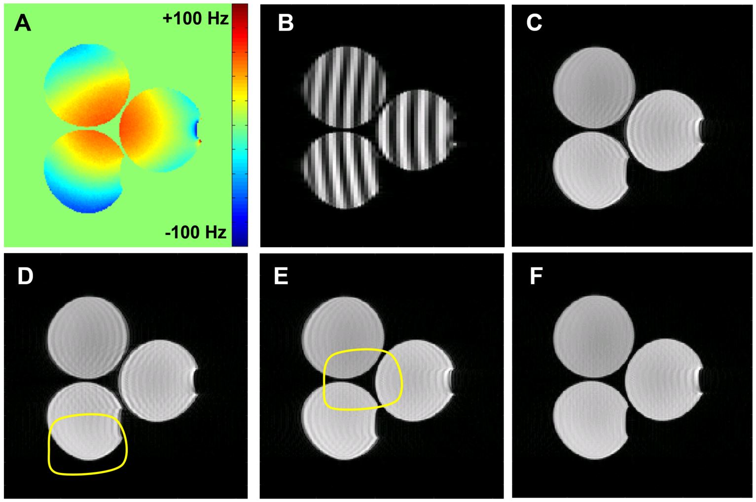
The resolution gain is thus maximally threefold when W is rectangular and k’ is set to its width. This, however, leaves Gibbs ringing on the low-resolution images, which propagates to the final SR image. Results are better with a sine-trapezoidal window and the three bands properly overlapping to produce a flat top of the final filter. The trade-off is a slightly reduced resolution gain. Due to magnetic field inhomogeneity, the sinusoidal tagging pattern is distorted with a local phase shift equal to the spin phase accrual between the two RF pulses. This phase shift propagates to the reconstructed k-space sidebands and causes ringing artefacts. However, the shift can be calculated based on a magnetic field map and taken into account during the reconstruction. For that purpose, the phase of partial images reconstructed from each band is corrected by the calculated position-dependent shift (with opposite sign for opposite-side bands) prior to the addition of the images.
Results and Discussion
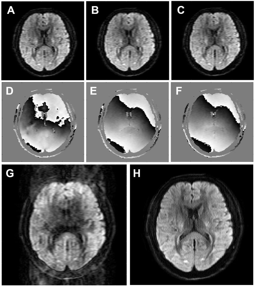
The advantage of partial band overlapping is demonstrated in Fig.2. With adjacent bands (and maximum resolution enhancement) the low resolution images must be reconstructed without apodization and therefore show Gibbs ringing, which propagates to the final SR image. An attempt to reduce ringing by k-space apodization (hamming window) makes the image even worse due to periodicity of the effective filter caused by the replication in Eq. 1. However, with partial band overlapping (and a slightly reduced resolution gain), a filter can be applied that combines to a flat-top widow upon replication and effectively reduces the ringing phenomenon. The artefact caused by field-inhomogeneity and the related tagging distortion, as well as the advantage of the field map based reconstruction is shown in Fig. 3. Without correction, ringing appears in off-resonance regions. The correction is effective everywhere except for regions where local field gradients are strong enough to cause intra-voxel dephasing. The utility of phaseless subpixel encoding in demonstrated by a diffusion-weighted (b=1000 s/mm2) EPI acquisition carried out on a 3T whole body system (Philips Achieva) equipped with 30 mT/m gradients (Fig. 4). A healthy male adult volunteered for the study in compliance with the institution guidelines. Single-shot images with resolution 3.2 mm (readout direction, horizontal) and 1.2 mm (phase encoding, vertical) were required with three shifts of the tagging pattern and used to reconstruct a super-resolved image with isotropic resolution of 1.2mm. This reconstruction is artefact-free despite apparent phase fluctuations between the shots. With classic segmented (interleaved) EPI the same phase fluctuation pattern would cause significant ghosting.
Conclusion
The k-space analysis of the tagging-based phaseless superresolution encoding, inspired by the methodology of structured illumination optics, reveals the source of the resolution gain of this technique, which is the mixing of three k-space bands. It allows for a compromise between the resolution gain and the strength of the anti-ringing filter and for a correction of tag distortions caused by an inhomogeneous magnetic field. EPI images of a resolution attainable only by multi-shot methods could be obtained without the phase sensitivity characteristic to multi-shot EPI.Acknowledgements
No acknowledgement found.References
- Ropele S, Ebner F, Fazekas F, Reishofer G. Super-resolution MRI using microscopic spatial modulation of magnetization. Magn Reson Med. 2010; 64:1671–5 2.
- Frohn JT, Knapp HF, Stemmer A. True optical resolution beyond the Rayleigh limit achieved by standing wave illumination. Proc Natl Acad Sci. 2000; 97:7232–6 3.
- Gustafsson MGL. Surpassing the lateral resolution limit by a factor of two using structured illumination microscopy. J Microsc. 2000; 198:82–7 4.
- Hennel F and Pruessmann KP, MRI with phaseless encoding, Magn Reson Med 2016, DOI: 10.1002/mrm.26497
Figures