1204
Fast Distortion-Free Diffusion Imaging using “tilted-CAIPI” PSF-EPI1Center for Biomedical Imaging Research, Department of Biomedical Engineering, School of Medicine, Tsinghua University, Beijing, China, 2A. A. Martinos Center for Biomedical Imaging, Department of Radiology, Massachusetts General Hospital, Charlestown, MA, United States, 3Harvard-MIT Health Sciences and Technology, MIT, Cambridge, MA, United States
Synopsis
Single-shot EPI is widely used for diffusion imaging, but suffers from susceptibility-induced distortion and T2* blurring, which limit its resolution and ability to detect detailed structures. PSF-encoded EPI with an added 2D-navigator has recently been developed to allow distortion- and blurring- free diffusion imaging. However, the extremely long acquisition of PSF-EPI makes it impractical for high angular resolution diffusion imaging. In this work, PSF-EPI is accelerated by >20x using a novel “tilted-CAIPI” approach, along with self-navigation strategy. This has enabled distortion- and blurring-free diffusion imaging at 1.2mm isotropic to be acquired with 48 diffusion directions in 21-minutes for whole-brain.
Introduction
Single-shot EPI (SS-EPI) is widely used for diffusion imaging, but suffers from susceptibility-induced distortion and T2* blurring, which limit its resolution and ability to detect detailed structures. To reduce distortion and blurring, SS-EPI with parallel imaging [1,2] has been developed to reduce the effective echo spacing (ESP). However, distortion/blurring mitigation is limited by achievable parallel imaging acceleration (RPE), where high accelerations lead to severe SNR loss. As an alternative, a number of multi-shot EPI approaches have been proposed to reduce the distortion while maintaining high SNR [3-7]. Nevertheless, to achieve imaging with very low/no distortion, a very large number of shots is required, and navigator acquisition might be necessary for phase correction.
Recently, a distortion- and blurring-free imaging technique termed, Point-Spread-Function encoded EPI (PSF-EPI) [8,9] has been applied to diffusion imaging with 2D phase navigator acquisition, allowing high-quality, high-resolution DWI [8]. Nonetheless, the acquisition time of PSF-EPI is extremely long. For example, fully-sampled PSF-EPI needs ~200 shots for 1mm-resolution brain imaging, and even with accelerated methods, ~40 shots are still necessary [8]. To achieve fast PSF-EPI, a novel method termed tilted-CAIPI is proposed in our work [10], which achieves high-quality distortion-free imaging at this resolution range in just 4-8 shots. In this work, we apply and further refine tilted-CAIPI PSF-EPI for diffusion imaging, to also allow for self-navigation of phase correction.
Methods
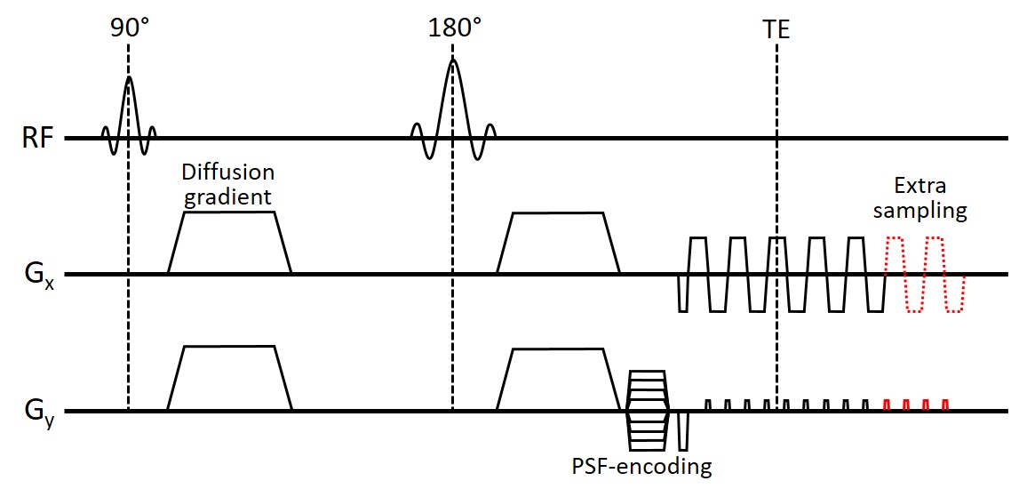
PSF-EPI utilizes an additional phase-encoding dimension, PSF-encoding (kpsf), to acquire a distortion-free spatial dimension. As shown in Fig.1, a spin-warp gradient along the phase-encoding (PE) direction is applied before EPI readout, and changes shot-to-shot. Since signals along PSF direction are acquired at the same echo time, there is no susceptibility-induced phase accumulation or T2* decay, thus, PSF-encoding dimension is distortion-free and blurring-free. To improve the acquisition efficiency, PSF-encoding dimension is under-sampled by a very large acceleration factor, and can be well-reconstructed with our tilted-CAIPI [10] acquisition and reconstruction. For further acceleration and TE shortening, partial Fourier acquisition along both PSF and PE dimensions was applied, and recovered by the POCS algorithm.
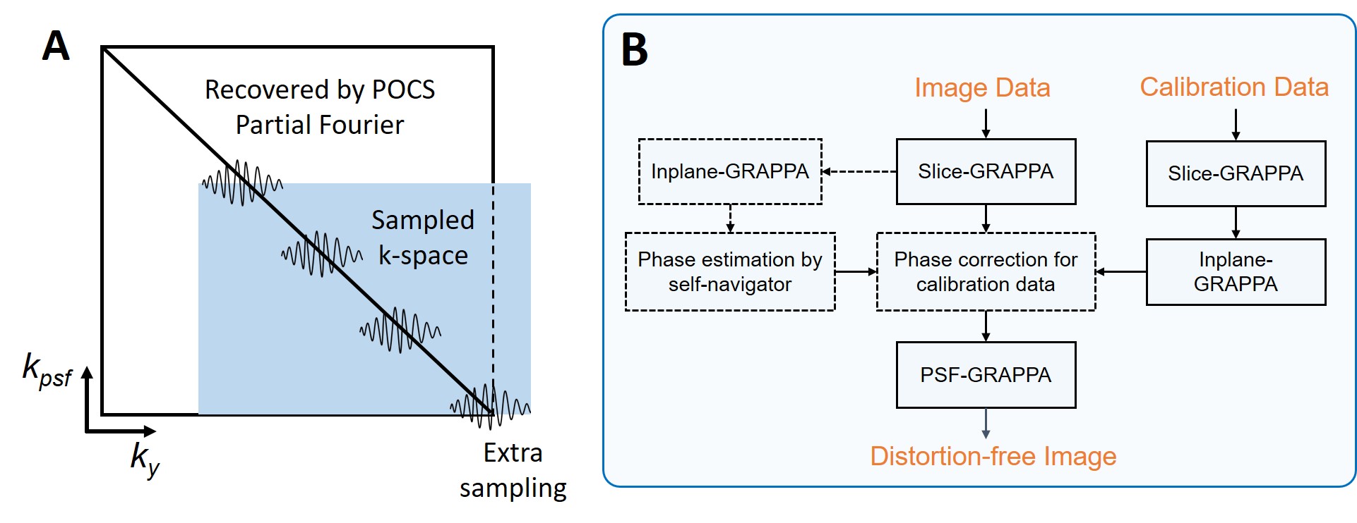
A big challenge for multi-shot DWI is the correction of shot-to-shot image phase corruption due to physiological motion during diffusion encoding. Here, a self-navigated method is developed for PSF-EPI. As shown in Fig.2A, each kpsf-encoding contains a portion of 2D low-frequency signals in kx-ky (marked by wiggly signals (kx is omitted)), which can be used to estimate the image phase of that shot. Since identical kpsf-encodings are used for non-DW and DW images, physiological motion-induced phase can be estimated by subtracting the phase of a non-DW image from one of DW image of the same kpsf-encoding. However, the kpsf-encoding shifts the k-space center which causes the last several kpsf-encoding steps to lose low-frequency information (Fig.2A). To compensate for the missing low-frequency signals, extra-sampling of PE lines is implemented as shown in Fig.1&2.
Since a joint PSF-PE GRAPPA reconstruction across kpsf-ky data is used in tilted-CAIPI to achieve high-quality reconstruction, shot-to-shot phase variation across kpsf needs to be accounted for prior to this reconstruction. Here, the phase corruption of each kpsf shot is estimated by first reconstructing each shot individually through conventional slice- and inplane-GRAPPA, and then comparing the reconstructed image phase to that of the non-DW data at same kpsf-encoding. The estimated phase is then added into calibration data for each PSF-encoding as an additional information to train PSF-GRAPPA kernel for each DWI volume [3,11]. The flowchart of the reconstruction is shown in Fig.2B.
Experiments: All experiments were performed on a 3T Siemens Prisma scanner. The first DWI experiment was performed at 1×1×3mm3 resolution to evaluate the performance of tilted-CAIPI PSF-EPI and the feasibility of phase correction using self-navigator. Two additional DWI experiments were then performed for HARDI acquisition at 0.8×0.8×3mm3 and 1.2mm-isotropic resolution, each with 48-diffusion directions.
Results
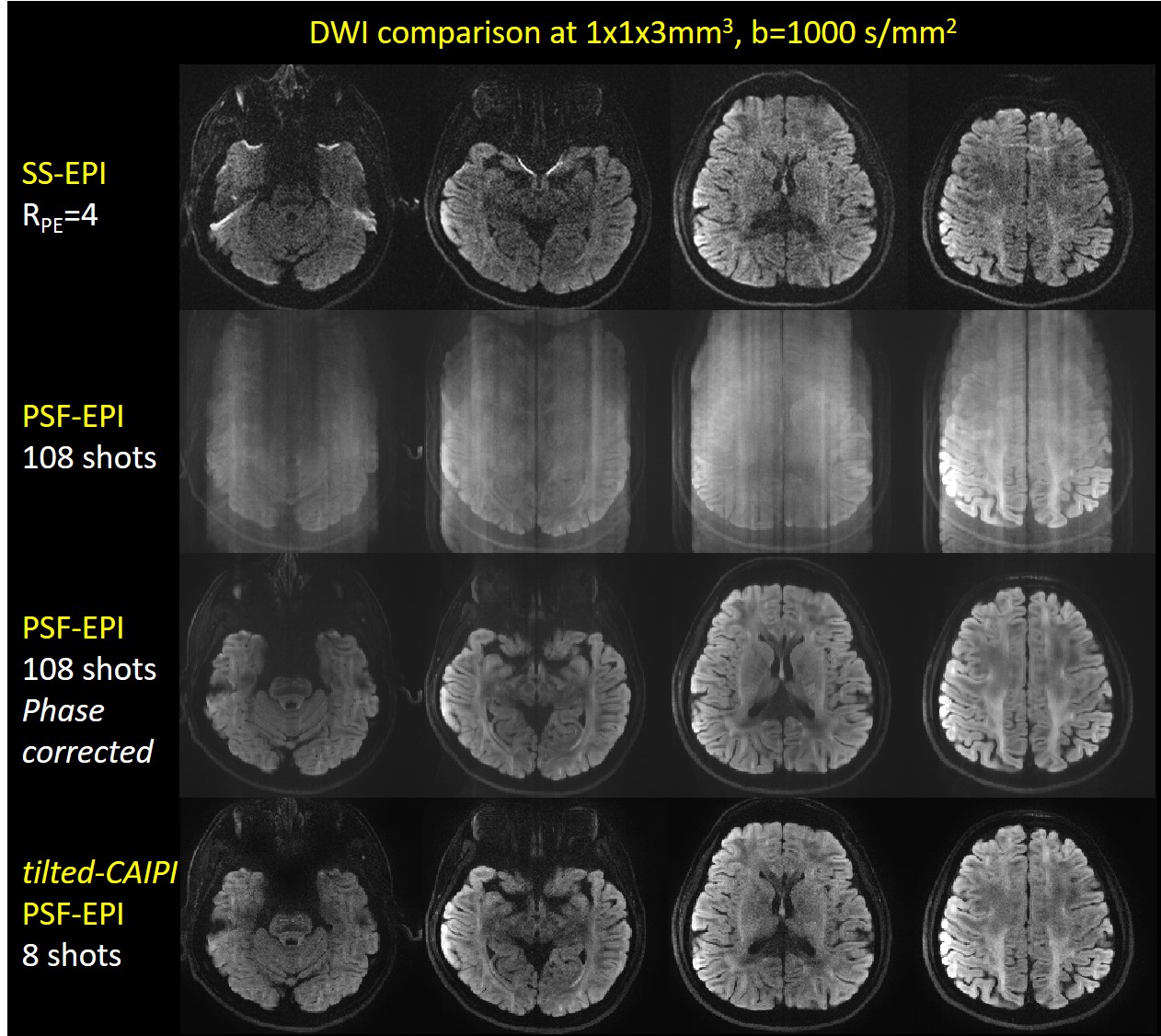
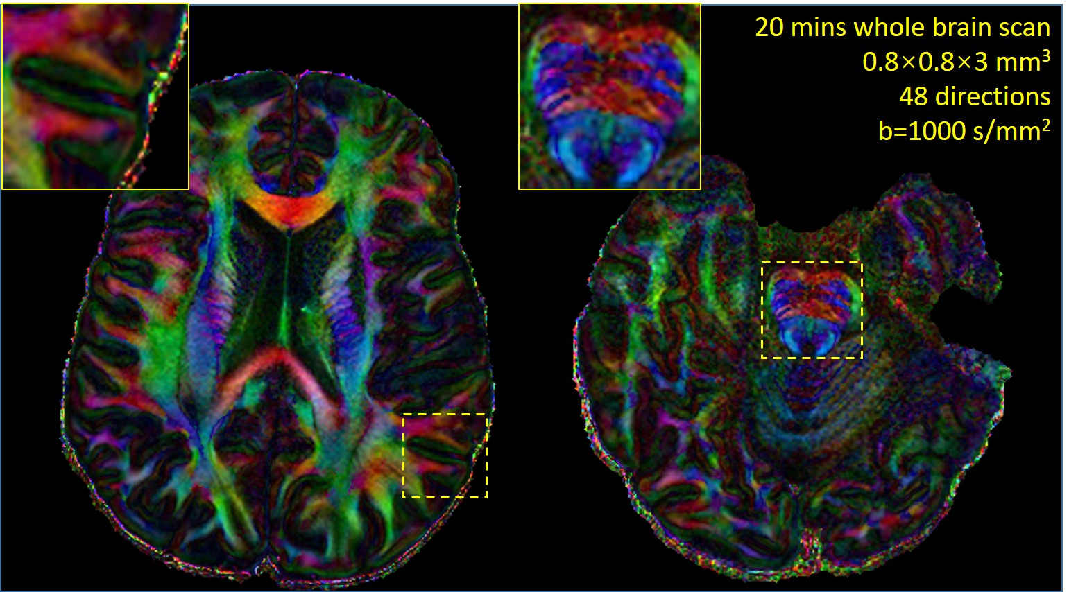
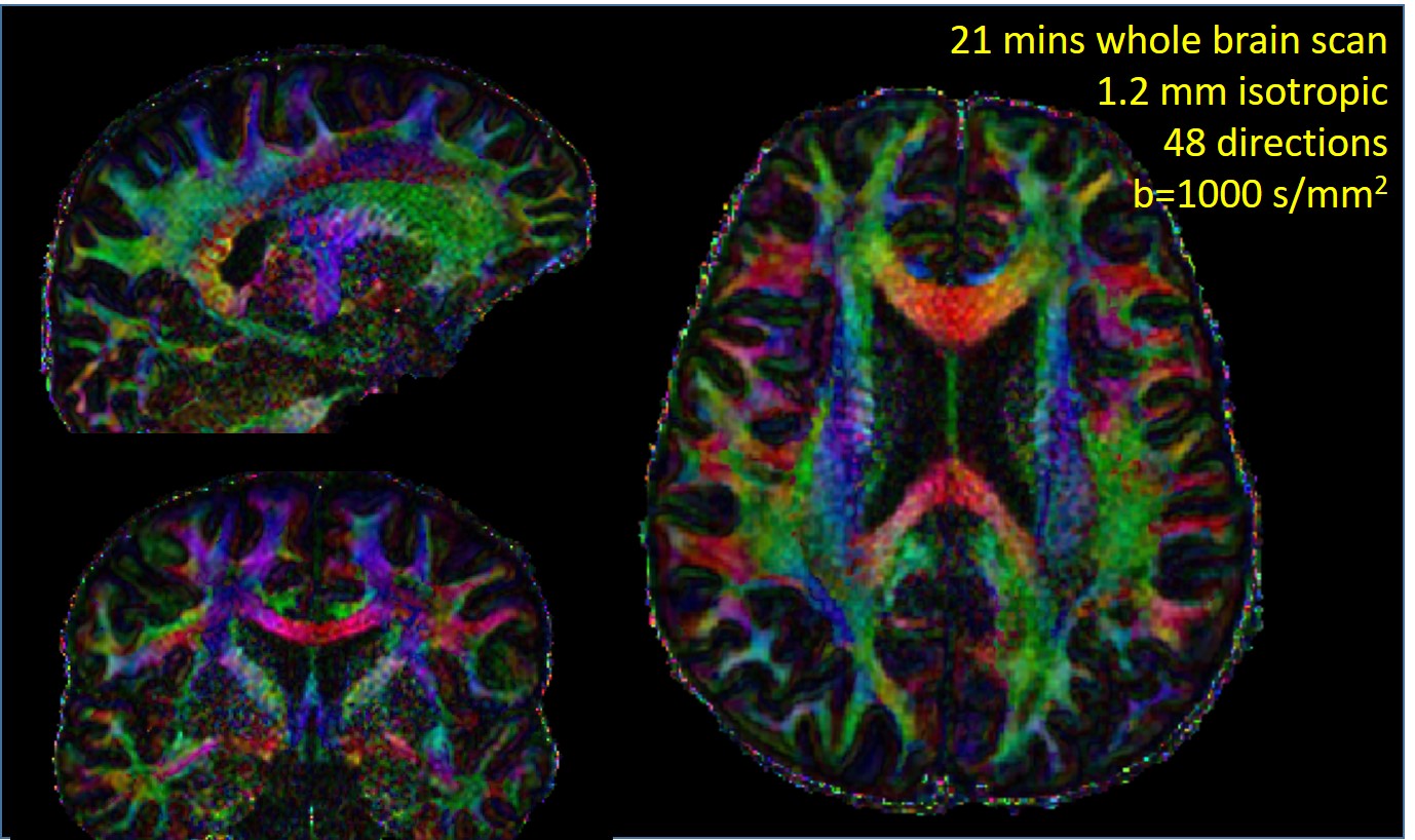
In Fig.3, SS-EPI shows severe distortions for 1×1×3mm3 acquisition, even at RPE=4 and an effective ESP of 0.24ms (top-row). On the other hand, the 108-shot PSF-EPI without phase correction suffers from strong artifacts (second-row). This issue was nicely mitigated through phase-corrected PSF-EPI with self-navigation to achieve high-quality DWIs (third-row). With tilted-CAIPI, acquisition burden of PSF-EPI is significantly reduced to just 8-shots (bottom-row). In Fig.4&5, color-coded fractional anisotropy maps are shown for whole-brain 0.8×0.8×3mm3 and 1.2mm-isotropic acquisitions, each acquired in ~20 minutes.Discussion and Conclusion
The proposed tilted-CAIPI PSF-EPI with self-navigation allows distortion-free and T2* blurring-free diffusion imaging in 4-8 shots with physiological motion correction. High-resolution, high-SNR DWI at 48 directions can be performed in ~20 minutes, allowing accurate diffusion imaging which should prove useful in neurosurgery and neuroimaging.Acknowledgements
This work was supported in part by NIH research grants: R01EB020613, R01EB019437, R24MH106096, P41EB015896, and the shared instrumentation grants: S10RR023401, S10RR019307, S10RR019254, S10RR023043.References
1. Pruessmann KP, Weiger M, Scheidegger MB, Boesiger P. SENSE: sensitivity encoding for fast MRI. Magn. Reson. Med. 1999;42:952-962.
2. Griswold MA, Jakob PM, Heidemann RM, Nittka M, Jellus V, Wang J, Kiefer B, Haase A. Generalized autocalibrating partially parallel acquisitions (GRAPPA). Magn. Reson. Med. 2002;47:1202-1210.
3. Dong Z, Wang F, Ma X, Zhang Z, Dai E, Yuan C, Guo H. Interleaved EPI diffusion imaging using SPIRiT‐based reconstruction with virtual coil compression. Magn. Reson. Med.
4. Chen NK, Guidon A, Chang HC, Song AW. A robust multi-shot scan strategy for high-resolution diffusion weighted MRI enabled by multiplexed sensitivity-encoding (MUSE). NeuroImage 2013;72:41-47.
5. Jeong HK, Gore JC, Anderson AW. High-resolution human diffusion tensor imaging using 2-D navigated multishot SENSE EPI at 7 T. Magn. Reson. Med. 2013;69:793-802.
6. Holdsworth SJ, Skare S, Newbould RD, Bammer R. Robust GRAPPA-accelerated diffusion-weighted readout-segmented (RS)-EPI. Magn. Reson. Med. 2009;62:1629-1640.
7. Wang FN, Huang TY, Lin FH, Chuang TC, Chen NK, Chung HW, Chen CY, Kwong KK. PROPELLER EPI: an MRI technique suitable for diffusion tensor imaging at high field strength with reduced geometric distortions. Magn. Reson. Med. 2005;54:1232-1240.
8. In MH, Posnansky O, Speck O. High-resolution distortion-free diffusion imaging using hybrid spin-warp and echo-planar PSF-encoding approach. NeuroImage 2017;148:20-30.
9. Zaitsev M, Hennig J, Speck O. Point spread function mapping with parallel imaging techniques and high acceleration factors: fast, robust, and flexible method for echo-planar imaging distortion correction. Magn. Reson. Med. 2004;52:1156-1166.
10. Dong Z, Wang F, Reese TG, Manhard MK, Bilgic B, Wald LL, Guo H, Setsompop K. Tilted-CAIPI for Highly Accelerated Distortion-Free EPI with Point Spread Function (PSF) encoding. Submitted to: In Proceedings of the 26th Annual Meeting of ISMRM 2018.
11. Ma X, Zhang Z, Dai E, Guo H. Improved multi-shot diffusion imaging using GRAPPA with a compact kernel. NeuroImage 2016;138: 88-99.
Figures