1203
Distortion-Free, High-Resolution Diffusion Imaging in a Clinically-Feasible Scan Time on a Compact 3T MRI with High-Performance Gradients1Department of Radiology, Mayo Clinic, Rochester, MN, United States, 2GE Global Research, Niskayuna, NY, United States
Synopsis
DIADEM (Distortion-free Imaging: A Double Encoding Method) is a hybrid, multi-shot approach using a spin-warp and echo-planar phase-encoding strategy. It is inspired by the point-spread-function mapping method to enable distortion-free high-resolution diffusion imaging, which has a great potential for clinical practice. However, its prolonged scan time poses an obstacle for its adoption. We demonstrate that DIADEM achieves high-resolution (1.4 mm3 isotropic or 0.86 mm2 in-plane), distortion-free, and whole-brain, diffusion tensor images under 9 minutes scan time with: i) sequence optimization and ii) the high-performance gradients (80 mT/m, 700 T/m/s) on a compact 3T MRI.
Introduction
Theory and Methods
The standard application of reduced field of view (rFOV) is to reduce the number of phase-encoding steps in the spin-warp phase-encoding (SW-PE) dimension, which is also used to accelerate the DIADEM scan3. To obtain a distortion-free image without wraparound (i.e., aliasing) artifacts from the DIADEM data, however, the maximum rFOV factor is limited by the extent of distortion in the EPI-PE dimension1,2. Assuming a maximum off-resonance frequency Δf(s)max, the maximum available rFOV factor can be determined by the effective echo-spacing Δtesp in the EPI-PE dimension of the DIADEM data1:
rFOVmax=1/(∆tesp×|∆f(s)max|×2) (1)
In addition to parallel imaging in the EPI-PE dimension, gradient hardware performance can be exploited to improve DWI image quality. High gradient slew-rate is a key factor to shorten the echo-spacing4, which decreases the geometric distortion and enables further DIADEM acceleration with a higher rFOV factor. In addition, high gradient amplitude can reduce TE and increase signal-to-noise ratio (SNR). The diffusion DIADEM sequence1 was implemented on the C3T2 (Fig. 1). The gradient coil has a 42 cm inner diameter, and employs an asymmetry design for the two transverse axes. Artifacts due to linear and spatially independent concomitant fields are eliminated by gradient pre-emphasis5 and frequency tracking6, respectively. To further minimize the scan time without sacrificing the correction fidelity of the motion-induced ghost artifacts, a four-fold reduction of the navigator data acquisition window covering the center of k-space was applied, as suggested in a previous study7. To explore the highest in-plane and isotropic resolution obtainable in under 10-minutes of acquisition time, phantom experiments were first performed to optimize the protocol. Under an IRB- approved protocol, two healthy volunteers were scanned using either an 8-channel (Invivo, Gainesville, FL) or a 32-channel coil (Nova Medical, Wilmington, MA) (Table 1). After off-line reconstruction and post-processing described previously1, these images were compared with anatomical images including T2-weighted fast-spin-echo (FSE) and fluid-attenuated-inversion-recovery (FLAIR).
Results and Discussion
Using the maximum slew rate of 700T/m/s and a parallel imaging factor of 3, a very short effective echo-spacing (209.3µs) and TE (42.5ms) were obtained on the C3T even with high in-plane imaging matrix of 256×256. This compares to 349.3µs achievable on a standard, whole-body scanner with 50mT/m, 200T/m/s gradient performance. Under the effect of off-resonance of up to ±250Hz, the maximum distortion was ±13.4 pixels, which enabled a maximum rFOV factor of up to 8 for the acceleration, rather than a maximum of 5 that was previously reported1. The total number of segments (or shots) for each non-DW and DW scan was further reduced with a partial Fourier acquisition of 85% in the SW-PE dimension (Table 1). Overall, the proposed approach was able to achieve in-plane resolution of 0.86mm2 with 4 mm slice thickness (Fig. 2) or isotropic resolution (1.4 mm3, Fig. 3), distortion-free, whole brain DWIs within 10-minute scan time on the C3T. As illustrated by the mean diffusivity map in Fig. 2A-d, high-resolution DWI without geometric distortions can provide detailed anatomic information even adjacent to the paranasal sinuses, where strong susceptibility effects usually appear. Due to the matching image resolution, direct one-to-one image comparison between the diffusion (Fig. 2A) and the anatomical images (Fig. 2B) is possible for clinical evaluation. By reducing the navigator data acquisition window from 13.4 to 3.3 ms, a shorter TE for the gradient-echo-based navigator-echo acquisition yields higher SNR, and the scan time was further reduced by up to 1 minutes 38 seconds (Table 1), without sacrificing any reconstruction fidelity (images are not shown here).Conclusion
The high-performance gradients of the C3T can reduce the geometric distortion in EPI, which enables further accelerations of the DIADEM scan by approximately a factor of 3 compared to a conventional 3T MRI system. In addition, both high gradient amplitude and slew rate are beneficial to reduce the TE, and consequently increase the SNR for high-resolution DWI. With the benefits of the high-performance gradients on the C3T and further sequence optimization, the proposed DIADEM approach was able to achieve high-resolution, distortion-free, and whole-brain diffusion images within a scan time that is feasible for clinical use.Acknowledgements
This work was supported by NIH U01 EB024450-01.References
1. In MH, Posnansky O, Speck O. High-resolution distortion-free diffusion imaging using hybrid spin-warp and echo-planar PSF-encoding approach. Neuroimage. 2017 Mar 1;148:20-30. doi: 10.1016/j.neuroimage.2017.01.008.
2. Lee S-K, Mathieu J-B, Graziani D, et al. Peripheral nerve stimulation characteristics of an asymmetric head-only gradient coil compatible with a high-channel-count receiver array. Magn Reson Med 2016;76:1939–1950. doi: 10.1002/mrm.26044.
3. Zaitsev M, Hennig J, Speck O. Point spread function mapping with parallel imaging techniques and high acceleration factors: fast, robust, and flexible method for echo-planar imaging distortion correction. Magn Reson Med. 2004 Nov;52(5):1156-66.
4. High slew-rate head-only gradient for improving distortion in echo planar imaging: Preliminary experience. Tan ET, Lee SK, Weavers PT, Graziani D, Piel JE, Shu Y, Huston J 3rd, Bernstein MA, Foo TK. J Magn Reson Imaging. 2016 Sep;44(3):653-64. doi: 10.1002/jmri.25210.
5. Tao S, Weavers PT, Trzasko JD, Shu Y, Huston J 3rd, Lee SK, Frigo LM, Bernstein MA. Gradient pre-emphasis to counteract first-order concomitant fields on asymmetric MRI gradient systems. Magn Reson Med. 2017 Jun;77(6):2250-2262. doi: 10.1002/mrm.26315.
6. Weavers PT, Tao S, Trzasko JD, Frigo LM, Shu Y, Frick MA, Lee SK, Foo TKF, Bernstein MA. B0 concomitant field compensation for MRI systems employing asymmetric transverse gradient coils. Magn Reson in Med 2017. doi:10.1002/mrm.26790
7. Skare S, Holdsworth S, Newbould RD, Bammer R. On the battle between Rician noise and phase-interferences in DWI. ISMRM 2009, p. 1409
Figures
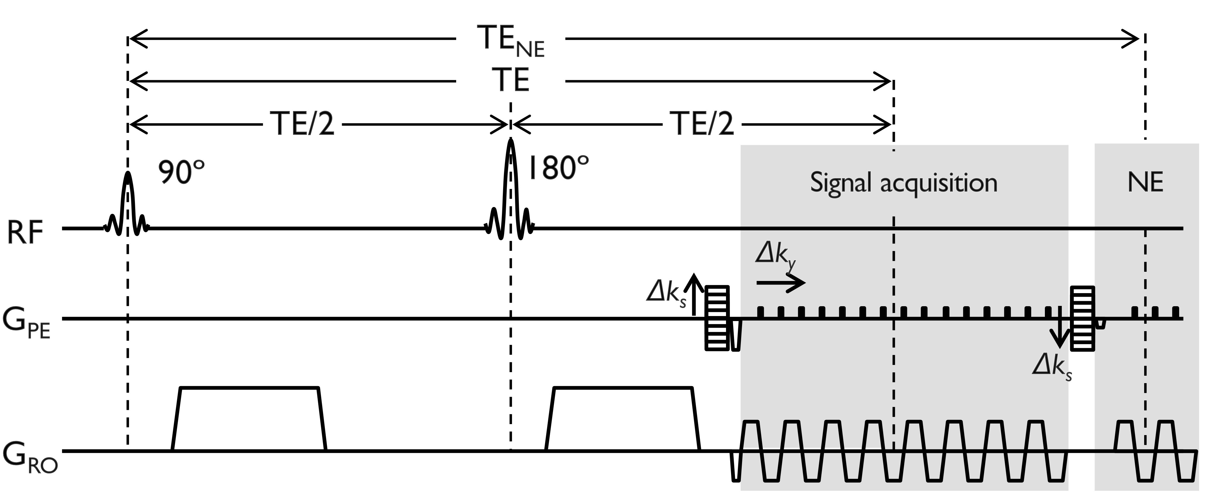
Figure 1. A diffusion DIADEM sequence diagram with a navigator-echo (NE). The NE acquisition window was reduced four times compared to the signal acquisition window. Note that spin-warp phase-encoded and corresponding rewinder gradients (see Δks) are applied before and after the spin-warp phase-encoded signal acquisition.
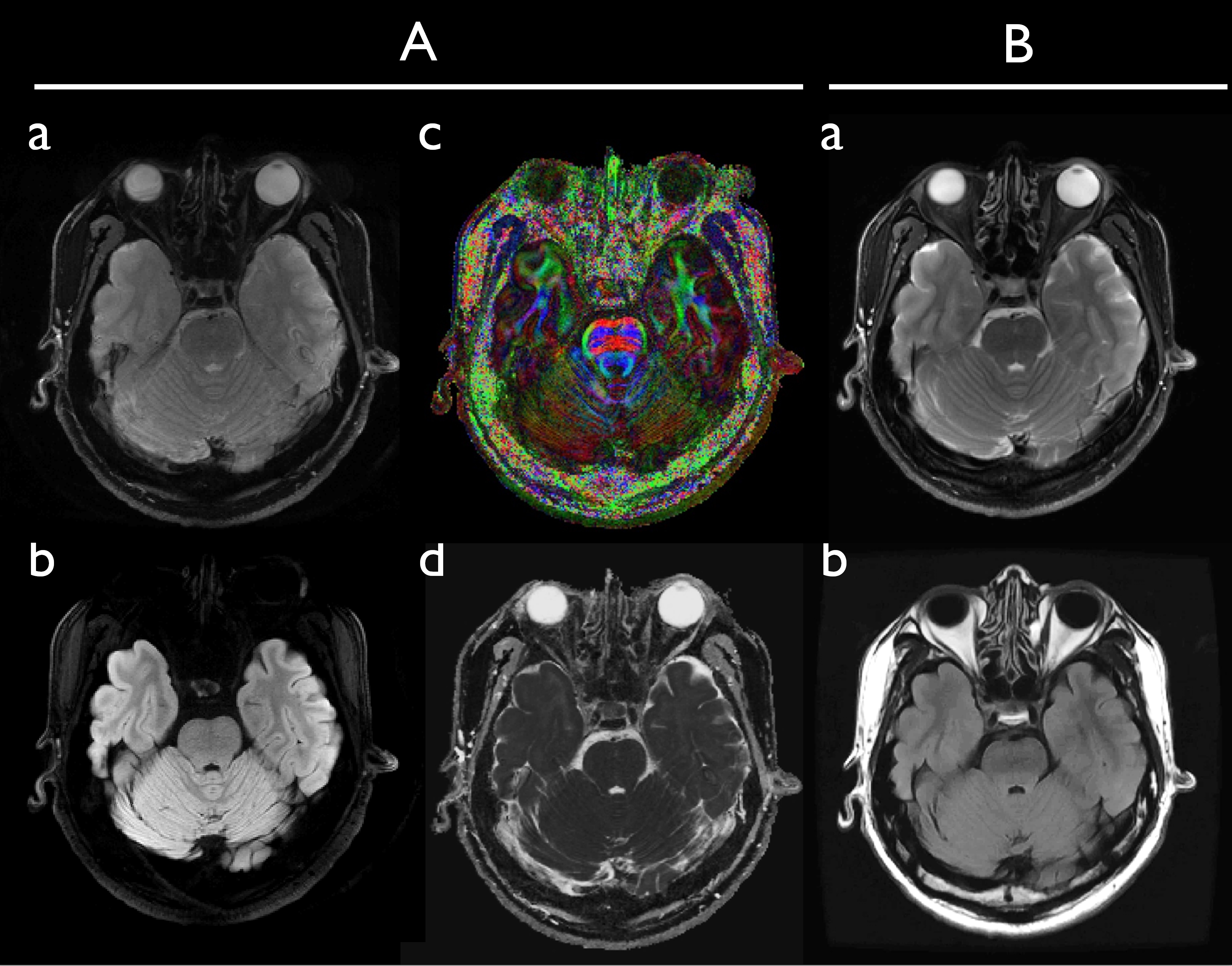
Figure 2. Distortion-free diffusion (A) and conventional anatomical images (B) with a high in-plane resolution. A slice from non-DWI (A-a), isoDWI (A-b), color-coded FA map (A-c), and mean diffusivity map (A-d) are displayed. Note that isoDWI is obtained by summation of all 6 DWIs. The corresponding slices from the anatomical 2D-FSE (B-a) and FLAIR data (B-b) are shown for comparison. The intrinsic image resolution is 0.86×0.86×4 mm3 for both diffusion and anatomical data.
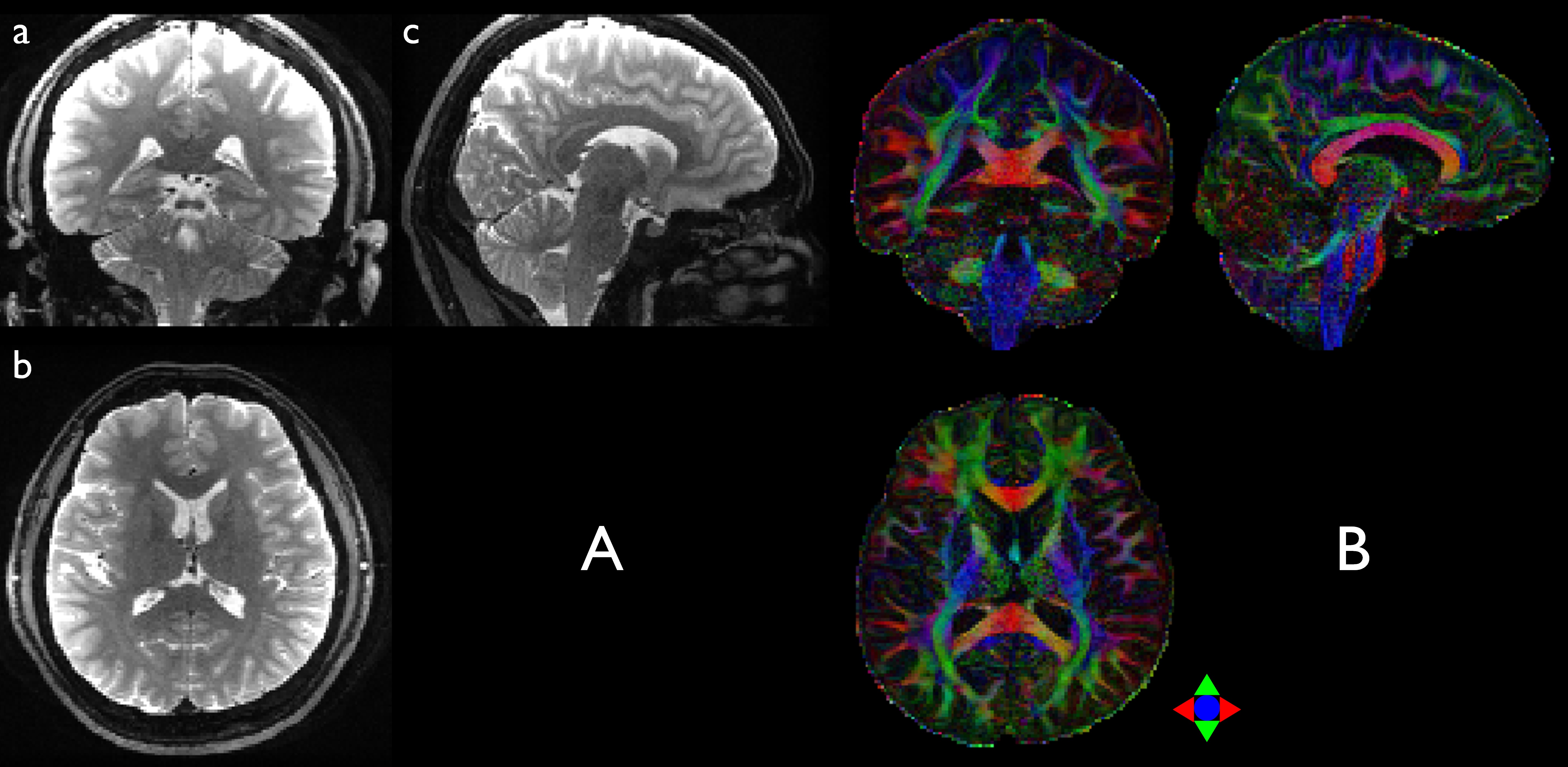
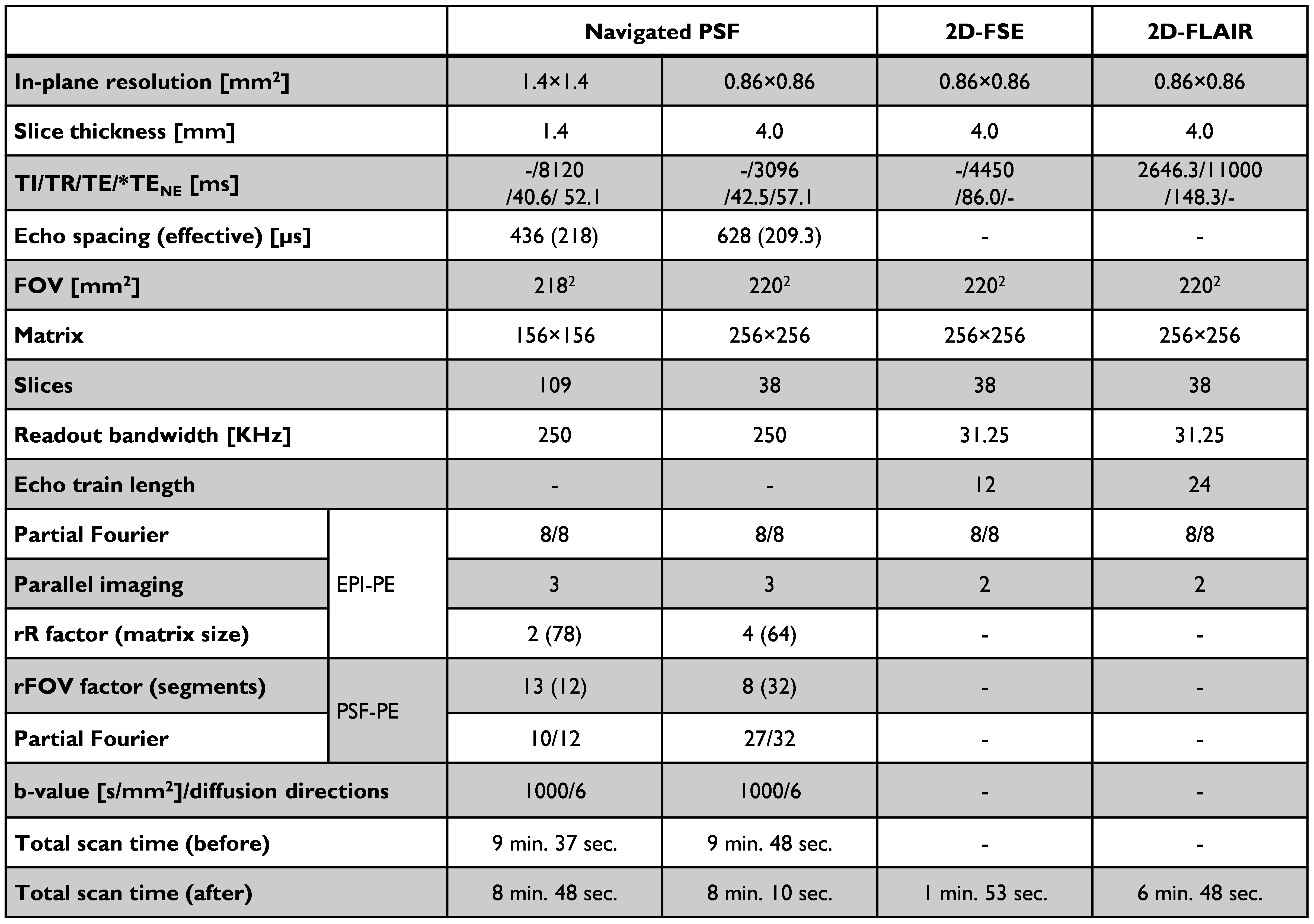
Table 1. Experimental imaging protocols. *Note that TENE is the echo time for navigator echo (NE) in the DIADEM acquisition. Two different total scan times measured before and after the reduction of the navigator echo acquisition window are shown in the last two rows.