1196
Evaluation of 2D simultaneous multi-slice EPI at 1.5T for MR-thermometry in presence of motion.1IHU Liryc, Electrophysiology and Heart Modeling Institute, Fondation Bordeaux Université, Bordeaux, France, 2Univ. Bordeaux, Centre de recherche Cardio-Thoracique de Bordeaux, U1045, Bordeaux, France, 3INSERM, Centre de recherche Cardio-Thoracique de Bordeaux, U1045, Bordeaux, France, 4Image Guided Therapy SA, Bordeaux, France, 5Institute of Mathematics of Bordeaux, UMR 674 5251, Bordeaux, France, 6Siemens Healthineers, Saint Denis, France, 7Siemens Healthcare, Saint Denis, France, 8Siemens Healthcare, Erlangen, Germany
Synopsis
Temperature mapping in presence of respiratory motion can accommodate intra-scan motion using fast 2D-EPI sequence but inter-scan motion remains a challenge during free-breathing acquisition. To address this limitation, we evaluated, in vitro on a mobile gel, a 2D simultaneous multi-slice EPI sequence with a slice acceleration factor up to 3 during radiofrequency ablation. Inter-scan motion and temperature elevation measured with accelerated sequences are compared to reference values using a non-accelerated sequence. Additionally, evidence or absence of potential false-positive heating are considered.
INTRODUCTION
Respiration-induced motion of abdominal organs poses significant challenges to measure accurate MR temperature maps [1] using the proton resonance frequency shift technique. Several strategies have been proposed using different MR-based information [2, 3], but existing MR sequences had to sacrifice either volume coverage or frame rate. Simultaneous multi-slice (SMS) echo-planar imaging (EPI) using parallel image reconstruction is an emerging technology that may be exploited to increase volume coverage or reduce acquisition duration of temperature mapping. Nevertheless, the use of SMS-EPI imaging combined with in-plane acceleration results in a reduction of image signal-to-noise ratio (SNR) [4]. Moreover, the presence of motion may induce artifacts on reconstructed images since operators used for image un-aliasing may imperfectly correct overlapping signals due to combined GRAPPA and SMS acceleration. In this work, we have investigated the benefits of SMS acquisitions for monitoring radiofrequency ablation (RFA) in a mobile gel. The performance of the sequence was evaluated in terms of temporal standard deviation of temperature σ(T) and potential false-positive heating at multiband (MB) factors of 1, 2, and 3.METHODS
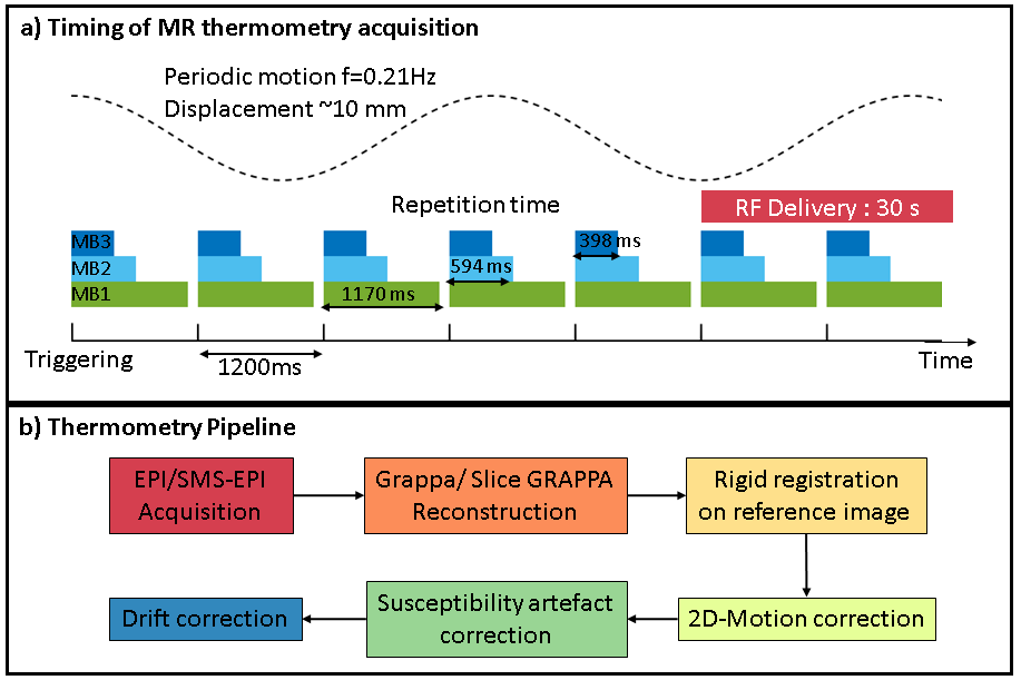
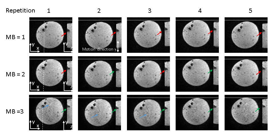
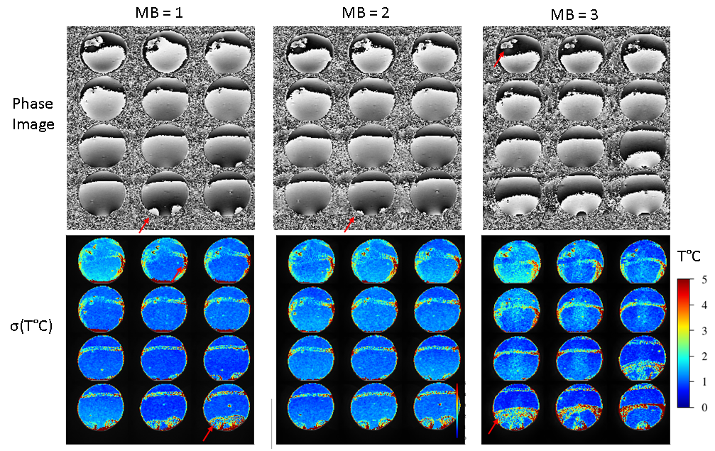
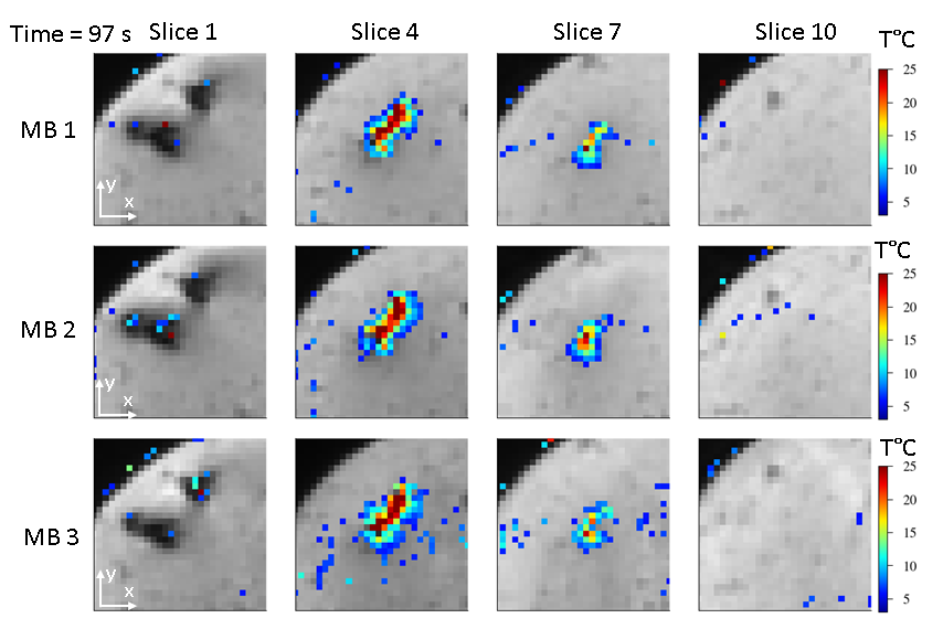
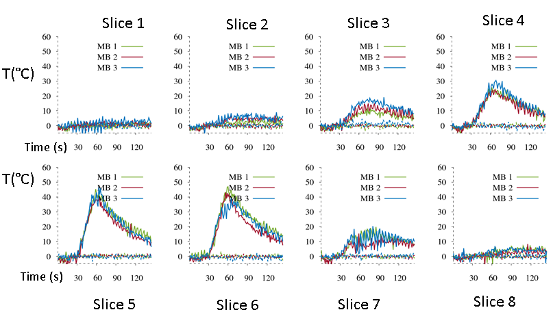
Acquisition: To simulate a respiratory motion, a gel phantom composed of 3 % agar was positioned on a motor-driven platform to generate a horizontal oscillating translation of the sample with an amplitude of 10 mm and a frequency of 0.21 Hz. Twelve slices were acquired using an interleaved pattern in a coronal orientation (motion included in the imaging plane) every 1.2 s, over a total duration of 3 minutes, on a 1.5 T clinical imaging system (MAGNETOM Aera, Siemens Healthcare, Erlangen, Germany) using a prototype 2D single-shot gradient echo blipped-CAIPI SMS echo planar imaging (Figure 1a). Three different MB factors (1, 2 and 3) were combined with in-plane parallel imaging (GRAPPA acceleration factor of 2 with 50% oversampling in the phase encoding direction). Other sequence parameters were: TE=26 ms, FOV=180x180 mm2, spatial resolution 1.6x1.6x3mm3 voxel size, FA = 90°, 6/8 partial Fourier and pixel bandwidth = 1565 Hz/pixel. The spine coil and two flexible coils surrounded the gel, allowing the activation of 24 receiver channels. RF-ablation device: Two MR-compatible RF electrodes were inserted into the gel and connected to a programmable RF generator (Image Guided Therapy, Pessac France) located outside the Faraday cage. RF energy was delivered at 15 W during 30 s. Thermometry: in-plane motion compensation, susceptibility correction, and spatial-temporal drift correction was implemented as previously described [5]. Each stack of slices of the reference frame was also automatically aligned in the slice direction using additional rigid registration (Figure 1b) to then reconstruct the stack of 2D thermometry data at each time point. To assess the precision of MR-thermometry, the temporal standard deviation of temperature σ(T) was computed in each pixel from all slices during 20 repetitions acquired before RF energy delivery and the distribution of σ(T) values was analyzed on a manually drawn ROI (~37000 voxels).RESULTS
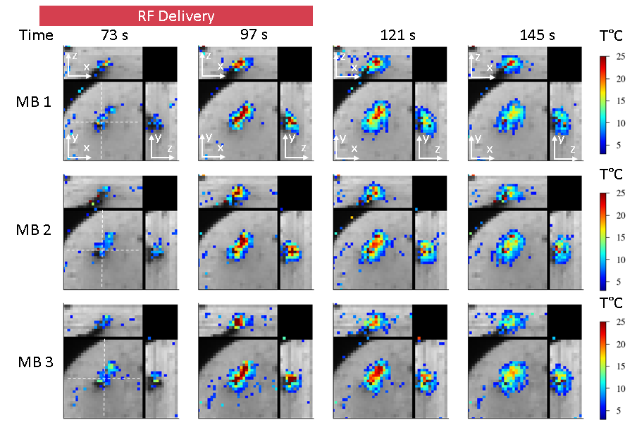
The temporal resolution for acquiring the 12 slices was 1179 ms, 594 ms and 398 ms for MB=1, 2 and 3, respectively. Figure 2 displays representative magnitude images acquired on the gel. Image quality was found satisfactory, although some remaining artifacts were visible for MB acceleration of 3. However, similar σ(T) (spatial mean ± standard deviation over the ROIs) values were observed for each slice with a mean value of 1.8/1.7/1.7 °C for MB 1/2/3 respectively (Figure 3). A temperature rise of 25°C was reached for each acquisition with a resulting heated region of 8x3 pixel at the end of energy delivery. No false-positive temperature spots due to potential signal leakage between simultaneously excited slices were observed under the tested conditions (Figure 4, 5 and 6). The temperature distribution in the (x, z) and (y, z) planes indicate that spatial homogeneity of the heating was preserved whatever the acceleration factor (Figure 5).DISCUSSIONS AND CONCLUSIONS
This study presents the first evaluation of MR-thermometry using SMS-EPI acquisition in presence of motion. While increasing the SMS factor to 3 induced some aliasing artifacts, it reduced displacement between adjacent slices and thus allowed to reconstruct a 3D volume (180x180x37 mm3) more precisely, with acceptable temperature uncertainty (<2°C). Further improvement in coil combination are expected to improve thermometry quality by removing spatial phase wraps on phase images before in vivo clinical evaluation on mobile organs. Pseudo-volumetric temperature imaging could thus be performed without compromises on acquisition time and resulting temperature images showed similar patterns independent of the SMS acceleration factor (up to 3). Such a strategy is expected to increase procedure safety by monitoring larger volumes more rapidly for MR guided thermotherapies on mobile organs.Acknowledgements
This work received financial support from the French National Investments for the Future Programs: ANR-10-IAHU-04 (IHU Liryc) and Laboratory of Excellence ANR-10-LABX-57 (TRAIL), and the research programs ANR-11-TecSan-003-01 (TACIT) and Equipex ANR-11-EQPX-0030 (MUSIC).References
1. Rempp H, Martirosian P, Boss A, Clasen S, Kickhefel A, Kraiger M, et al. MR temperature monitoring applying the proton resonance frequency method in liver and kidney at 0.2 and 1.5 T: segment-specific attainable precision and breathing influence. MAGMA. 2008;21(5):333-43.
2. Seror O, Lepetit-Coiffe M, Le Bail B, de Senneville BD, Trillaud H, Moonen C, et al. Real time monitoring of radiofrequency ablation based on MR thermometry and thermal dose in the pig liver in vivo. Eur Radiol. 2008;18(2):408-16.
3. Holbrook AB, Santos JM, Kaye E, Rieke V, Pauly KB. Real-time MR thermometry for monitoring HIFU ablations of the liver. Magn Reson Med. 2010;63(2):365-73.
4. Borman PT, Bos C, de Boorder T, Raaymakers BW, Moonen CT, Crijns SP. Towards real-time thermometry using simultaneous multislice MRI. Phys Med Biol. 2016;61(17):N461-77.
5. Ozenne V, Toupin S, Bour P, de Senneville BD, Lepetit-Coiffe M, Boissenin M, et al. Improved cardiac magnetic resonance thermometry and dosimetry for monitoring lesion formation during catheter ablation. Magn Reson Med. 2016.
Figures