1183
Diffusion-weighted magnetic resonance imaging in bladder cancer: comparison of readout-segmented EPI and single-shot EPI Techniques1Radiology, Changhai Hospital of Shanghai, Shanghai, China
Synopsis
Diffusion weighted imaging showed the potential to assess bladder cancer. This study aimed to investigate whether readout-segmented EPI can offer better image quality in imaging bladder patients in comparison with single-shot EPI, and to compare quantitative image parameters, derived from RS-EPI with those of SS-EPI. Thirty-five patients were examined using both diffusion techniques. There were significant differences in susceptibility artifacts, lesion detectability, image blurring, CNR and SIR values, except for motion artifacts, SNR and ADC values of the bladder lesions. This study found that the RS-EPI technique provides significant image quality improvement compared with SS-EPI in bladder at 3 Tesla.
Purpose
Bladder cancer is a common cancer worldwide1. Several studies have been reported that the clinical use of diffusion weighted imaging (DWI) for bladder cancer evaluation2,3. Single-shot echo planar imaging (SS-EPI) is commonly used method duo to its relative insensitive to motion-induced phase errors and short acquisition times4. However, SS-EPI images are characterized by blurring along the phase encoding direction due to T2* decay and are sensitive to off-resonance effects5. In recent years, a new multi-shot technique, referred to as readout-segmented echo planar imaging (RS-EPI), has been proposed to improve the spatial resolution, decrease the susceptibility based image distortion and T2* blurring6. We hypothesized that the RS-EPI could be a promising method for DWI in bladder cancer. Therefore, the aims of the present study were to evaluate whether RS-EPI can provide better image quality in imaging bladder cancer in comparison with SS-EPI, and to compare quantitative image parameters, derived from RS-EPI with those of SS-EPI.Materials and Methods
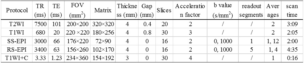
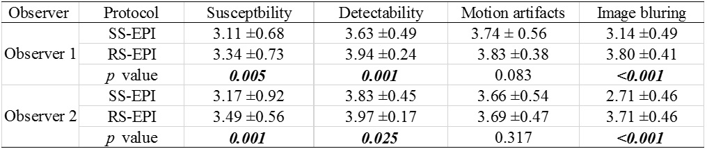
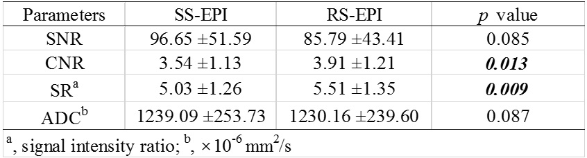
Subjects Thirty-five patients (range, 31-89 years; male/female, 34/1) with bladder cancer were enrolled in this prospective study, which was approved by the local institutional review board and written informed consent was obtained from each patient. MRI protocols Magnetic resonance imaging was performed on a 3T MR system (Skyra, Siemens medical solution, Germany) using a standard 18-channel phased-array soft coil. In all patients, the examination protocols of bladder cancer included an axial T1WI, T2WI, two axial DWI with RS-EPI and SS-EPI techniques and contrast enhanced T1WI. The parameters of these protocols were summarized in Table 1. The total scan time of each subject was approximately 12 minutes. Image analysis RS-EPI and SS-EPI DWI images were evaluated by two independent observers (with 4 and 5 years of experience, respectively) for identification of susceptibility, detectability, motion artifacts and image blurring of the lesions using quantitative 4-point scale. Signal-to-noise ratio (SNR), contrast-to-noise ratio (CNR), signal intensity ratio (SIR) and ADC values of bladder lesions were measured and compared. The regions of interest (ROI) of the lesions were manually outlined on ADC maps with the sizes of ROIs ranged from 0.6 mm2 to 17.0 mm2. Statistical analysis Significant differences between RS-EPI and SS-EPI for visual scores, and quantitative parameters were assessed by using Wilcoxon signed rank tests.Results
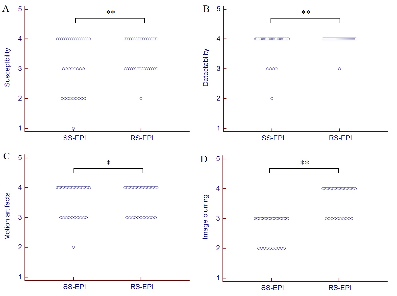
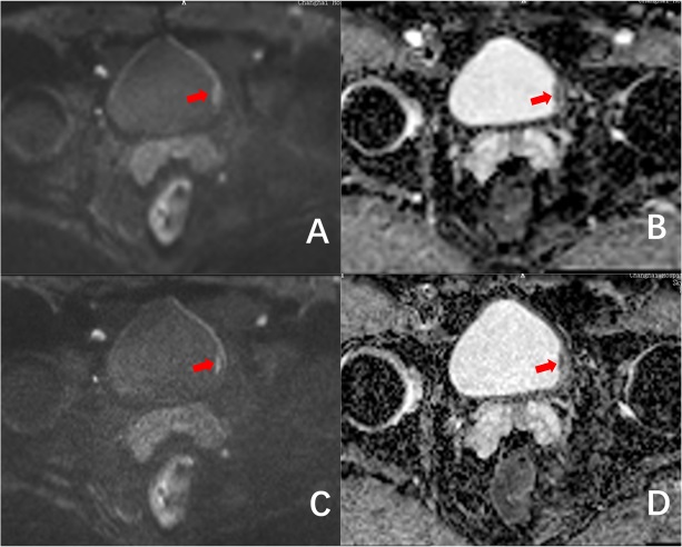
The statistical results of quantitative scores and quantitative parameters in assessing bladder lesions were summarized in Tables 2 and 3. There were significant differences in susceptibility, detectability and image blurring (all p < 0.05), except for motion artifacts of bladder lesions (p > 0.05, Figure 1). RS-EPI technique is superior to SS-EPI technique in image quality (Figure 2). No significant difference was observed for SNR and ADC values of bladder lesions between SS-EPI and RS-EPI techniques (96.65±51.59 vs.85.79±43.41, p=0.085 and 1239.09±253.73×10⁻⁶ mm2/s vs 1230.16±239.60×10⁻⁶ mm2/s, p=0.087, respectively). There were significant differences in CNR and SIR values of the lesions between both techniques (3.54±1.26 vs. 5.51±1.35, p=0.013 and 5.03±1.26 vs. 5.51±1.35, p=0.009, respectively).Discussion and Conclusion
In the present study, we demonstrated significantly higher image quality and better image contrast of bladder DWI using RS-EPI than that using SS-EPI technique, in addition no significant difference in SNR and ADC values, which showed that RS-EPI is a promising method for reducing image artifacts and improving image quality in bladder cancer. The image blurring and lesion detectability of bladder cancer was significantly improved with RS-EPI. The RS-EPI technique provides significant image quality improvement compared with SS-EPI at 3 Tesla, and can potentially provide better image quality in patients with bladder cancer.Acknowledgements
No acknowledgement found.References
[1] Chavan S, Bray F, Lortet-Tieulent J, Goodman M, Jemal A. International variations in bladder cancer incidence and mortality. Eur Urol. 2014; 66:59–73.
[2] Takeuchi M, Sasaki S, Naiki T, Kawai N, Kohri K, Hara M, and Shibamoto Y. MR imaging of urinary bladder cancer for T-staging: a review and a pictorial essay of diffusion-weighted imaging. J Magn Reson Imaging. 2013; 38, 1299–1309.
[3] Yoshida S, Koga F, Masuda H, Fujii Y, and Kihara K. Role of diffusion-weighted magnetic resonance imaging as an imaging biomarker of urothelial carcinoma. Int J Urol. 2014; 21, 1190–1200.
[4] Turner R, Lebihan D. Single-Shot Diffusion Imaging at 2.0 Tesla. J Magn Reson. 1990;86: 445–452.
[5] Taviani V, Nagala S, Priest AN, et al. 3T diffusion-weighted MRI of the thyroid gland with reduced distortion: preliminary results. Br J Radiol. 2013;86(1028):20130022.
[6] Porter DA, Heidemann RM. High resolution diffusion-weighted imaging using readout-segmented echo-planar imaging, parallel imaging and a two-dimensional navigator-based reacquisition. Magn Reson Med. 2009;62(2):468-475.
Figures