1178
MR R2* values in diagnosing the stage Ia and Ib endometrial carcinoma1Radiology, the First Affiliated Hospital of Dalian Medical University, Dalian, China, 2GE Healthcare, Beijing, China
Synopsis
Endometrial carcinoma(EC) patients staged stage Ia or Ib have different operation methods and prognosis. To evaluate the value of enhanced T2*-weighted angiography(ESWAN) in the differential diagnosis between stage Ia and Ib of endometrial carcinoma. In this work, we found it is feasible that ESWAN sequence derived R2* value can be applied in the differential diagnosis between stage Ia and Ib of endometrial carcinoma, which can provide detailed information for clinical treatment.
Introduction
Endometrial carcinoma (EC) is one of the common gynecologic malignancies. Tumors confined to the endometrium and those invading the superficial myometrium are designated as stage Ia, and tumors invading the deep myometrium are designated as stage Ib [1]. Depth of myometrial invasion is the most important morphologic prognostic factor [1]. The incidence of lymph node metastases increases from 3% with superficial myometrial invasion to 46% with deep myometrial invasion. Preoperative information about the depth of myometrial invasion is therefore essential in tailoring the surgical approach for patients in stage Ia or Ib [2]. For accurate staging of endometrial carcinoma and guiding clinical treatment, this study investigated R2* values generated from enhanced T2*-weighted angiography(ESWAN) sequence in diagnosing the stage Ia and Ib of endometrial carcinoma.Methods
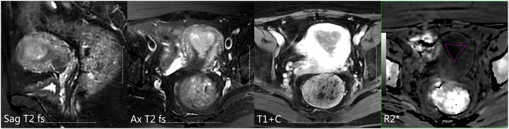
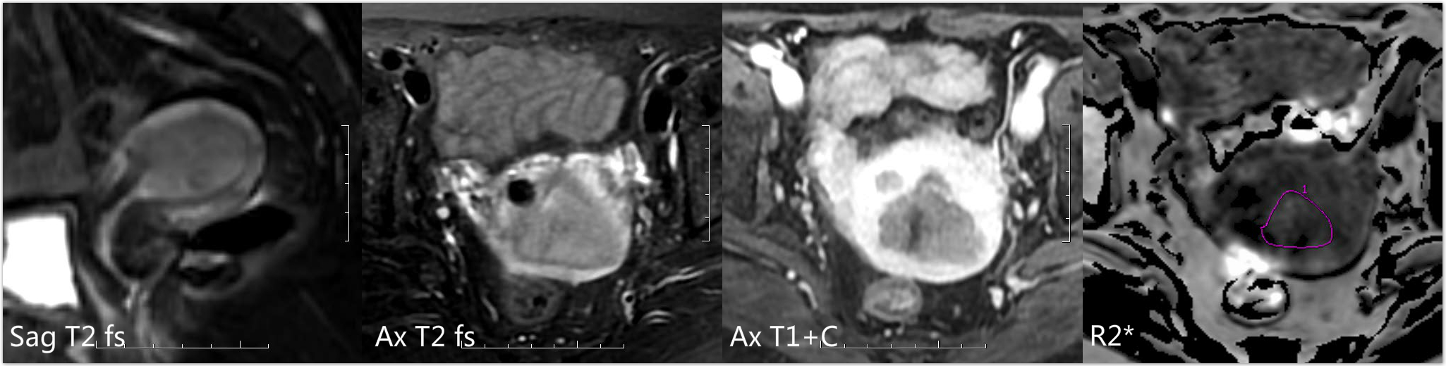
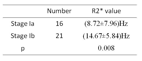
Thirty seven pathologically proved patients (16 in stage Ia and 21 in stage Ib) were enrolled in the study. Images were acquired with ESWAN sequence on 1.5T MR scanner. R2* maps were reconstructed and mean R2* values were calculated by two observers. The inter-observer consistency was tested. The ROI covered the lesion excluding the hemorrhage and necrosis on the largest slice. R2* values were compared between different stages. ROC analysis was used to evaluate the diagnostic performance of R2* value in staging endometrial carcinoma and to discover the optimal threshold.Results
The inter-observer reliability was verified (ICC=0.964). Mean R2* values of stage Ia and stage Ib were (8.72±7.96)Hz, (14.67±5.84)Hz, respectively. Significant difference of R2* values was found between two groups (p=0.008). The area under ROC curve of R2* value of different stages was 0.756, with sensitivity of 62.5% and specificity of 90.5% when the threshold was 10.39Hz.Discussion
R2* value derived from the ESWAN sequence is extremely sensitive to the concentration change of the paramagnetic substance like blood metabolites. Accordingly, it can perform as a quantitative index to evaluate the change of oxygen content in local tissue [3]. It is associated with tumor oxygenation, hemoglobin levels, blood flow and blood volume [4]. This study showed that endometrial carcinoma at stage Ib had higher R2* value than those at stage Ia, which is consistent with [4] which indicated that the tumor at low stage had lower R2* value than the tumor at high stage. It might be due to that the tumor at high stage are prone to have abnormal tumor vessel, hemorrhage, and thus hypoxia increases.Conclusion
ESWAN sequence derived R2* value can be used to distinguish stage Ia from stage Ib of endometrial carcinoma and thereby perform as a non-enhancement quantitative index for staging this cancer.Acknowledgements
No acknowledgement found.References
[1] Beddy P, O'Neill A C, Yamamoto A K, et al. FIGO staging system for endometrial cancer: Added benefits of MR imaging. Radiographics, 2012, 32(1):241.
[2] Beddy P,Moyle P,Kataoka M,et al. Evaluation of depth of myometrial invasion and overall staging in endometrial cancer: Comparison of diffusion-weighted and dynamic contrast-enhanced MR imaging.Radiology,2012,262(2):530-537.
[3] Ning N, Zhang L, Gao J, et al. Assessment of iron deposition and white matter maturation in infant brains by using enhanced T2 star weighted angiography (ESWAN): R2* versus phase values. Plos One, 2014, 9(2):e89888.
[4] Li X S, Fan H X, Fang H, et al. Value of R2* obtained from T2*-weighted imaging in predicting the prognosis of advanced cervical squamous carcinoma treated with concurrent chemoradiotherapy. Journal of Magnetic Resonance Imaging, 2015, 42(3):681.
Figures