1170
Prospective Motion Correction in Multiband fMRI Using Multislice-to-Volume Image Registration1Fraunhofer MEVIS, Bremen, Germany, 2University of Bremen, Bremen, Germany, 3Imaging Centre of Excellence, College of Medical, Veterinary & Life Sciences, University of Glasgow, Glasgow, Scotland
Synopsis
This study introduces an image-based technique for prospective motion correction in multiband fMRI utilizing multislice-to-volume image registration. Motion detection is based on a single multiband excitation and readout, which allows a high temporal resolution and does not depend on the repetition time or the total number of slices. Results show high accuracy in correcting involuntary as well as intentional head movements in typical fMRI experiments. Residual motion parameters were observed to be within the ranges ±0.2mm/±0.2° without and ±0.5mm/±0.5° with intentional head movements. Comparison with volume-to-volume based prospective motion correction demonstrated an improved performance for high-frequency components of motion.
Introduction
Head motion is among the most common reasons for image artifacts and slice misalignments in functional Magnetic Resonance Imaging (fMRI). Prospective motion correction can reduce the motion to be corrected by post processing algorithms. Volume-to-volume based prospective motion correction1 comes with the disadvantages of low temporal resolution and missing intra-volume correction. Temporal resolution can be increased with multiband sequences and simultaneous multislice (SMS) image reconstruction2,3. In earlier work we introduced the use of multislice-to-volume image registration for navigated prospective motion correction in spin-echo sequences (MS-PACE)4. In this work, a self-navigated multiband version of the method is introduced to fMRI that achieves prospective motion correction using a single, simultaneous multislice excitation and further increases temporal resolution of the motion correction.Methods
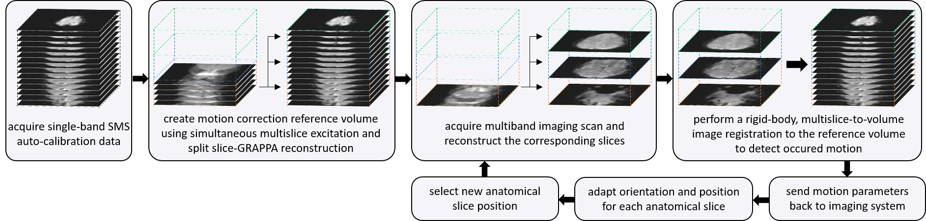
Figure 1 shows the MS-PACE technique for a multiband factor of three. First, a single-band reference volume is acquired to provide auto-calibration data for separating slices in subsequent multiband acquisitions; this slice separation is performed using the split slice-GRAPPA technique5. The first volume of multiband scans is then used as reference volume for prospective motion correction. The next separated slices create the input for the first multislice-to-volume image registration to the reference volume, based on the Mattes’ Mutual Information metric6 (ITK). The detected motion parameters are then used in real time to adapt the slice position and orientation of subsequent scans.
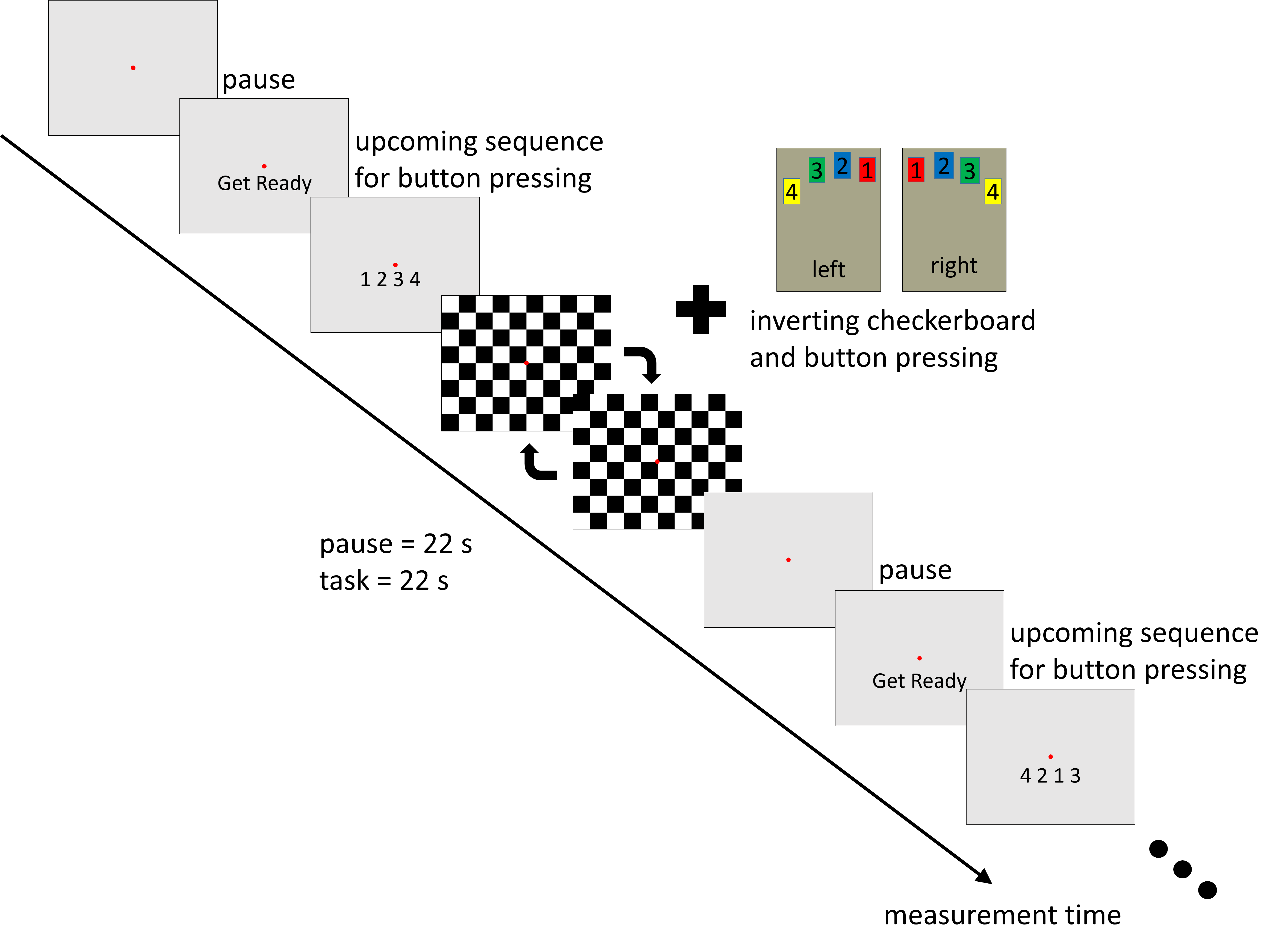
The method was evaluated at 3T (MAGNETOM Skyra, Siemens Healthcare GmbH) using a well-established fMRI paradigm (fig. 2) without head restraint. The repeating task consisted of a 22-second pause followed by an inverting checker board pattern, together with a button-pressing instruction. The button order was changed between the stimulation phases and introduced immediately before the task began.
The measurement protocol was performed without any, with multislice-to-volume (MS-PACE) and with the manufacturer’s volume-to-volume based prospective motion correction, both with and without intentional head motion. The protocol parameters of the dedicated T2*-weighted EPI sequence were: TR=1100ms, TE=30ms, 42 slices, 3mm slice thickness, no slice gap, 64x64 matrix with 192x192mm FOV, 275 volumes, flip angle of 64°, and multiband factor 3.
Results
The average time to separate three simultaneously excited slices was 40ms; MS-PACE image registration took about 70ms; each slice position was corrected using the most recent motion update available.
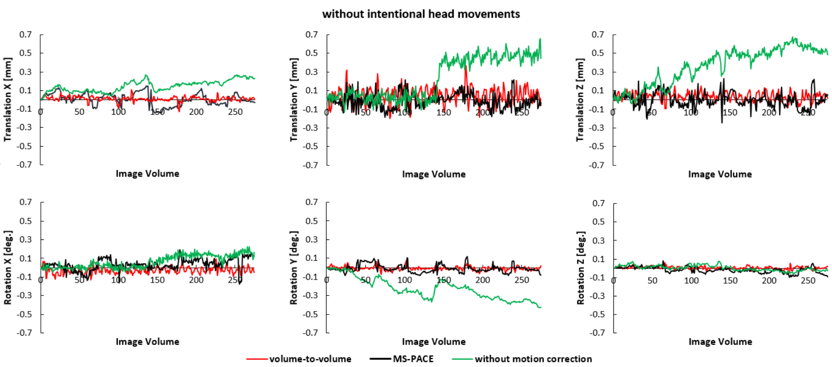
Figures 3 and 4 show residual motion parameters, detected by SPM127. The motion estimates of measurements without intentional head movements (fig. 3) show that, without prospective motion correction, there is a small amount of motion due to the missing head restraint and the effect of the fMRI tasks performed. Both motion correction techniques were able to reduce this motion to below ±0.2mm for translations and ±0.2° for rotations.
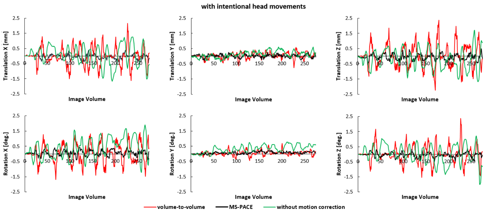
The motion parameter estimates of measurements with deliberate head motion (fig. 4) indicate that the MS-PACE motion correction reduced the residual motion to below ±0.5mm/±0.5° despite the significant motion shown in the curves of the measurement without prospective motion correction. This type of motion could not be fully corrected by the volume-to-volume based technique.
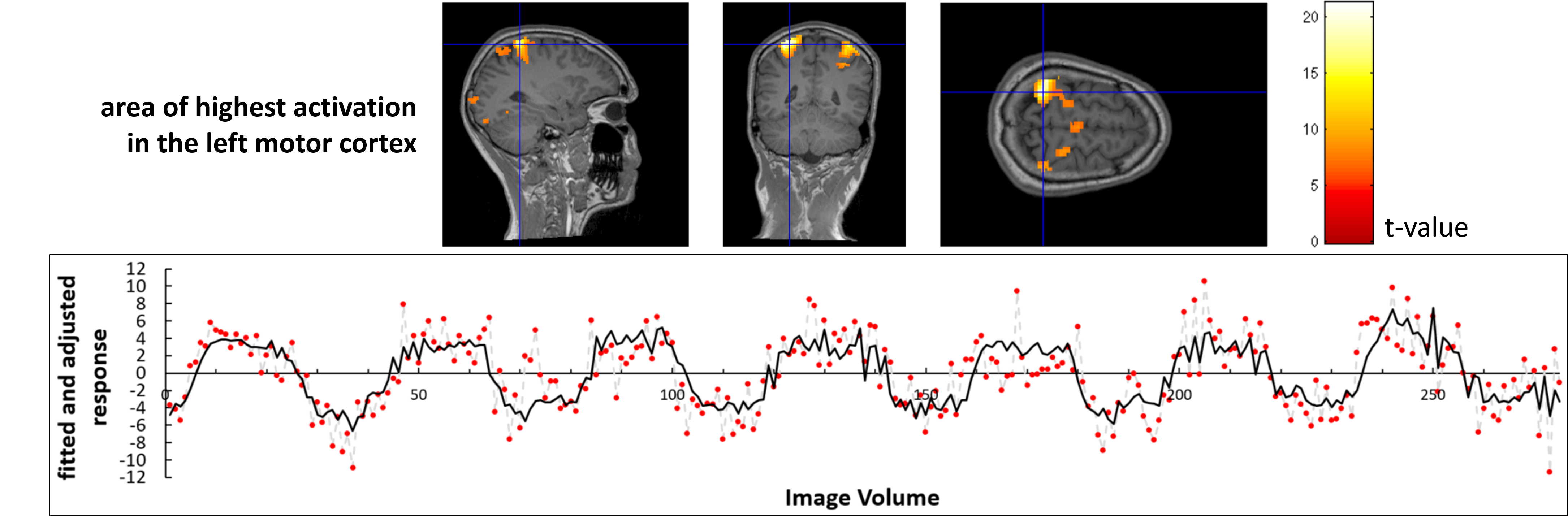
Preliminary results of the SPM12 analysis of the MS-PACE corrected data can be seen in figure 5. The point of highest activation in the left motor cortex is marked in the images and the corresponding fitted and adjusted response function is shown graphically.
Discussion
The images from measurements with voluntary head movements show typical motion effects, often seen when scanning uncooperative subjects. Volume-to-volume based prospective motion correction is not able to sufficiently correct for these motion events due to low temporal resolution, even with a short TR of 1100ms. The MS-PACE technique, however, offers fast motion-correction updates and can reduce the residual motion significantly. Results of measurements without intentional head movements indicate that there is a small increase in residual motion using MS-PACE compared to volume-to-volume based motion correction.
In this work, a multiband factor of three was applied. However, the MS-PACE technique could be combined with other multiband factors with a slight adaption of image registration parameters. An increased number of simultaneously excited slices would increase the accuracy of motion detection, but also the computing time for separating the multiband slices and image registration.
Conclusion
This study has extended the application of MS-PACE prospective motion correction to multiband fMRI. MS-PACE uses separated slices from a single multiband readout to perform a multislice-to-volume image registration to a reference volume. It offers higher temporal resolution compared to volume-to-volume based methods and is able to correct intra-volume motion. The method can be used with a range of multiband factors and shows a substantial reduction of residual motion parameters in the presence of significant subject motion.Acknowledgements
The Authors are grateful to Dr Robert Frost and the Welcome Centre For Integrative Neuroimaging at Oxford University for providing source code for their slice-GRAPPA implementation. Thanks are also due to Klaus Eickel for his contributions to the SMS methods used in this study.
All funding for this study was provided by the internal Attract funding program of the German Fraunhofer-Gesellschaft.
References
1. Thesen S, Heid O, Mueller E, Schad LR. Prospective acquisition correction for head motion with image-based tracking for real-time fMRI. Magn Reson Med 2000;44:457–465.
2. Larkman DJ, Hajnal JV, Herlihy AH, Coutts GA, Young IR, Ehnholm G. Use of multicoil arrays for separation of signal from multiple slices simultaneously excited. J Magn Reson Imaging 2001;13:313–317.
3. Setsompop K, Gagoski BA, Polimeni JR, Witzel T, Wedeen VJ, Wald LL. Blipped-controlled aliasing in parallel imaging for simultaneous multislice echo planar imaging with reduced g-factor penalty. Magn Reson Med 2012;67:1210–1224.
4. Hoinkiss DC, Porter DA. Prospective motion correction in 2D multishot MRI using EPI navigators and multislice-to-volume image registration. Magn Reson Med 2017. Early View. DOI: 10.1002/mrm.26951.
5. Cauley SF, Polimeni JR, Bhat H, Wald LL, Setsompop K. Interslice leakage artifact reduction technique for simultaneous multislice acquisitions. Magn Reson Med 2014;72:93-102.
6. Mattes D, Haynor DR, Vesselle H, Lewellen TK, Eubank W. PET-CT image registration in the chest using free-form deformations. IEEE Trans Med Imaging 2003;22:120–128.
7. Friston KJ, Ashburner J, Kiebel SJ, Nichols TE, Penny WD, editors. Statistical parametric mapping: The analysis of functional brain images. Academic Press, 2007.
Figures