1147
Correlating Proteoglycan Concentration in Intervertebral Discs Using 1H MR Spectroscopy with Lumber Lordotic Angle1Radiological Science, Brighton and Sussex University Hospitals NHS Trust, Brighton, United Kingdom, 2Clinical Imaging Sciences Centre, Brighton Sussex Medical School, Brighton, United Kingdom, 3Computing, Engineering and Mathematics, University of Brighton, Brighton, United Kingdom
Synopsis
Both proteoglycan (PG) concentration and lumbar lordotic (LL) angle are linked with the health of spinal discs. Previously, it has been shown that PG concentration can reliably be measured using 1H MR Spectroscopy at 1.5T and that PG concentration is higher in female discs compared with male discs. This study has now been extended to show a correlation between PG and LL angle in a group of female participants.
Introduction
Intervertebral discs (IVDs) are fibrocartilaginous cushions that act to protect both the vertebrae and the brain by working as a shock absorbing system for the spine. Proteoglycan molecules (PGs) are an important component of IVDs, serving to both attract and retain water. Thus, they contribute to the hydration of the tissue, and also serve the purpose of regulation of matrix assembly. The loss of PG from the disc is the main chemical change observed in intervertebral disc degeneration [1]. Previous studies have shown the feasibility of reliably quantifying PG concentration using 1H magnetic resonance spectroscopy (1HMRS) at 1.5T [2], and found a gender difference in concentration [3], with women having a significantly higher concentration (p = 0.008).
It has been suggested that lumbar lordosis (LL) is linked with health of the lumbar region of the spine and ageing [4], and that women have evolved to have a higher LL angle [5]. Here it is hypothesised that a higher proteoglycan concentration is linked with a higher value for LL angle.
Methods
Scans were performed on a group of 10 healthy volunteers (age = 27.4± 3.4 yo; 5F, 5M) using a 1.5T Siemens Avanto with the inbuilt spine coil. All participants gave written informed consent and the study was approved by the local research ethics committee.
1HMRS was performed using the following parameters: TE = 30ms, TR = 1500ms, and 256 averages. Voxels were 25mmx20mm in the axial plane and 8mm in thickness, so as to be contained fully in the disc (figure 1). Saturation bands were placed surrounding the voxel to reduce signal from external tissues. Voxels were placed within lumbar discs, either between L3 and L4 or L4 and L5, whichever was the thickest. Water unsuppressed data was also collected (16 averages) to allow for eddy current correction and quantification. Shimming was performed manually, to obtain the best possible linewidth.
Peaks arising from the N-acetyl resonance (2.04ppm) associated with PG were fitted, using the jMRUI processing tool with AMARES [6]. All results are quoted as mean±sd.
Additional high resolution sequences were performed in the sagittal plane to allow for measurement of LL. LL was measured using the 2-line Cobb method outlined by Harrison et al [7] in ImageJ (figure 2); data was analysed blind and repeated 5 times to reduce error.
Results and Discussion
Of the 10 participants, all had corresponding good quality 1HMRS data (line width of 8.0±2.0Hz and SNR of 11.2±6.8) and a corresponding Sagittal MRI dataset. All of the spectra showed a clear singlet at 2.04ppm relating to PG (figure 3).
The calculated PG concentration (/water) of 308.0±60.2 is in good agreement with a study performed at 3T (299±62) [8]. Additionally, female participants show a significantly higher (p=0.008) PG concentration (358.1±15.4) compared with male participants (257.3±23.3).
The mean measured LL for female and male participants was very similar: 47.2±1.6 and 42.3±5.3 respectively. There was no correlation with age seen (R2 = 0.25). When assessing a correlation between LL angle and PG concentration (figure 4), none was seen in the male group (R2 = 0.28), however there was a correlation seen within the female data (R2 = 0.86).
Both concentration of PG [1] and LL angle [3, 4] are linked with health of IVDs, here it has been shown that, for women, there is a correlation to be seen between these 2 measures.
Conclusion
Our hypothesis, that LL angle and PG concentration would be correlated, was shown by the results of the female participants in this study. This correlation is supported by evidence that both these measures are linked with spinal health, especially in women. A correlation was not observed in the male cohort. The study was limited by small numbers, but the aim is now to extend this further.Acknowledgements
I would like to acknowledge the help and support of the team at the Clinical Imaging Sciences Centre, in particular the radiographers.References
[1] Johannessen W, Auerbach JD, Wheaton AJ et al. Assessment of Human Disc Degeneration and Proteoglycan Content Using T1ρ-weighted Magnetic Resonance Imaging. Spine. 2006; 31(11): 1253–1257
[2] Harris LM, Hodder E, Cercignani M et al. Proteoglycan concentration in intervertebral discs assessed by 1HMRS at 1.5T. ISMRM 2016. Singapore
[3] Harris LM, Hodder E, Cercignani M et al. Proteoglycan concentration in intervertebral discs assessed by 1HMRS at 1.5T. ESMRMB, 2016. Vienna
[4] Skaf GS, Ayoub CM, Domloj NT, et al. Effect of age and lordotic angle on the level of lumbar disc herniation. Adv Orthop. 2011
[5] Whitcome KK1, Shapiro LJ, Lieberman DE. Fetal load and the evolution of lumbar lordosis in bipedal hominins. Nature. 2007; 450(7172):1075-1078
[6] Vanhamme L1, van den Boogaart A, Van Huffel S. Improved method for accurate and efficient quantification of MRS data with use of prior knowledge. J Magn Reson. 1997;129(1):35-43
[7] Harrison DE1, Harrison DD, Cailliet R et al. Radiographic analysis of lumbar lordosis: centroid, Cobb, TRALL, and Harrison posterior tangent methods. Spine. 2001;26(11):235-242
[8] Zuo J1, Joseph GB, Li X et al. In vivo intervertebral disc characterization using magnetic resonance spectroscopy and T1ρ imaging: association with discography and Oswestry Disability Index and Short Form-36 Health Survey. Spine. 2012; 37(3):214-221
Figures
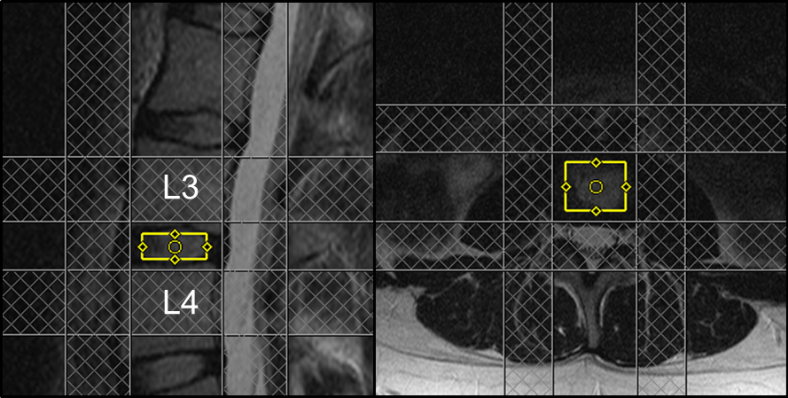
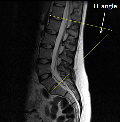
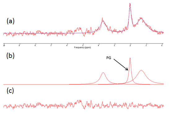

Figure 4: Graph showing correlation between PG (x-axis) and LL angle (y-axis)
Blue - female participants; Red - male participants
R2 values are quoted.