1145
Impact of the glymphatic system on the kinetic and distribution of gadodiamide in the rat brain: Observation by dynamic MRI and effect of circadian rhythm on tissue gadolinium concentrations.1Nagoya University, Nagoya, Japan, 2MR&CT Contrast Media Research, Bayer AG, Berlin, Germany
Synopsis
We tried to elucidate the role of the glymphatic system on the distribution and kinetics of a linear gadolinium based contrast agent in the rat brain. Dynamic MRI signal changes of the brain and the cerebrospinal fluid (CSF) indicated that intravenously administrated gadodiamide enter from blood into the CSF, indicating the involvement of the glymphatic system. The long term presence of gadolinium in the brain after repeated administration of gadodiamide quantified by ICP-MS indicates the involvement of the glymphatic system on the clearance of gadolinium from brain tissue in terms of the influence of circadian rhythm and anesthesia during administration.
Purpose
We tried to elucidate the role of the glymphatic system on the distribution and kinetics of the linear gadolinium based contrast agents (GBCA) gadodiamide in the rat brain by two experiments. One is a short term imaging experiment to evaluate MRI signal changes of the brain and the cerebrospinal fluid (CSF) after a single intravenous administration. The other is a long term experiment to quantify the brain tissue gadolinium (Gd) concentration after repeated intravenous administration at different times of the day and under the impact of anesthesia. This was to investigate the effect of the circadian rhythm and anesthesia which influence glymphatic system activity (1,2).Subjects and Methods
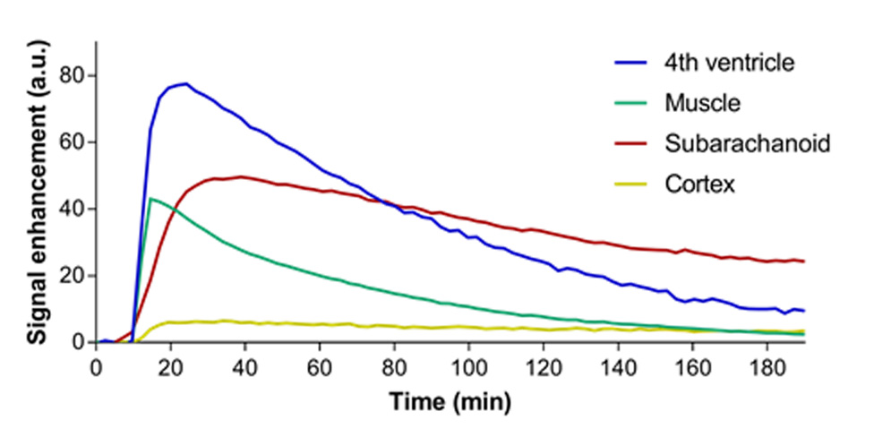
Imaging experiment: 6 Han-Wistar rats received single intravenous injection of gadodiamide (1.0 mmol/kg), and T1-weighted dynamic scans (4.7T, RARE TR/TE =1016/18 ms) were made for 3 hours with 2 minutes intervals. Signal intensity (SI) curves were acquired for the cortex, fourth ventricle, subarachnoid space of prepontine cistern and the temporal muscle.
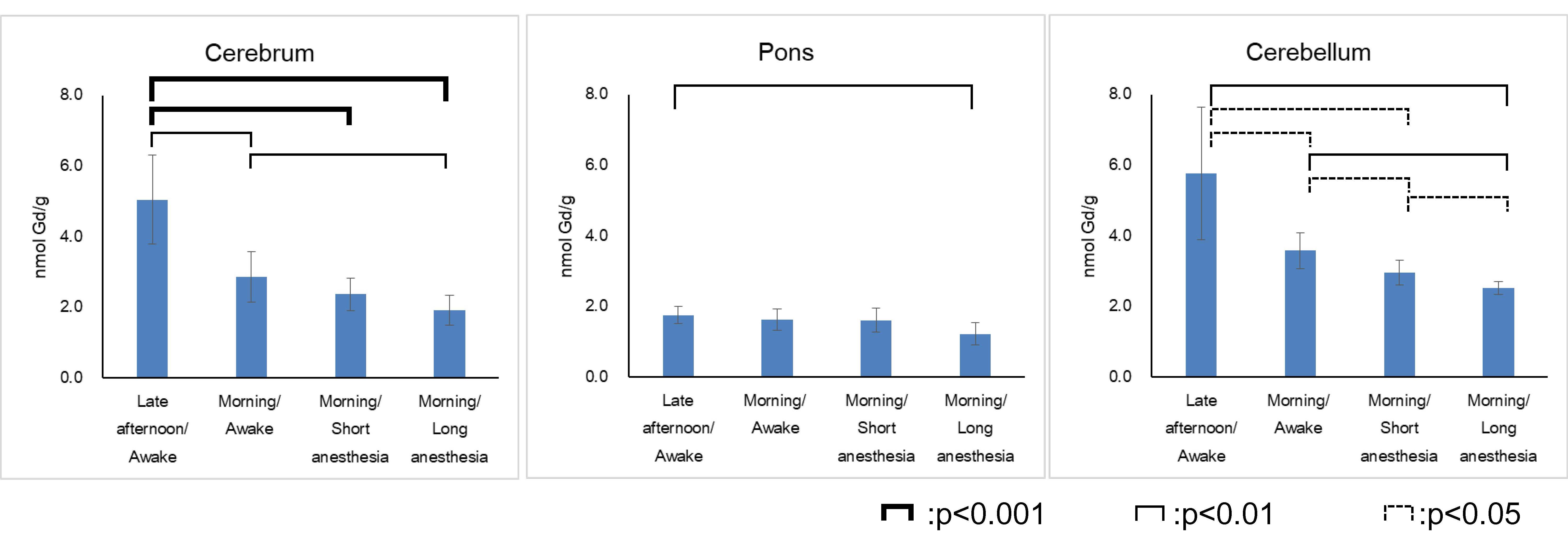
Gadolinium quantification in brain tissue: 24 Han-Wistar rats were divided into four groups: Group 1; morning injection, Group 2; evening injection, Group 3; morning injection under short duration anesthesia and Group 4; morning injection under long duration anesthesia. Each animal received 1.8 mmol/kg gadodiamide (8 times over 2 weeks). 5 weeks after the last administration the brains were dissected and the Gd concentration in the cerebellum, pons and cerebrum were quantified by inductively coupled plasma mass spectrometry (ICP-MS).
Results
Imaging experiment: Time intensity curves are shown in Fig 1. The SI of the fourth ventricle increased immediately after injection followed by a continuous decrease. The SI profile showed a similar kinetics than that of the temporal muscle. In the cortex a slight but fast SI increase after the administration and a slow decrease almost to the baseline level within 3 hours was observed. The signal of the prepontine subarachnoid space showed gradual increase followed by a less pronounced decrease compared to the 4th ventricle and the muscle.
Gadolinium quantification in brain tissue (Fig. 2): In the cerebellum, mean concentration were 3.58, 5.77, 2.95 and 2.51 (nmol Gd/g tissue) in groups 1, 2, 3 and 4 respectively. Likewise, mean concentration were 2.86, 5.04, 2.37 and 1.92 (nmol Gd/g) in cerebrum, and 1.63, 1.76, 1.61 and 1.22 (nmol Gd/g) in pons.
Discussion
The recently introduced glymphatic system hypothesis postulate that the perivascular space serves as a conduit for CSF flow into the brain parenchyma (1). The perivascular space around the arteries allows the CSF to exchange with the interstitial space of the brain tissue. According to this hypothesis, the initial pathway of the GBCA into the brain tissue can be either from blood via the circulation system or from CSF via the glymphatic system. Both systems may be involved in the distribution and clearance of GBCA in the brain tissue.
In the dynamic MRI, the signal of the cortex showed fast SI increase which may be primarily due to the vasculature of the brain tissue. Thus, we could not observe the passage of gadodiamide from CSF into the brain tissue as reported in preexisting report of intracisternal administration of GBCA that indicate the glymphatic transmission (3). However, the SI of the CSF in the fourth ventricle, which contains choroid plexus, increased immediately after injection, which may indicate that the GBCA enters instantaneous from blood to the CSF most likely via the choroid plexus. While, subarachnoid space of the prepontine cistern, which does not contain choroid plexus, showed a different time course compared to that of the fourth ventricle.
After repeated administrations of gadodiamide Gd concentrations in the cerebellum and cerebrum were highest in the group that received the GBCA in the evening, followed by the group with the administration in the morning. Lower concentrations were found for the injections during anesthesia. As rats are nocturnal animals, glymphatic system is expected to be more active during daytime (2). The results might be explained by the higher glymphatic clearance following the morning injection, while a reduced glymphatic activity is present after the injection in the evening. Anesthesia seemed to facilitate the glymphatic clearance of GBCA especially for longer durations.
Conclusion
A fast transition of gadodiamide from blood to CSF via choroid plexus in the rat brain was demonstrated by dynamic MR imaging. The further distribution from CSF into the brain tissue remained unclear. While, for the clearance of the gadodiamide from the brain tissue, involvement of the glymphatic system was indirectly indicated in terms of the previously described impact of the circadian rhythm and anesthesia on the glymphatic system.Acknowledgements
No acknowledgement found.References
1. Iliff JJ, Wang M, Liao Y, Plogg BA, Peng W, Gundersen GA, et al. A paravascular pathway facilitates CSF flow through the brain parenchyma and the clearance of interstitial solutes, including amyloid beta. Science translational medicine 2012;4(147):147ra11.
2. Xie L, Kang H, Xu Q, Chen MJ, Liao Y, Thiyagarajan M, et al. Sleep drives metabolite clearance from the adult brain. Science 2013;342(6156):373-7.
3. Gaberel T, Gakuba C, Goulay R, Martinez De Lizarrondo S, Hanouz JL, Emery E, et al. Impaired glymphatic perfusion after strokes revealed by contrast enhanced MRI: a new target for fibrinolysis? Stroke 2014;45(10):3092-6.
Figures