1095
Mean lung alveolar dimension mapping with hyperpolarized 3He and 129Xe diffusion-weighted MRI1Academic Unit of Radiology, University of Sheffield, Sheffield, United Kingdom
Synopsis
Lung morphometry parameters can be derived from multiple b-value hyperpolarized gas diffusion-weighted (DW)-MRI using the cylinder (CM) and stretched exponential (SEM) models. Mean alveolar diameter (LAlv) and diffusive length scale (LmD) are calculated from the CM and SEM, respectively. This work compares the parameters LAlv and LmD derived from a range of subjects with both 3He and 129Xe DW-MRI. Excellent linear agreement is observed between the LAlv and LmD parameters for both gases. This indicates these parameters are equivalently representative indices of mean alveolar diameter dimension within the range of experimental conditions considered.
Introduction
Theoretical models of gas diffusion have been proposed to derive lung morphometry parameters from the hyperpolarized gas diffusion-weighted (DW)-MRI signal. The cylinder model (CM) is based upon infinitely long non-connected cylinders, and has been used to obtain estimates of acinar airway radius (R) and mean chord length (Lm) from a model of anisotropic diffusion. 1,2 The stretched exponential model (SEM) is a mathematical model of the non-Gaussian diffusion signal that derives estimates alveolar dimensions (LmD) within each voxel from a probability distribution of diffusion length scales. 3,4
Previously, in vivo comparisons of Lm and LmD have demonstrated that the two lung morphometry parameters are related but not identical. 5,6 The mean alveolar diameter (LAlv) is an alternative CM parameter that is defined as the chord length of one alveolus in the CM cylindrical airway geometry. 2 This work evaluates and compares SEM-derived LmD and CM-derived LAlv from a range of subjects using 3He and 129Xe DW-MRI.
Methods
3D multiple b-value DW-MRI datasets were acquired on a 1.5 T GE HDx scanner in 51 and 31 subjects with 3He and 129Xe, respectively. 3He and 129Xe DW-MRI (3He Δ=1.6 ms, b=[0, 1.6, 4.2, 7.2 s/cm2]; 129Xe Δ=8.5 ms, b=[0, 12, 20, 30 s/cm2]) were acquired using compressed sensing as described previously. 4,7 Healthy volunteers, ex-smokers, idiopathic pulmonary fibrosis (IPF), and COPD patients were recruited, representing a range of acinar microstructural length scales. The 3He and 129Xe diffusion signal decay within each voxel was fitted to both the CM and SEM to derive maps of R 2 and LmD 3, respectively. 129Xe DW-MRI was acquired with Δ=8.5 ms; therefore, 3He CM expressions 2 were applied to both 3He and 129Xe DW-MRI to derive maps of R, 7 and LAlv was subsequently calculated from LAlv=2Rsin(π/8).Results and Discussion
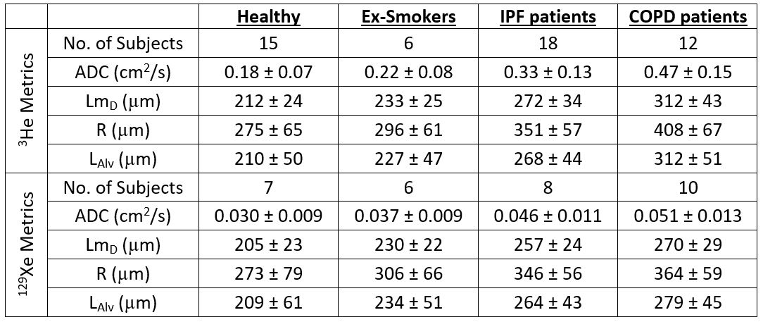
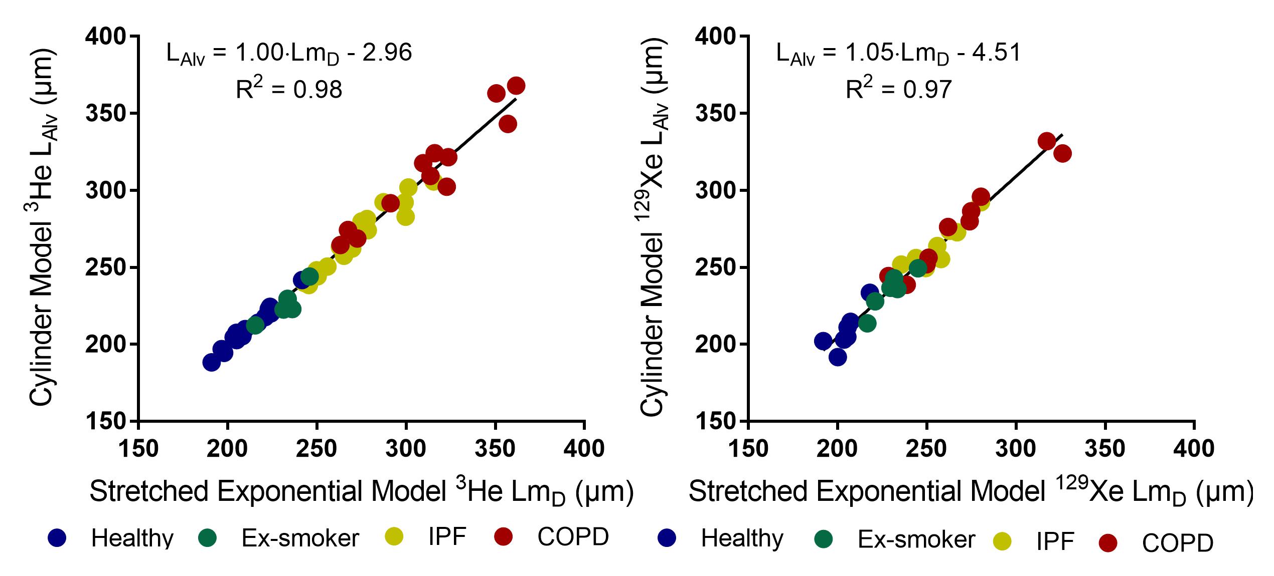
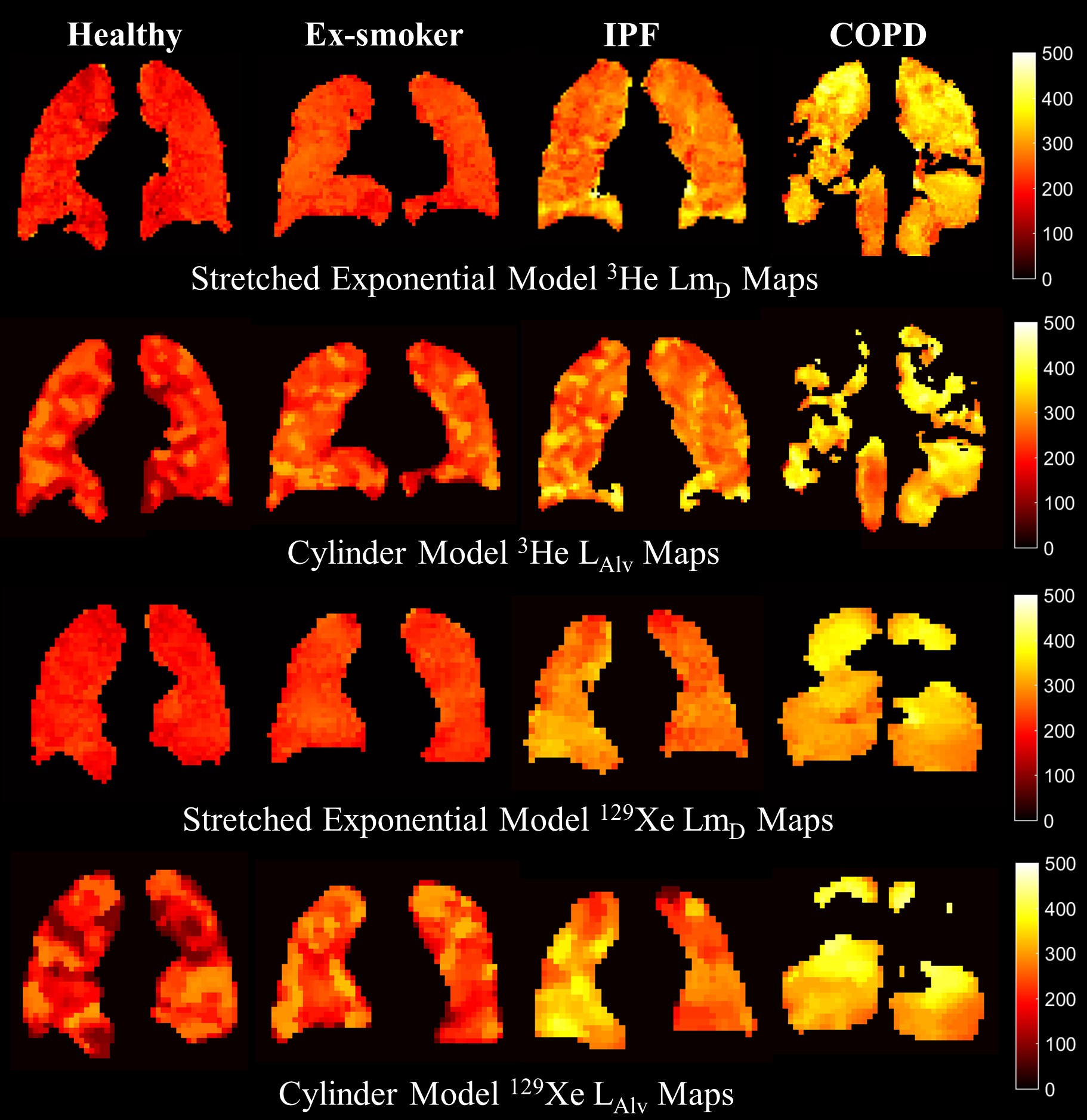
A summary of global 3He and 129Xe lung morphometry metrics is provided in Table 1. All metrics for each patient group were larger than those for healthy subjects reflecting the respective changes to acinar microstructure related to fibrosis in the IPF patients, and emphysema for the ex-smokers and COPD patients. A statistically significant linear correlation (P<0.001) between LmD and LAlv was observed for both 3He and 129Xe DW-MRI (Figure 1). The linear regression fit (3He β1=1.00, 129Xe β1=1.05) suggests excellent agreement between the two parameters for both gases, and Bland-Altman analysis confirmed this with small mean bias of +1.0% and -2.6% towards LmD for 3He and 129Xe, respectively. In the representative LAlv maps from 3He and 129Xe measurements in the same patients (Figure 2), regions of elevated values appear to qualitatively match those observed in corresponding LmD maps. The analogous linear correlation and lung morphometry maps demonstrates the same microstructural information can be obtained with both 3He and 129Xe.
In a previous comparison, 3He LmD was related to Lm by a non-linear power relationship; 6 however, the results in this comparison suggests that LAlv is more comparable to LmD than Lm. The two distinct relationships observed between LmD with Lm and LAlv can be associated with the different sections of the acinar airway geometry that is measured by each parameter.
In the CM geometry, eight alveoli units surround the acinar airway such that the chord length of an alveolus matches typical alveolar diameter measurements from histology. 2 While in the SEM, LmD represents the average distance that 3He or 129Xe gas atoms diffuse within the acinar airspace. The maximum LmD is constrained by the theoretical free diffusion length of the respective DW-MRI experiments (~500 µm). According to histology, the mean alveolar diameter in healthy adult lungs is approximately 200-250 µm. 8 Therefore, in these experiments, gas atoms are predominantly restricted by the alveolar geometry and as such LmD, like LAlv, is reflective of mean alveolar diameter dimensions.
In contrast, the CM-derived Lm is calculated through an inferred relationship between the volume (alveolus and alveolar duct) and surface area of a single alveolus unit. Therefore, the inclusion of the alveolar duct volume in the calculation might allows Lm values that are beyond the theoretical free diffusion lengths of the diffusion experiments, and probably accounts for the mismatch in LmD and Lm observed previously. 5,6
Conclusion
This is the first comparison of mean alveolar diameter parameters derived from the CM and SEM with 3D multiple b-value 3He and 129Xe DW-MRI acquired in a range of subjects with a range of severity of microstructural lung changes. LAlv and LmD demonstrated excellent agreement with both gases indicating that these parameters derived from different theoretical gas diffusion models are equivalent.Acknowledgements
This work was supported by NIHR grant NIHR-RP-R3-12-027 and MRC grant MR/M008894/1. The views expressed in this publication are those of the author(s) and not necessarily those of the NHS, the National Institute for Health Research or the Department of Health.References
1. Yablonskiy, D. A., et al. (2002). Proc Natl Acad Sci USA 99(5): 3111-3116.
2. Yablonskiy, D. A., et al. (2009). J Appl Physiol (1985) 107(4): 1258-1265.
3. Parra-Robles, J., et al. (2014). Proc ISMRM:3529
4. Chan, H.-F., et al. (2017). Magn Reson Med 77(5): 1916-1925.
5. Ouriadov, A., et al. (2017) Magn Reson Med. doi:10.1002/mrm.26642.
6. Chan, H.-F., et al. (2017). Proc ISMRM:876
7. Chan, H.-F., et al. (2017). Magn Reson Med. doi:10.1002/mrm.26960.
8. Weibel, E. R. (1963). Morphometry of the human lung, Springer-Verlag.
Figures