1063
Dynamic Spatially Selective Dephasing for Outer Volume Suppression1Biomedical Engineering, University of Cincinnati, Cincinnati, OH, United States, 2Radiology, University of Pittsburgh, Pittsburgh, PA, United States
Synopsis
A novel dynamic spatially selective dephasing (DSSD) for outer volume suppression (OVS) is introduced. This technique uses high-order shim pulses to accomplish OVS by creating a low and high field gradient within and outside of region of interest. The effectiveness of DSSD is investigated with phantom and human using an 8-channel parallel transmit 7 Tesla Siemens system. Similar suppression performance was obtained in phantom and human brain using DSSD with a circular and an elliptical shape, respectively. The results show that OVS can be achieved by using a high degree shim insert to generate a steep B0 gradient at periphery of target region.
Introduction
Outer volume suppression (OVS) is commonly used in MR spectroscopic imaging (MRSI) to reduce contamination from strong lipid and water signals of peripheral regions. Traditionally, these signals are suppressed using slice-selective pulses that are placed around the brain to approximate the elliptical shape of the skull (1). The transverse magnetization generated is then dephased with large crusher gradients. Recently, an in-plane OVS was introduced using RF shimming and an eight-element transceiver array which excited a ring about the periphery of the head and left the central brain regions largely unaffected (2). Another approach for OVS introduced the so-called gradient rotating outer volume excitation method which uses a single RF pulse together with a varying gradient to produce a frequency-swept excitation of a desired region (3). In this work, we introduce a novel OVS method, dynamic spatially selective dephasing (DSSD), which uses high-order shim pulses to create a low and high field gradient within and outside of region of interest (ROI), respectively. Thus, OVS is accomplished by de-phasing the transverse magnetization outside of ROI due to the high shim field gradient. Transverse magnetization within ROI is maintained in phase since overall spatial derivative of B0 distribution is small.Methods
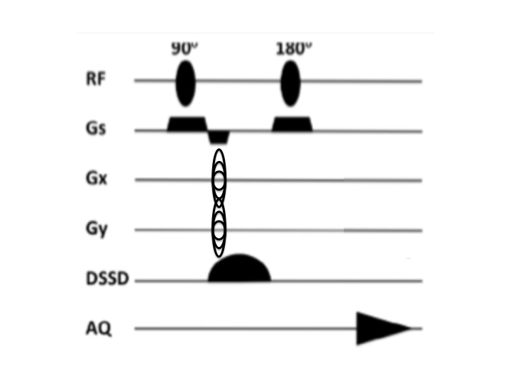
High-order shim gradient pulses required for OVS were carried out using custom-written spherical harmonic gradient waveforms optimization software in Matlab (The MathWorks, Inc., Natick, MA, USA). The program calculated and stored multiple shim gradient strengths so that the required shims could be effectively transferred to scanner and examined. The OVS performance was initially tested and optimized using a 17cm water-filled spherical MRI phantom. Healthy volunteers (n = 5) participated in the study after giving informed consent according to procedures approved by the Institutional Review Board of the University of Pittsburgh. All scans were conducted on an 8-channel parallel transmit 7 Tesla (T) Siemens system (Erlangen, Germany). An 18-channel shim insert coil with 38-cm ID, 43cm OD, and 70-cm length, 18.5cm from patient end to magnetic center, consisting of 3rd, 4th, and two 5th-degree shims (ZC4 and ZS4) with 10A shim supplies (Resonance Research, Billerica, MA USA) was used for high-degree shimming. The B0 field used for OVS optimization was obtained using the B0 loop encoded readout (Bolero) method (4). Shimming was performed by optimizing the shim current values using a least-squares algorithm based on a calibrated spherical harmonic model (4) to minimize the standard deviation of the B0 field over the target ROI. The pulse sequence used for acquiring 2D MRSI is shown in Figure 1. The B0 shims are pulsed during a spin echo sequence either single waveform or paired waveform of opposite sign or potentially distribution so as to enhance dephasing.Results
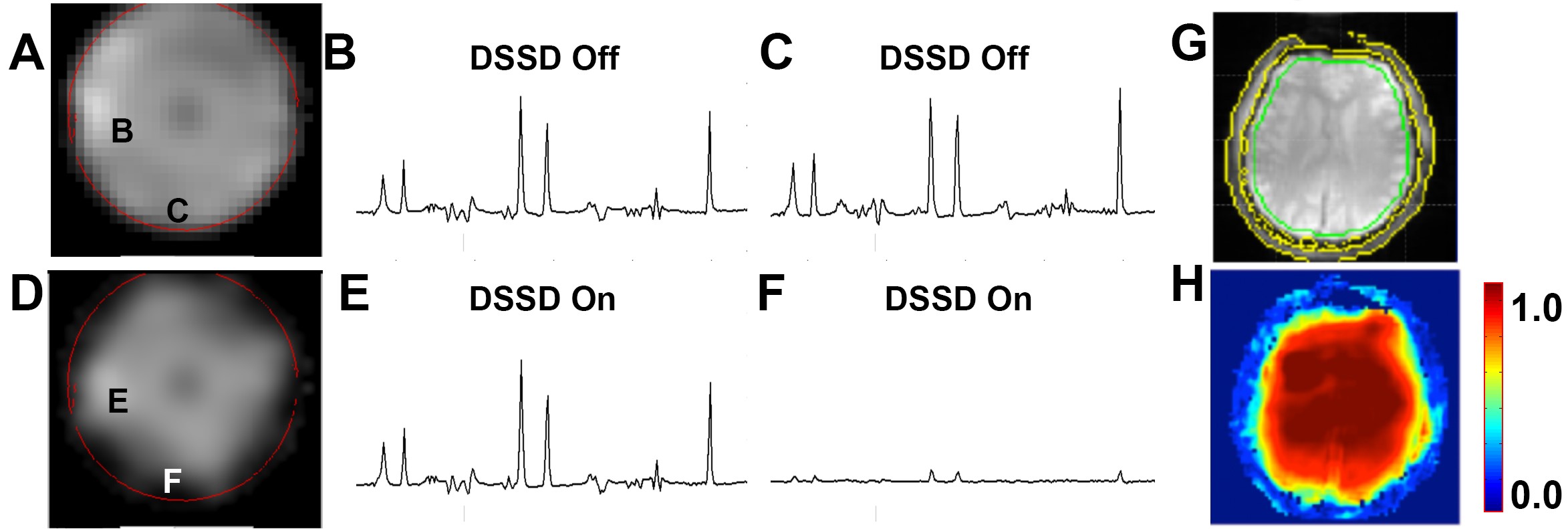
The results show that the DSSD pulse with optimized amplitudes can selectively suppress the target outer volume . Figures 2A and 2D show the images of a phantom from NAA without and with the DSSD pulse, respectively. The corresponding spectra from the phantom were acquired with the DSSD pulse off (Fig. 2B and 2C) and on (Fig 2E and 2F). Figures 2G and 2H display the retention of NAA magnetization for the human brain with red=1.0 for full signal retention and blue=0.0 for full suppression.Discussion
The results show that OVS can be achieved by using a high degree shim insert to generate a steep B0 gradient at periphery of the target region. The DSSD pulse is a transient change in B0 shim values, and is added onto the optimal setting only during the shim pulse. Results also show that DSSD requires less SAR and time than traditional OVS schemes.Conclusion
This report outlines the theory and experimental verification of the novel DSSD approach. The efficacy of this new technique is demonstrated both in phantom and human subjects.Acknowledgements
No acknowledgement found.References
1. Duyn JH, Gillen J, Sobering G, et al. Radiology. 1993; 188:277.
2. Hetherington HP, Avdievich NI, Kuznetsov AM, et al. Magn Reson Med. 2010; 63:9.
3. Powell NJ, Jang A, Park JY, et al. Magn Reson Med. 2015;73:139.
4. Hetherington HP, Chu WJ, Gonen O, Pan JW. Magn Reson Med 2006;56:26–33.
Figures