1061
Elliptical Localization with Pulsed Second-Order Fields (ECLIPSE) for Robust Lipid Suppression in Proton MRSI1MRRC, Yale University, New Haven, CT, United States
Synopsis
Proton MRSI has great clinical potential for metabolic mapping of healthy and pathological human brain. However, technical challenges related to poor spectral quality caused by magnetic field inhomogeneity, limited RF transmit power and incomplete lipid suppression have dampened the utility of MRSI. Here a novel method for lipid suppression is presented based on localization of an elliptical region-of-interest using pulsed second-order magnetic fields. A high-amplitude gradient setup was designed and constructed, containing coils to generate Z2, X2Y2 and XY magnetic fields. Simulations, phantom MRI and MRSI on human brain in vivo demonstrate robust localization and suppression of extracranial lipids.
Introduction
Despite its potential, proton MRSI is not a widespread clinical imaging modality because the method is hampered by a number of technical challenges related to magnetic field inhomogeneity, limited RF transmit amplitude and lipid contamination. Popular lipid suppression methods include inner volume selection (IVS) of a large cuboidal volume based on single-volume MR techniques and outer volume suppression (OVS) of the skull region with up to 12 slice-selective excitation pulses. Most existing methods have limitations in terms of attainable lipid suppression, peak and average RF power deposition and brain coverage. Here we present a novel method for IVS or OVS based on high-amplitude, second-order spherical harmonic (SH) magnetic fields. The method is referred to as ‘elliptical localization using pulsed second-order fields’ or ECLIPSE. Experimental MR images show the principle of elliptical volume selection using Z2 and X2Y2 magnetic fields, as well as rotation and translation with XY and linear fields, respectively. Proton MRSI of the human brain in vivo was performed to demonstrate high-quality lipid suppression at low RF power deposition.Methods
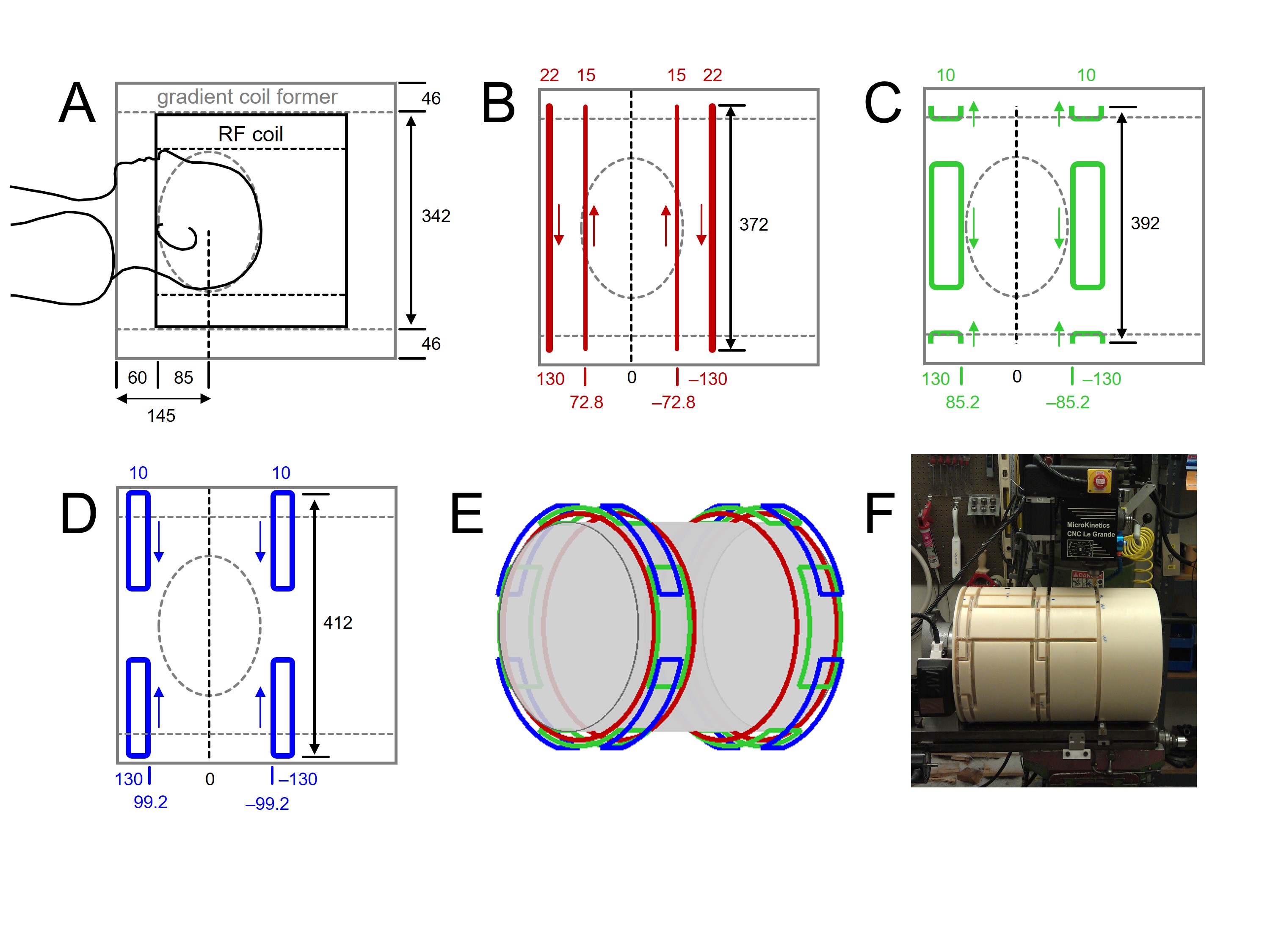
A short, high-amplitude gradient coil (Fig. 1) was constructed on a nylon cylinder (ID 342 mm, OD 434 mm) capable of accommodating a 16-element volume Tx/Rx RF coil (Fig. 1A). Target field amplitudes for the Z2, X2Y2 and XY coils were set to 2.5, 1.25 and 1.25 Hz/mm2 for a 50 A input current, respectively. Coils were manually wound from 12 AWG (Ø 2.1 mm) polyurethane-coated copper wire and placed within the wire tracks milled in the nylon former. Following placement of PT100 thermal sensor probes the wires were permanently fixed with a two-part epoxy/hardener adhesive. The final gradient coils were characterized by inductances of 859, 182, and 154 μH and resistances of 475, 240 and 238 mΩ for the Z2, X2Y2 and XY coils, respectively.Results
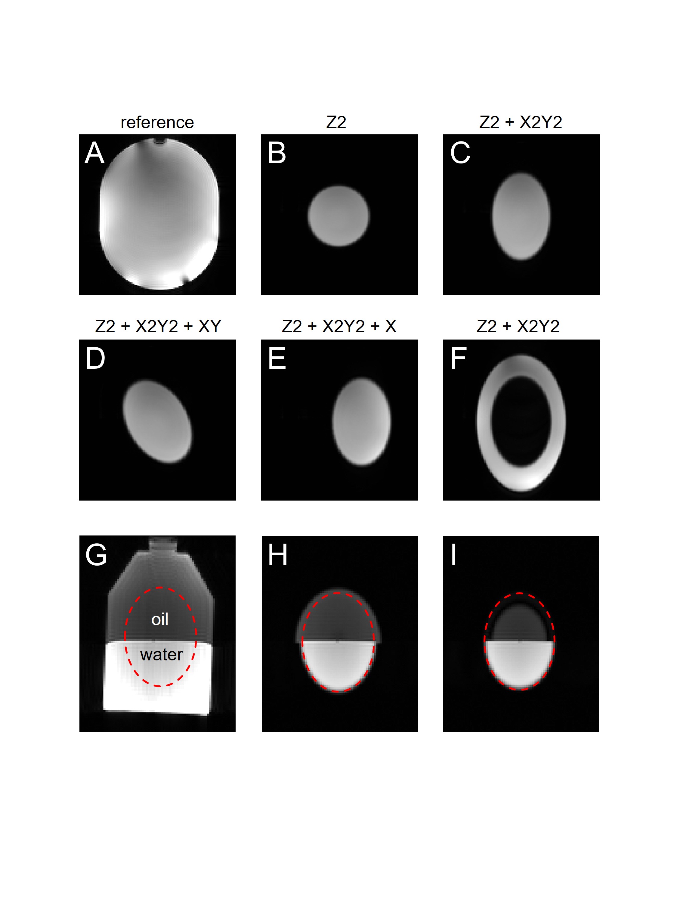
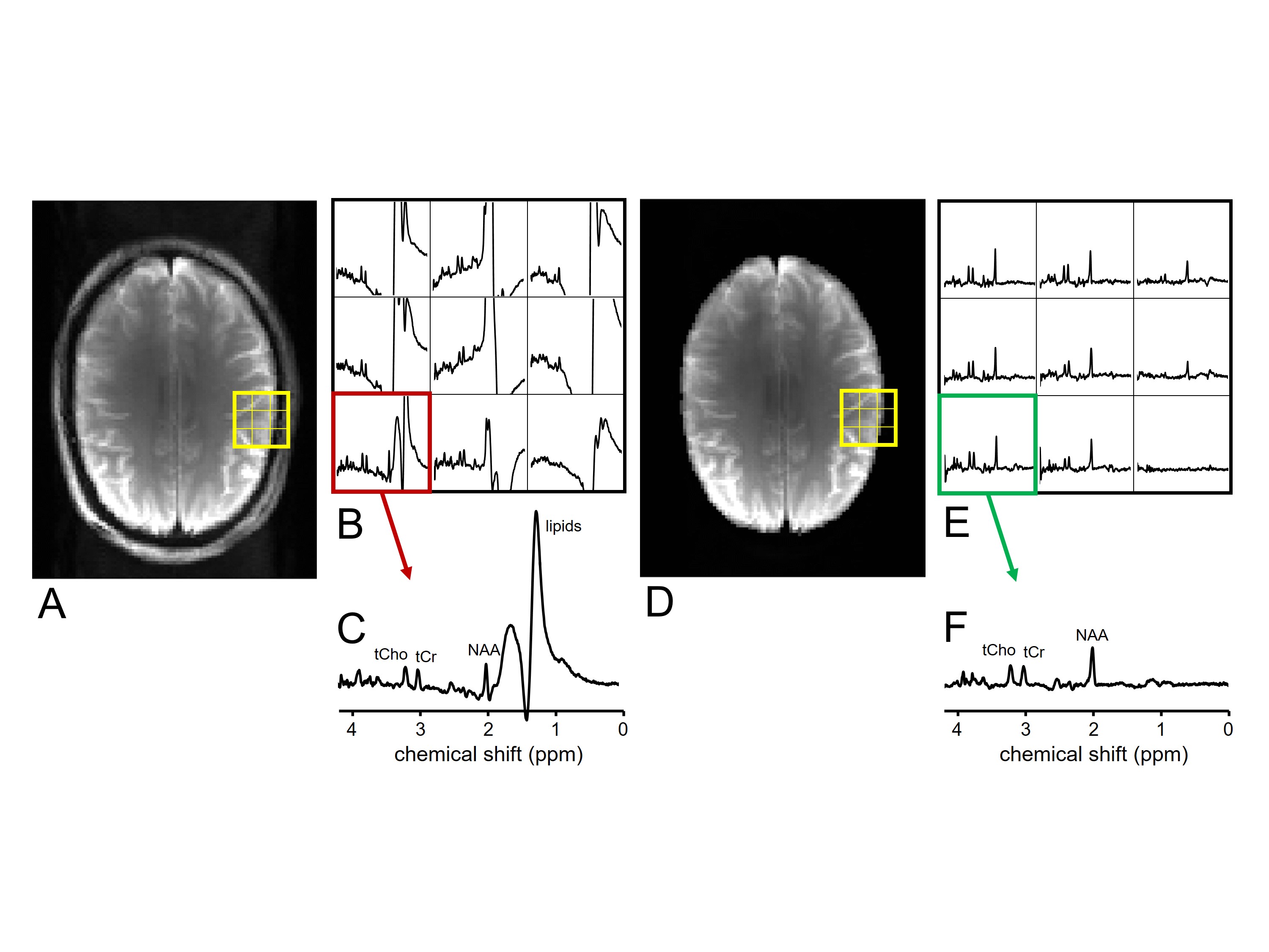
Fig. 2 demonstrates the localization capabilities of ECLIPSE. In the presence of a Z2 magnetic field, a frequency-selective RF pulse selects a 2D circle (Fig. 2B). Adding an X2Y2 magnetic field results in the selection of a 2D ellipse (Fig. 2C). The elliptical volume can be rotated (Fig. 2D) and shifted (Fig. 2E) by adding XY and X magnetic fields, respectively. By adjusting the RF pulse center frequency, the outside of the ellipse shown in Fig. 2C can be selected (Fig. 2F). The presence of different chemical shifts leads to a change in ellipse size, rather than a shift in spatial localization (Fig. 2G-I). Fig. 3 shows 1H MRSI on the human brain in vivo (A-C) without and (D-F) with 2D in-plane localization using ECLIPSE. Without OVS the extracranial lipid signals dominate each of the MR spectra in the 21 x 21 MRSI dataset, including spectra well within the brain (Fig. 3C). The MR image acquired with ECLIPSE (Fig. 3D) demonstrates the high-quality elliptical localization similar to that achieved in phantom studies (Fig. 2). The localization performance is confirmed by the 1H MRSI data (Fig. 3E/F) showing the absence of lipid signals in all voxels while retaining the metabolite signals at full intensity.Conclusions
Here we have presented a novel method for 2D volume selection for lipid suppression in proton MRSI based on frequency-selective RF pulses in the presence of a second-order SH magnetic field. The principle of using second-order SH magnetic fields for field-of-view restriction in MRI was first described by Cho and co-workers [1-3] using a dedicated Z2 gradient coil. More recent reports have used standard second-order SH shim coils to achieve field-of-view restriction in MRI [4-6]. The introduction of multi-coil (MC) shimming [7, 8] has provided complex magnetic field shaping that is ideally suited for 2D localization [9, 10]. However, the relatively small magnetic field strengths generated by SH or MC shim setups become a significant limitation for spectroscopic localization, where a small magnetic field gradient is synonymous with a large chemical shift displacement. The success of ECLIPSE for spectroscopic applications therefore rests on the availability of high-amplitude gradients. In this study a dedicated, home-build second-order gradient coil was designed and constructed to achieve high-quality 1H MRSI without lipid contamination. Whereas the ECLIPSE method has been demonstrated for lipid suppression in MRSI, it can also find application in single-volume MRS and reduced field-of-view MRI. For MRS the main advantages would be related to lower SAR and shorter echo-times due to the reduced minimum number of RF pulses. In addition, ECLIPSE can be beneficial in pathologies (tumors, multiple sclerosis) where an elliptical VOI often better mimics the shape of the lesion under investigation.Acknowledgements
This research was supported by NIH grant R01- EB014861.References
[1] S.Y. Lee, Z.H. Cho, Localized volume selection technique using an additional radial gradient coil, Magn Reson Med, 12 (1989) 56-63.
[2] C.H. Oh, S.K. Hilal, Z.H. Cho, I.K. Mun, New spatial localization method using pulsed high-order field gradients (SHOT: Selection with High-Order gradienT), Magn Reson Med, 18 (1991) 63-70.
[3] E.X. Wu, G. Johnson, S.K. Hilal, Z.H. Cho, A new 3D localization technique using quadratic field gradients, Magn Reson Med, 32 (1994) 242-245.
[4] R.A. de Graaf, D.L. Rothman, T.W. Nixon, Spatial localization with pulsed second-order shims, Proc Int Soc Magn Reson Med, 15 (2007) 1350.
[5] C. Ma, D. Xu, K.F. King, Z.P. Liang, Reduced field-of-view excitation using second-order gradients and spatial-spectral radiofrequency pulses, Magn Reson Med, 69 (2013) 503-508.
[6] H. Islam, G.H. Glover, Reduced field of view imaging using a static second-order gradient for functional MRI applications, Magn Reson Med, 75 (2016) 817-822.
[7] C. Juchem, P.B. Brown, T.W. Nixon, S. McIntyre, D.L. Rothman, R.A. de Graaf, Multicoil shimming of the mouse brain, Magn Reson Med, 66 (2011) 893-900.
[8] C. Juchem, T.W. Nixon, S. McIntyre, V.O. Boer, D.L. Rothman, R.A. de Graaf, Dynamic multi-coil shimming of the human brain at 7T, J Magn Reson, 212 (2011) 280-288.
[9] C. Juchem, T.W. Nixon, P.B. Brown, S. McIntyre, D.L. Rothman, R.A. de Graaf, Spatial selection through multi-coil magnetic field shaping, Proc Int Soc Magn Reson Med, 19 (2011) 385.
[10] U.S. Rudrapatna, R.A. de Graaf, T.W. Nixon, C. Juchem, Multi-dimensional reduced field-of-view excitation by integrated RF pulse and DYNAMITE B0 field design, Proc Int Soc Magn Reson Med, 24 (2016) 1010.
Figures