1040
Fully-Automated One-Button-Push 10-min 3D CAIPIRINHA SPACE TSE MRI of the Knee: A Multi-Center Multi-Reader Multi-Field-Strength Validation Study1Ospedale Regionale di Lugano, Lugano, Switzerland, 2Siemens Healthcare GmbH, Erlangen, Germany, 3The Johns Hopkins University School of Medicine, Baltimore, MD, United States, 4Bond Business School, Gold Coast, Australia
Synopsis
2D TSE MRI is widely used for the evaluation of internal knee derangement but is time-consuming. Recently introduced 3D TSE acceleration strategies, such as CAIPIRINHA allow for fast sampling, and together with AutoAlign technology enable now fully automated one-button-push 3D MRI protocols in 10 minutes total scan time. In a prospective study of 150 subjects, we analyzed the frequencies of structural abnormalities, inter-reader reliability, inter-method concordance, diagnostic definitiveness, and interchangeability of 10-min 3D CAIPIRINHA SPACE TSE protocols and 20-min 2D TSE standard-of-reference protocols. Our results indicated that that 10-min 3D TSE protocols are at least equivalent to 20-min 2D TSE protocols for the diagnosis of internal derangement of the knee.
Introduction
MRI protocols for the evaluation of internal knee derangement include pulse sequence that are tailored for the morphological assessment of anatomic structures and fluid-sensitive pulse sequences that are tailored to maximize the conspicuity of findings with long T2 constants, such as inflammation, collections, and edema. 2D TSE pulse sequences can be acquired with a high in-plane spatial resolution, but a mandatory slice thickness of 2-4 mm prevents multiplanar reformations and requires the separate acquisition of multiple series of images in axial, sagittal and coronal orientation, which can be a time-consuming process and often requires total protocol acquisition times of 20 minutes. 3D TSE techniques yield substantially more MR signal and provide an opportunity to image which substantially higher spatial resolution.1 The two phase-encoding directions of 3D SPACE provides an opportunity for bidirectional acceleration, which in combination with a shifted Controlled Aliasing In Parallel Imaging Results IN Higher Acceleration (CAIPIRINHA) sampling pattern2 results in excellent image quality.3 CAIPIRINHA-accelerated 3D SPACE TSE with integrated anatomical landmark-based AutoAlign Knee technology4 provides automatic field-of-view and slice positioning and is capable of fully automated, one-button-push, high-resolution, 3D isotropic diagnostic knee MRI with intermediate- and T2SPAIR-weighted image contrasts and total acquisition times of approximately 10 minutes.
Purpose
To test the hypothesis that MRI of the knee with 10-min 3D CAIPIRINHA SPACE TSE protocols is at least equivalent to 20-min 2D TSE standard-of-reference protocols for the diagnosis of internal derangement.Methods
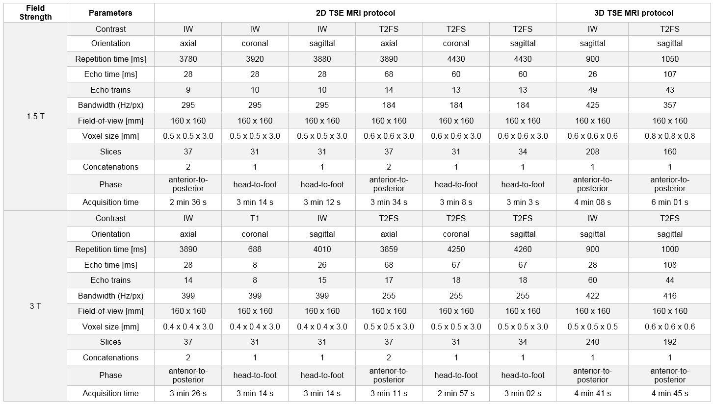
Following internal review board approval and prospective informed consent, 100 symptomatic subjects underwent MRI of the knee at 3T and 50 symptomatic subjects underwent MRI of the knee at 1.5 T, consisting of 10-min 3D CAIPIRINHA SPACE TSE and 20-min standard-of-reference 2D TSE protocols (Figure 1). The corresponding 2D and 3D TSE datasets were separated, anonymized and randomized into individual studies. Two fellowship-trained musculoskeletal radiologists assessed the studies independently for structural abnormalities of the menisci, ligaments, cartilage, and bone. Descriptive statistics, inter-reader reliability, inter-method concordance, diagnostic definitiveness, interchangeability comparison tests were applied. A p-value of <0.01 was considered significant.Results
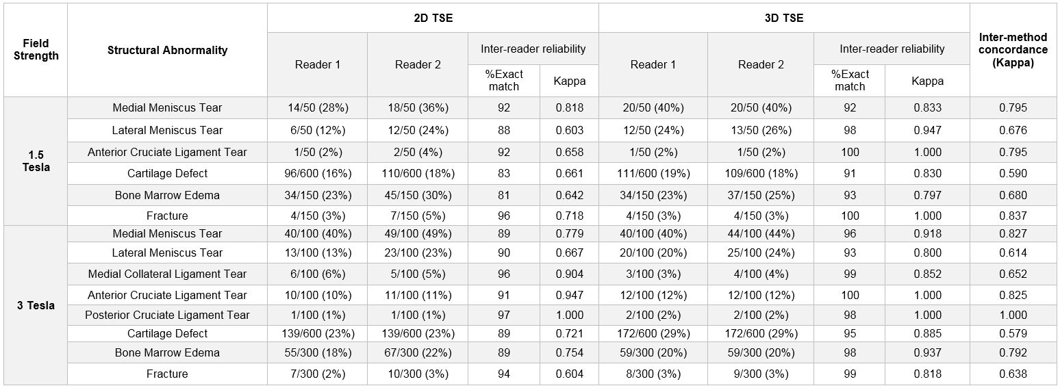
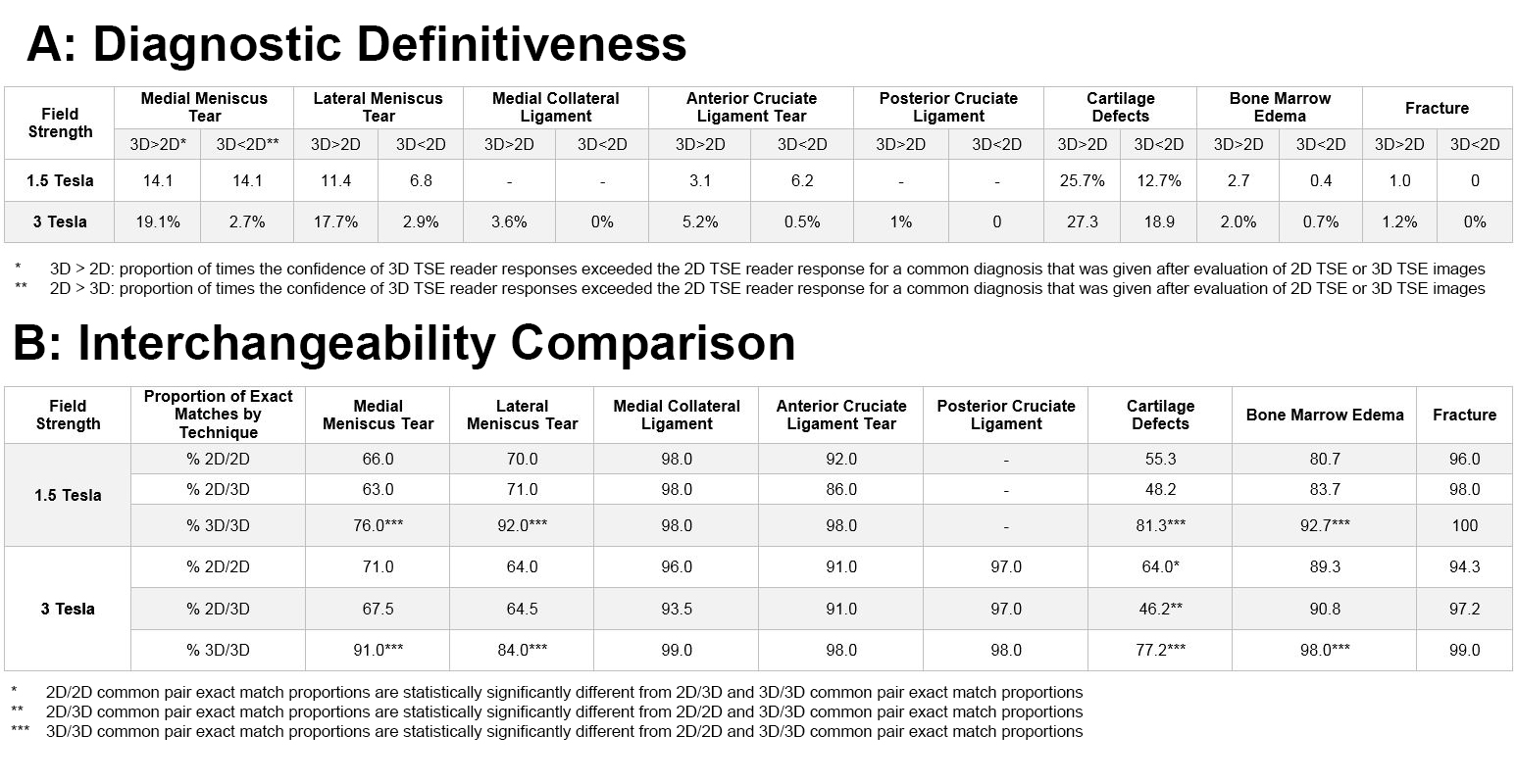
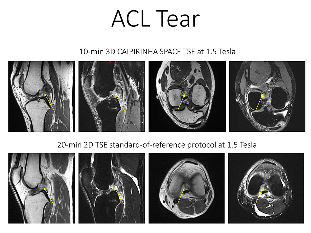
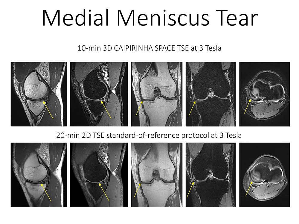
All MRI studies were successfully performed. Both readers diagnosed abnormalities with similar proportions regardless of the MRI technique or field strength (Figure 2). The inter-method concordance agreement (Figure 2) for matched diagnoses between 2D and 3D TSE was good to very good (kappa, 0.614 – 0.837) for meniscal and anterior cruciate ligament tears, areas of bone marrow edema, and fractures; whereas the concordance agreement was moderate for the detection and differentiation of partial- and full-thickness defects (kappa, 0.597 - 0.590). The readers' diagnostic definitiveness in diagnosing meniscal tears and cartilage defects was higher on 3D than 2D studies, and similar for 2D and 3D for the diagnosis of anterior cruciate ligament tears, bone marrow edema, and fractures (Figure 3A). In an interchangeability comparison using common pair exact match proportions (Figure 3B), 2D and 3D MRI were interchangeable for the diagnosis of medial collateral ligament tears, anterior cruciate ligament tears (Figure 4), and fractures; whereas the common pair exact match proportions were significantly (p < 0.01) higher for 3D TSE MRI when readers diagnosed medial and lateral meniscal tears (Figure 5) and bone marrow edema. The common pair exact match proportions for diagnosing cartilage defects was different for all combinations.
Discussion
Owing to the rising economic constraints and the demand of MRI for non-invasive diagnosis of internal knee derangement, there is a need for faster MRI acquisition without comprising diagnostic performance. 10-min 3D CAIPIRINHA SPACE TSE knee protocols offer an opportunity to shorten the table time for patients, increase institutional throughput, and potentially a path to compensate for decreasing reimbursements. The 10-min acquisition time was achieved through 2x2 acceleration with a shifted, bidirectional CAIPIRINHA undersampling in phase and partition encoding directions. This approach helped to minimize aliasing artifacts, reduce image noise, and provided a path to 4-fold acceleration without the need for image quality compromising acceleration strategies, including long echo trains, partial Fourier undersampling, and anisotropic data acquisition. Analyses of the frequencies of structural abnormalities, inter-reader reliability, inter-method concordance, diagnostic definitiveness, and interchangeability comparison indicate that 1.5 and 3T 3D CAIPIRINHA SPACE TSE protocols are at least equivalent to 2D TSE standard-of-reference protocols for the diagnosis of internal derangement of the knee.
Conclusion
10-min 3D CAIPIRINHA SPACE TSE MRI protocols are interchangeable and at least equivalent to 20-min 2D TSE standard-of-reference protocols for the diagnosis of internal derangement of the knee.Acknowledgements
No acknowledgement found.References
- Kijowski R, Davis KW, Woods MA, et al. Knee joint: comprehensive assessment with 3D isotropic resolution fast spin-echo MR imaging--diagnostic performance compared with that of conventional MR imaging at 3.0 T. Radiology 2009;252(2):486-495.
- Breuer FA, Blaimer M, Mueller MF, et al. Controlled aliasing in volumetric parallel imaging (2D CAIPIRINHA). Magn Reson Med 2006;55(3):549-556.
- Fritz J, Fritz B, Thawait GG, Meyer H, Gilson WD, Raithel E. Three-Dimensional CAIPIRINHA SPACE TSE for 5-Minute High-Resolution MRI of the Knee. Invest Radiol. 2016 Oct;51(10):609-17.
- Fagundes J, Longo MG, Huang SY, Rosen BR, Witzel T, Heberlein K, Gonzalez RG, Schaefer P, Rapalino O. Diagnostic Performance of a 10-Minute Gadolinium-Enhanced Brain MRI Protocol Compared with the Standard Clinical Protocol for Detection of Intracranial Enhancing Lesions. AJNR Am J Neuroradiol. 2017 Sep;38(9):1689-1694.
Figures