1028
Optimization of advanced high-resolution diffusion-weighted imaging (DWI) techniques in lower neck imagingTong Su1, Yu Chen1, Tianyi Qian2, Wei Liu3, Huadan Xue1, Zhengyu Jin1, Zhuhua Zhang1, and Hailong Zhou1
1PUMCH, Beijing, China, 2Siemens, Beijing, China, 3Siemens, Shenzhen, China
Synopsis
Readout-segmented echo-planar imaging (rs-EPI) could significantly reduce magnetic susceptibility artifacts in head and neck regions. In this study, the images were qualitatively evaluated among three types of rs-EPI: with and without readout partial Fourier (RPF), and with simultaneous multi-slice (SMS) technique. The SNRs and CNRs were compared with the additional use of special surface coils (SC). There was no significant differences in the image quality, SNRs, or CNRs among three rs-EPI acquisition methods of all the 31 volunteers and 9 hypopharyngeal carcinoma patients. Markedly, the special surface coils offered better image quality for the evaluation of lower neck lesions.
Introduction
Readout-segmented EPI (RESOLVE) could shorten the echo time and solve the susceptibility artifacts problem with high-resolution diffuse weighted imaging (DWI)[1]. Recently, some improvements have been made on RESOLVE to shorten total acquisition time, including combining with simultaneous multi-slices (SMS) technique and read-out partial Fourier (PRF) method. The purposes of this study were to qualitatively investigate the applications of different DWI techniques in lower neck imaging, and the performance of the special surface coils.Methods
Data were collected on a MAGNETOM Skyra 3T MR scanner (Siemens Healthcare, Erlangen, Germany) with a 20-channel head coil, and a pair of 4-channel special surface coils (SC). In the first part of this study, 31 healthy volunteers (12 males; mean age, 45 years; range, 29 – 77 years) were enrolled. In the second part, nine patients (8 males; mean age, 62 years; range, 39 – 82 years) with histologically confirmed hypopharyngeal carcinoma were recruited. Both the conventional head and special surface coils were used on 13 volunteers and five patients. The SMS technique was implemented in a prototype sequence based on a clinical rs-EPI sequence (RESOLVE, Siemens Healthcare), referred to as SMS-RESOLVE. DWI images from all volunteers and patients were acquired with RESOLVE, RESOLVE-RPF and SMS-RESOLVE. The imaging parameters were as follows: TR/TE = 6540/58 ms, slice thickness = 3 mm; voxel size = 1.5 × 1.5 × 3.0 mm3; FOV = 160 × 160 mm2; readout segments = 5; b value = 0 and 800 s/mm2. Three segments with readout partial Fourier factor 6/8 were used in RESOLVE-RPF. The TR/TE in SMS-RESOLVE was 4560/59 ms with slice acceleration factor 2. The acquisition time for RESOLVE, RESOLVE-RPF, and SMS-RESOLVE was 3:37 min, 2:58 min, and 2:47 min, respectively. The image quality was scored according to a scale from 1 (poor) to 5 (excellent). Circular ROIs were drawn on the muscle tissue, spine, and background at the epiglottis level of all the images. The signal intensity and standard deviations of all the ROIs were generated automatically and recorded. The signal-to-noise ratio (SNR) was defined as the ratio between the mean signal intensity of the muscle tissue and the standard deviation of the background noise. The contrast-to-noise ratio (CNR) was defined as the difference between the signal intensity of the muscle and spine divided by the standard deviation of the background noise. One-way ANOVA analysis was used to compare the image quality differences among RESOLVE, RESOLVE-RPF, and SMS-RESOLVE. T-tests were used to compare the differences between the same MR sequences acquired with and without SC.Results
The sample images from two volunteers are shown in Figure 1. There was no significant difference in the image quality scores, SNRs, or CNRs among RESOLVE, RESOLVE-RPF, and SMS-RESOLVE for all the volunteers and patients (Table 1). Moreover, the ADC values of the hypopharyngeal lesions of nine patients among RESOLVE, RESOLVE-RPF, and SMS-RESOLVE showed no statistical differences. Use of the special surface coils markedly improved the image quality, and the SNRs and CNRs of all three DWI techniques with SC were significantly higher than those without SC (Figure 2, 3). Both RESOLVE-RPF and SMS-RESOLVE can shorten the acquisition time by around 30% without loss of image quality. The results suggested combining the SMS-RESOLVE technique with special surface coils could have obtained the excellent image quality with shorter acquitision time.Discussion
Our study investigated the performances of different RESOLVE acquisition methods in the lower neck regions, including RESOLVE, RESOLVE-RPF, and SMS-RESOLVE. The study showed the excellent feasibility of combining SMS-RESOLVE technique with special surface coils in lower neck region. There are many obstacles limiting the evaluation of the lesions in head/neck DWI due to magnetic susceptibility artifacts and a long scan time. SMS or RPF technique markedly shortened the acquisition time, providing a great advantage in clinical practice. Moreover, the additional use of SC can further improve image quality. In this study, we recruited 31 healthy volunteers and nine hypopharygeal tumor patients and obtained good and stable quality images with reduced artifacts and higher image resolution using SC. This coil was placed much closer to the lower neck regions and was therefore complementary with conventional head or neck coils. The clinical values of these techniques on differentiating benign and malignant tumors, the surveillance and follow-up of treatments or relapse will be further studied and clarified by employing a larger sample size. In conclusion, we have demonstrated the excellent feasibility of fast imaging techniques, including RPF and SMS, with SC to perform high-quality MR images with short acquisition time for the lower neck region evaluation.Acknowledgements
No acknowledgements found.References
1. Zhao, M., Z. Liu, Y. Sha, et al., Readout-segmented echo-planar imaging in the evaluation of sinonasal lesions: A comprehensive comparison of image quality in single-shot echo-planar imaging. Magn Reson Imaging, 2016. 34(2): p. 166-72.Figures
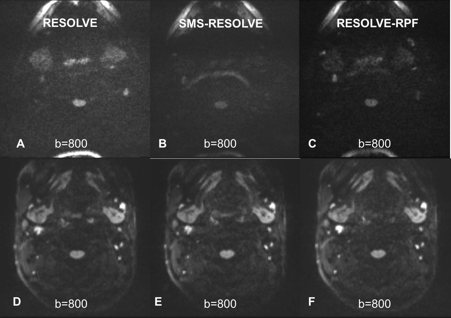
Figure 1. RESOLVE (A, D), SMS-RESOLVE (B, E), and RESOLVE-RPF
(C, F) images acquired from two healthy volunteers. Images D, E, F were scanned
with a special surface coil which clearly showed important anatomic structures.
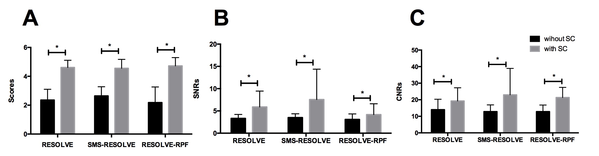
Figure 2. Image quality scores (A), the SNRs (B), and CNRs
(C) of the three DWI sequences: RESOLVE, SMS-RESOLVE, and RESOLVE-RPF, with and
without special surface coils. *P<0.05.

Figure 3. Parameters and qualitative evaluation of three
different DWIs with and without special surface coils.