1024
Accelerated Distortion-Free Diffusion Imaging at 7T – by Fusing PSF and VAT1Department of Biomedical Magnetic Resonance, Otto-von-Guericke-University Magdeburg, Magdeburg, Germany, 2Department of Radiology, Mayo Clinic, Rochester, MN, United States, 3Siemens Healthcare, San Francisco, CA, United States, 4Site Magdeburg, German Centre for Neurodegenerative Diseases (DZNE), Magdeburg, Germany, 5Leibniz Institute for Neurobiology, Magdeburg, Germany, 6Center for Behavioral Brain Sciences, Magdeburg, Germany
Synopsis
In this study, combining view angle tilting (VAT) with the novel PSF diffusion EPI sequence demonstrates the feasibility to further accelerate distortion-free diffusion weighted imaging at high field.
INTRODUCTION
Severe geometric distortions induced by susceptibility and eddy-currents pose a substantial obstacle in single-short echo-planar imaging (EPI), especially for high-resolution diffusion imaging at ultra-high field (UHF). A multi-shot EPI approach1 using a hybrid spin-warp (SW-PE) and echo-planar phase-encoding (EPI-PE) strategy inspired by the point-spread function (PSF) mapping technique serves as an alternative to enabling high-resolution imaging without distortions and T2 blurring, but requires a prolonged scan time through effective averaging. Although reduced field-of-view (rFOV) in the SW-PE (i.e. multi-shot) dimension can reduce the scan time, it is generally limited by the distortion extent in the EPI-PE dimension2.
Here we combine this approach with view angle tilting (VAT)3 to further accelerate the scan. Recently, VAT-EPI was suggested to reduce geometric distortions by introducing blipped VAT gradients on the slice-selection axis during the EPI acquisition2. Due to severe image blurring in the EPI-PE dimension, induced by this additional gradient, however, it was not widely adapted in single-shot EPI-based applications. In contrast, we demonstrate that the combination of the proposed approach with VAT can accelerate the scan without image blurring and fold-over artifacts in the reconstructed distortion-free image.
METHODS
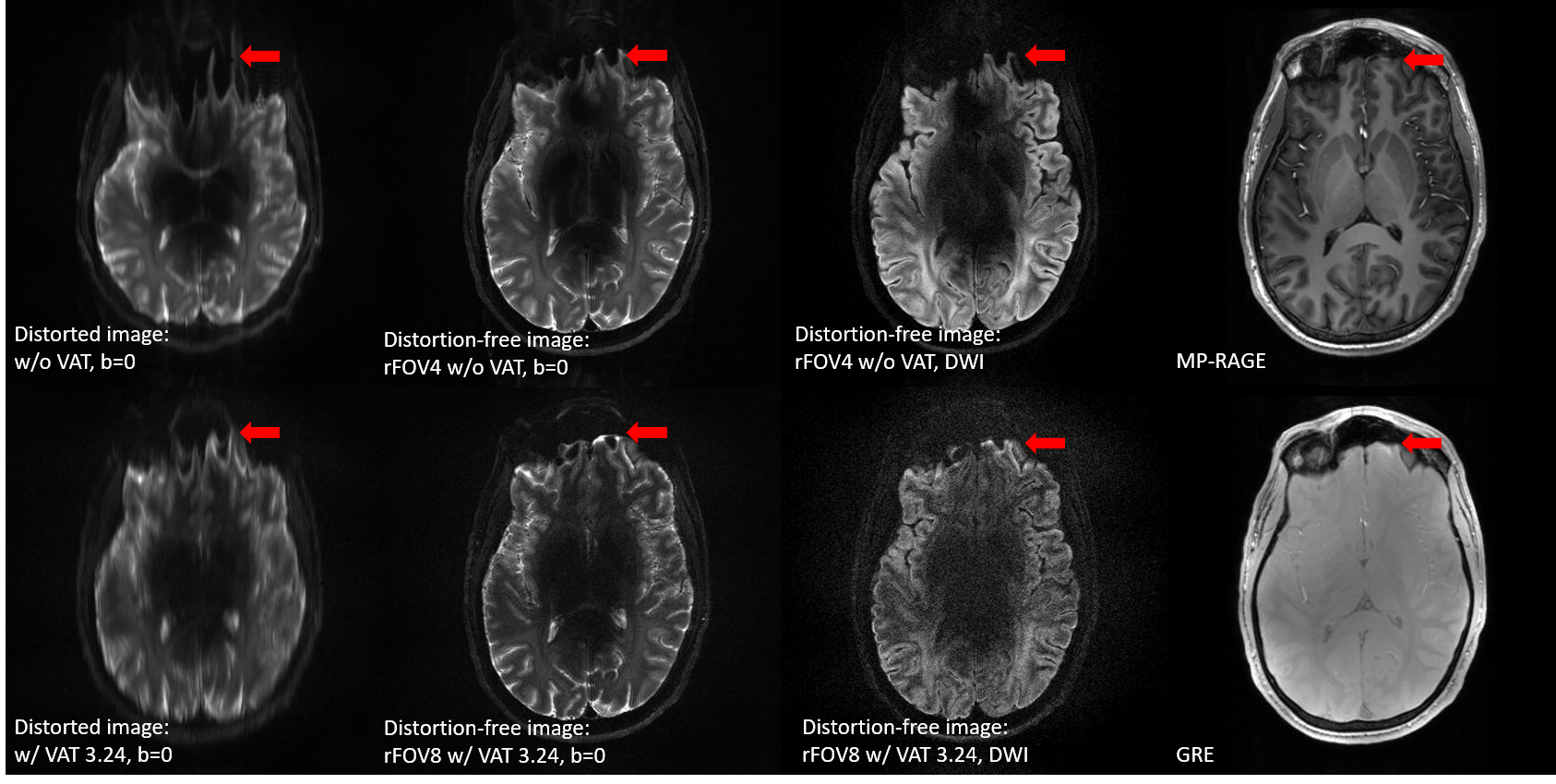
The VAT scheme is added to the navigated PSF diffusion sequence1. Phantom and in-vivo scans were performed on a 7T scanner (Siemens Healthcare, Erlangen, Germany) using a 32 channel head coil (Nova Medical, Wilmington MA, USA). The experimental parameters were: FOV=224×224 mm2, in-plane resolution=0.7×0.7 mm2, slice thickness=2.8 mm, 10 or 16 slices, TR=2000 ms, reduced resolution (rR) in the EPI-PE dimension=4, in-plane GRAPPA acceleration=2 or 3, and partial-Fourier acquisition=1 or 0.75. In the in-vivo experiment, TEs were 59 and 63 ms, respectively for rFOV4 without VAT and rFOV8 with VAT. The acquisition time for rFOV 4 without VAT and 8 with VAT were 2m48s and 1m27s respectively.
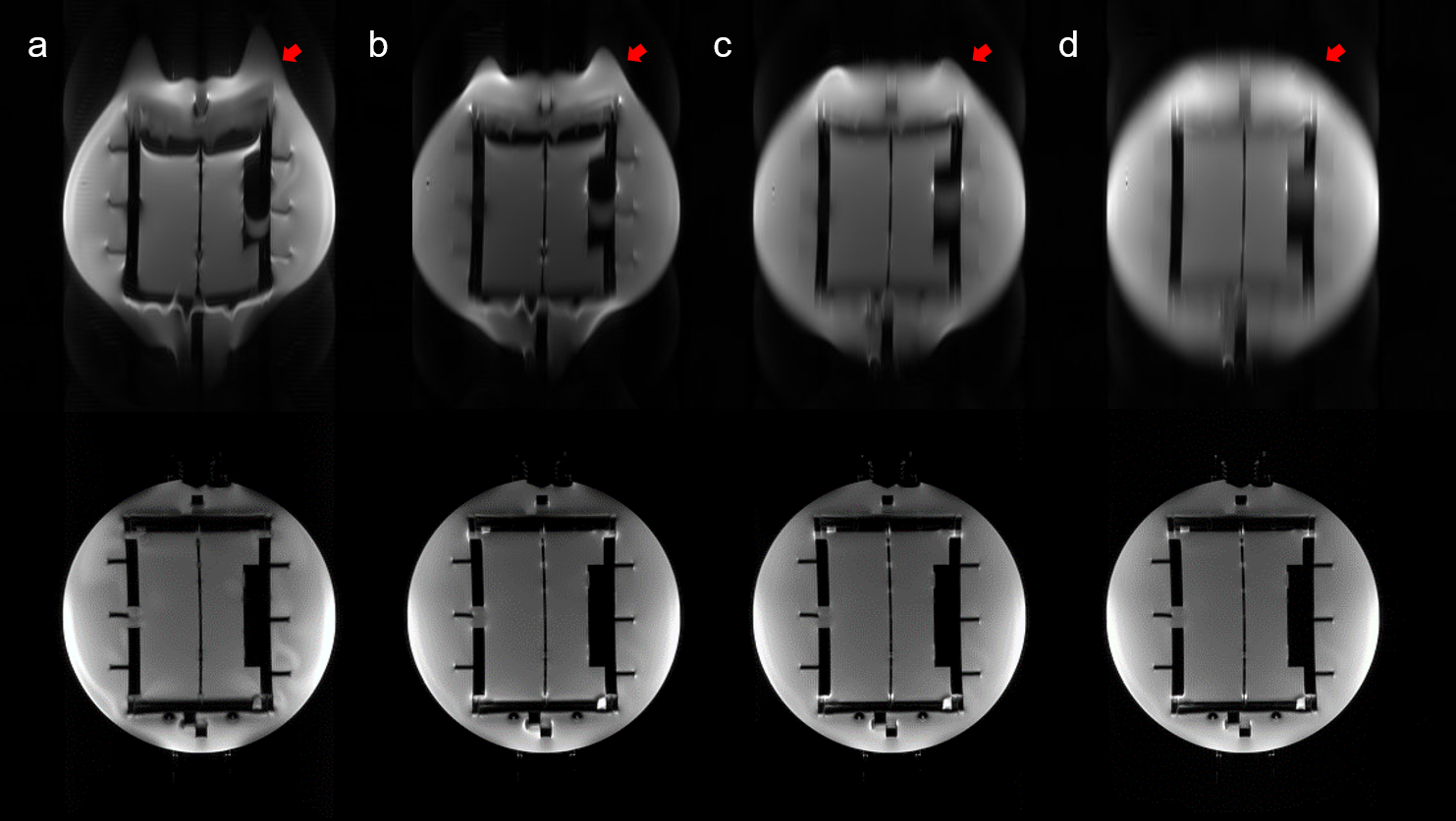
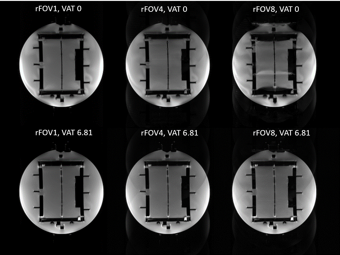
Fully-sampled PSF scans were performed in phantom with four different VAT gradient amplitudes (Gvat), which include 0, 3.4, 6.8, and 10, as a ratio compared to the EPI-PE gradient amplitude (Gpe). Both distorted images and distortion-free images were reconstructed from the measured PSF data by integration of the SW-PE and EPI-PE coordinate, respectively to evaluate the distortion reduction and image blurring introduced by the VAT gradient. In addition, a distortion-free image was additionally calculated from the PSF data with three different rFOV factors (1, 4, and 8) to investigate the maximum rFOV value without yielding fold-over artifact.
RESULTS and DISCUSSION
While geometric distortion in the EPI-PE dimension was reduced by increasing the VAT gradient amplitude, image blurring increased considerably (Fig. 1). Nevertheless, this blurring did not appear in the distortion-free images even with a strong VAT gradient. Since the image blurring induced by the VAT gradients appears only in the EPI-PE dimension, but not in the SW-PE dimension, the blurring effect can be avoided in the distortion-free image calculated from the PSF data. Due to the reduced distortion in the EPI-PE dimension with VAT, it was possible to apply even higher rFOV factor of up to 8 compared to 4 available without VAT, which resulted in 50% shorter scan time. PSF-fold-over artifacts appeared in the highly accelerated image without VAT. Similar results were achieved in in-vivo. Geometric distortions were significantly reduced with VAT in the EPI-PE dimension, especially in the frontal area and the reduced distortion could minimize the deviation of effective TE from the intended TE. Therefore, a reduced intensity distortion was also observed with VAT in the proposed distortion-free image near the areas of high susceptibility changes (arrows in Fig. 3). The signal-to-noise ratio decreased in the images with high acceleration due to the reduced number of shots (effective averaging).
The study demonstrated the complementary benefit of VAT and PSF to further accelerate the distortion-free DTI scan. Although a high VAT gradient amplitude is beneficial to minimize the geometric distortion, this increases the image blurring. In addition, additional geometric distortion is caused when Gvat > Gpe, leading to an increased echo spacing. Therefore, searching for an optimal VAT amplitude is required.
CONCLUSION
In this study, we demonstrated the benefit of combining VAT with PSF to further accelerate the proposed DTI approach. The reduced distortion with VAT enables highly accelerated PSF acquisition and the image blurring induced by VAT can be separated into the EPI-dimension. Therefore, the combination can be an attractive and complementary approach.Acknowledgements
This study was supported by the German Research Foundation, DFG (SP632-4).References
1. In MH, Posnansky O, Speck O. High-resolution distortion-free diffusion imaging using hybrid spin-warp and echo-planar PSF-encoding approach. Neuroimage. 2017 Mar 1;148:20-30.
2. Zaitsev M, Hennig J, Speck O. Point spread function mapping with parallel imaging techniques and high acceleration factors: fast, robust, and flexible method for echo-planar imaging distortion correction. Magn Reson Med. 2004 Nov;52(5):1156-66.
3. Ahn S, Hu XP. View angle tilting echo planar imaging for distortion correction. Magn Reson Med. 2012 Oct;68(4):1211-9.
Figures