1019
Improving uniqueness of Magnetic Resonance Fingerprinting (MRF) signal evolutions using spatio-temporal variation of non-linear ΔB0 shim coil.1Radiology, Case western reserve university, Cleveland, OH, United States, 2Quality Electrodynamics, Mayfield Village, OH, United States
Synopsis
Magnetic Resonance Fingerprinting (MRF) framework uses variation in acquisition parameters to generate unique signal evolutions, which can be treated as “fingerprints”. The iPRES coil concept provides independent and dynamic variations of multiple magnetic fields, which can be used to improve the uniqueness of signal evolutions through spatio-temporal variations of multiple fields. In this study, we present a proof-of-concept implementation illustrating spatio-temporal variations of local non-linear ΔB0 fields improve the uniqueness of signal evolutions for MRF. Our phantom results show reduction in variation of estimated tissue properties for data with 500frames, thereby illustrating the capability of acceleration using these field variations.
Purpose:
The purpose of this work is to explore the feasibility of using spatio-temporal variations of ΔB0 field with non-linear shim coils to improve the uniqueness of magnetic resonance fingerprinting (MRF)[1] signal evolutions.Introduction:
MRF is a recently developed framework where one of the primary goals is to generate unique signal evolutions, which can be treated as “fingerprints”. Since the identification of the underlying tissue properties is performed using pattern recognition of these fingerprints, their uniqueness is a critical property of the MRF acquisition. For example, a more unique fingerprint would be easier to separate from the overlapping signals due to undersampling, thereby providing higher acceleration rates and/or improved spatial resolution. Additionally, a more unique fingerprint will have higher tolerance to signal errors, which in turn will provide higher precision and/or motion tolerance. In general, the variation in the signal evolution can be achieved by varying acquisition and system related parameters. Here we propose to expand on our ability to generate unique signal evolutions using local non-linear shim fields. This may be performed using a recently developed iPRES coil[2,3] assembly, which provides manipulation of all three available magnetic fields (B1-, B1+, and ΔB0), with requisite electronics located locally[4]. In this study, we explore the potential of using spatio-temporal variations of ΔB0 to improve the uniqueness of signal evolutions for the MRF framework.Methods:
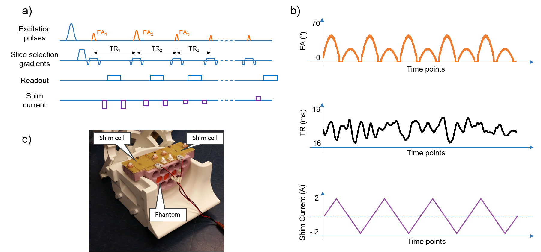
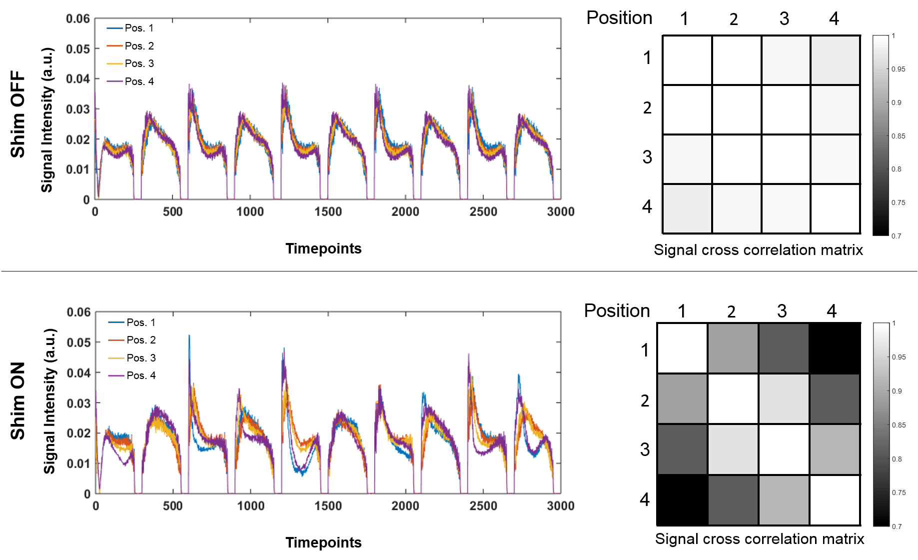
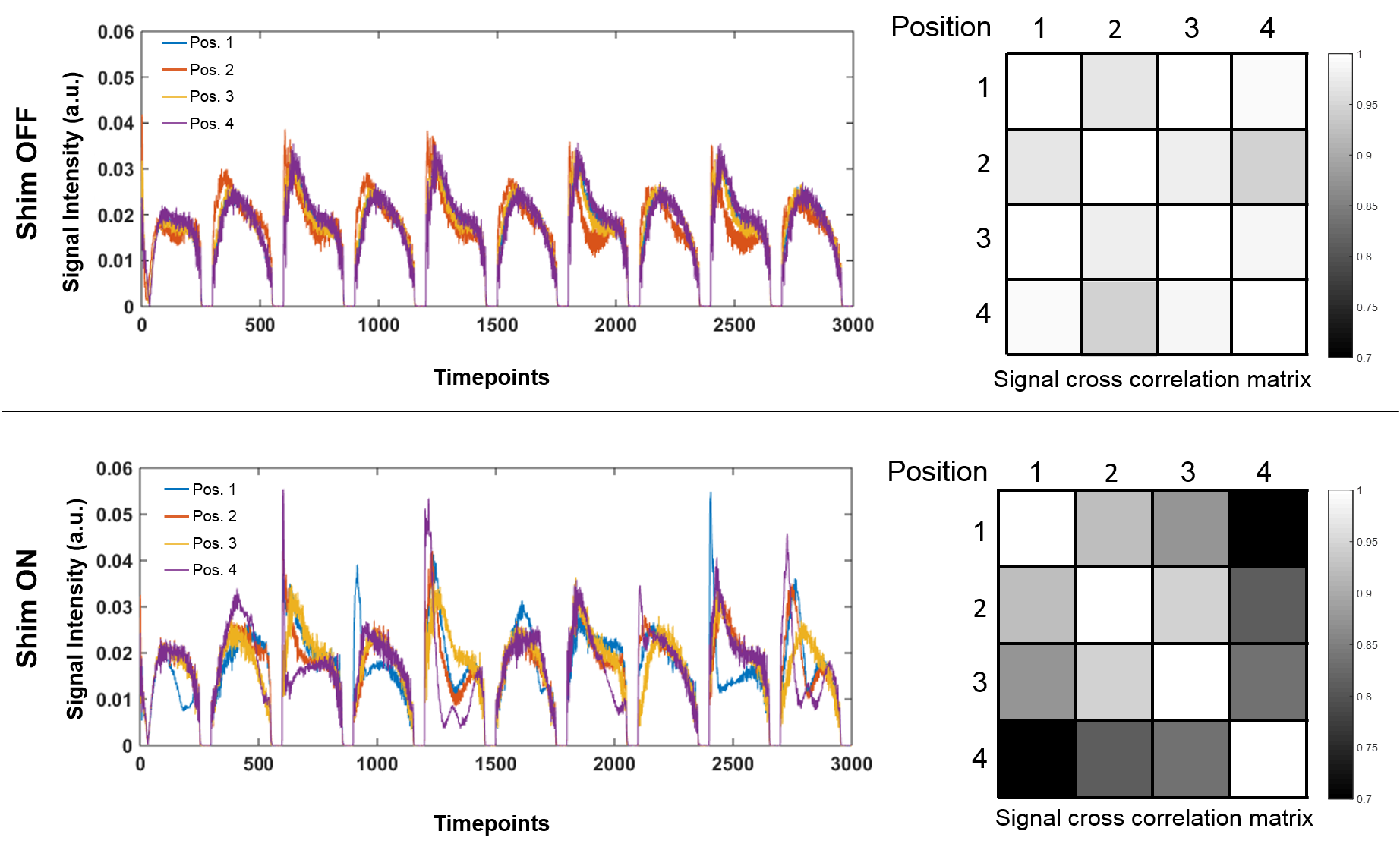
In our previous study[4], we showed through simulations that spatio-temporal variations of non-linear ΔB0 fields can improve the uniqueness of fingerprints. In this work, we explore the feasibility of these variations in phantom experiments. All imaging was performed using a Siemens Skyra 3T scanner. Figure 1c shows the experimental setup. Four gadolinium doped agar gel phantom vials with a variation in T1 and T2 values were imaged in the coronal plane. Two shim coils above the phantoms were driven with equal but opposite current to produce the shim field. For this study, two bSSFP based MRF[1] acquisitions were performed. One with shim field switched off for reference data. The other acquisition switched the shim current dynamically from TR to TR. Figure 1a illustrates a schematic version of the pulse sequence timing diagram and figure 1b shows the variation in sequence parameters used in this study. For each acquisition, two datasets were acquired. The first was low resolution (4.7x4.7x5mm3) fully sampled data (6 spirals for each time frame) for comparing signal evolutions, while the second was high resolution (2.4x2.4x5mm3) undersampled data (1 variable density spiral for each time frame) for comparing property mapping. The generated dictionary contained four properties, namely, T1, T2, the background off-resonance (δf) value which is constant from TR to TR, and the ΔB0 sensitivity of the dynamic shim field at that location. For matching of the data from acquisition with active shim current, randomized SVD and interpolation based approach[5] was used to reduce the dictionary size and matching time. Cross-correlation of the signals from fully sampled dataset at four locations on two different vials were used to quantitatively evaluate the signal uniqueness. To evaluate the feasibility of acceleration, maps were also generated from undersampled data using a reduced number of frames (500 time-points out of a total of 3000 time-points). For comparison, mean and standard deviations of estimated T1 and T2 were measured over an ROI roughly covering the entire third vial.Results & Discussions:
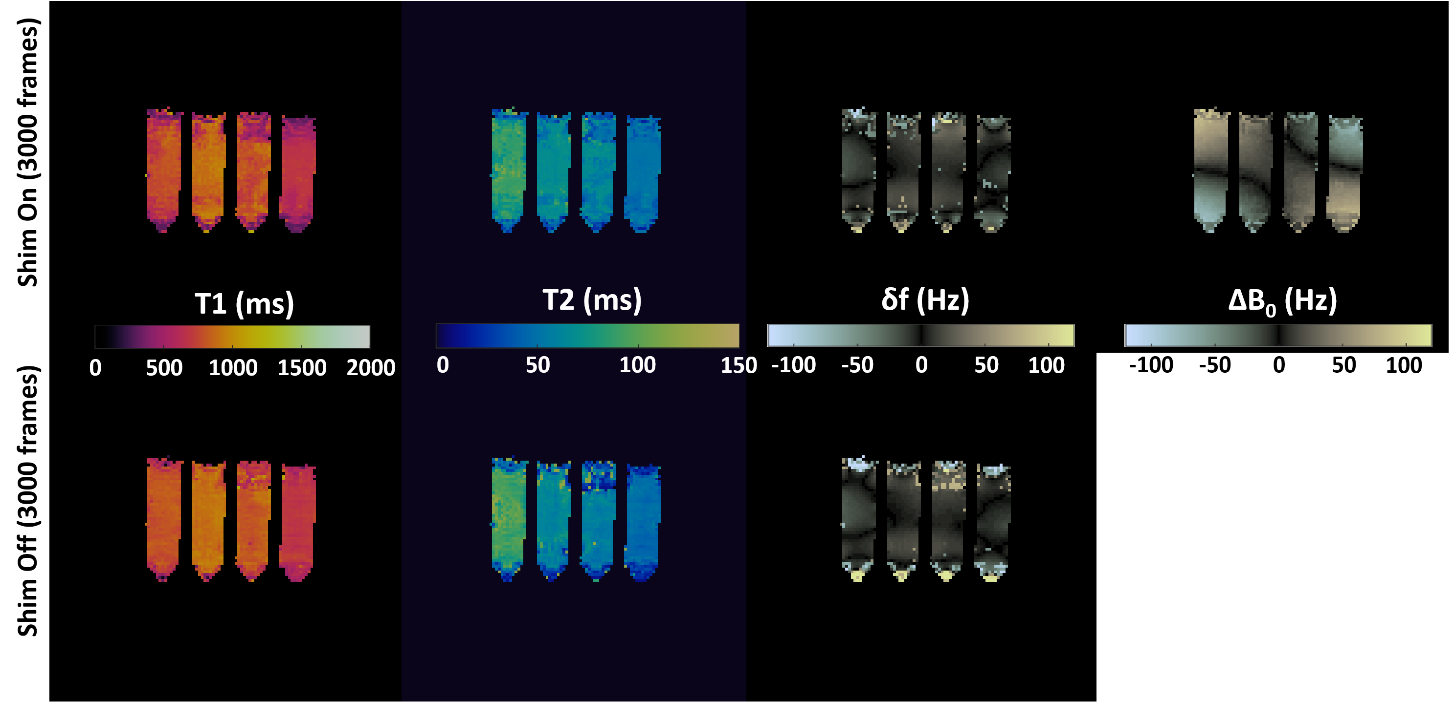
Figure 2 shows property mapping results from undersampled data using all 3000 frames. We see a close agreement in estimated properties between the two acquisitions (T1ShimOn=730±139ms; T1ShimOff=794±85ms; T2ShimOn=58±11.0ms; T2ShimOff=54±18.5ms). Figure 3 and 4 show signal evolutions and cross-correlation matrix from example locations for both the acquisitions. We see that the addition of the spatio-temporally varying ΔB0 field to the MRF sequence decreases the correlation between signals at different locations, thereby illustrating the improvement in the uniqueness of fingerprints. Figure 5 shows property mapping results from undersampled data using only 500 frames. We see the estimated T1 and T2 maps from the acquisition with shim off (T1ShimOff=857±131ms; T2ShimOff=66±26.7ms) present higher variations compared to maps from the acquisition with shim on (T1ShimOn=852±116ms; T2ShimOn=68±13.9ms), thereby illustrating the capability of acceleration using these field variations.Conclusion:
Spatio-temporal variations of local non-linear ΔB0 fields can be used to improve the uniqueness of signal evolutions for MRF. The improved uniqueness can potentially be used for improvement in various aspects such as precision, spatial resolution, acceleration rate, etc.Acknowledgements
The authors would like to acknowledge funding from Siemens Healthcare and NIH grants NIH 1R01EB016728 and NIH 5R01EB017219. This work made use of the High Performance Computing Resource in the Core Facility for Advanced Research Computing at Case Western Reserve University.References
1. D. Ma et al., “Magnetic resonance fingerprinting,” Nature, Mar. 2013.
2. H. Han et al., “Integrated parallel reception, excitation, and shimming (iPRES),” Magnetic Resonance in Medicine, Jul. 2013.
3. D. Darnell et al., “Integrated parallel reception, excitation, and shimming (iPRES) with multiple shim loops per radio-frequency coil element for improved B 0 shimming: Higher-order iPRES B 0 shimming,” Magnetic Resonance in Medicine, May 2016.
4. M. Twieg et al., “Compact iPRES coil assembly for Magnetic Resonance Fingerprinting,” In proceedings of 25th ISMRM Annual meeting, Honolulu, HI, 2017.
5. M. Yang et al., “Low rank approximation methods for MR fingerprinting with large scale dictionaries,” Magnetic Resonance in Medicine, August 2017.
Figures