1008
Fiber optic mediated extracellular glutamate and intracellular calcium recording with simultaneous fMRI1Max Planck Institute for Biological Cybernetics, Tübingen, Germany, 2University of Tübingen, Tübingen, Germany
Synopsis
Genetically encoded fluorescent reporter iGluSnFR for extracellular glutamate (Glu) sensing and genetically encoded calcium indicator GCaMP6f for calcium sensing are applied with two channel fiber optic recording system in combination with BOLD fMRI. The intracellular calcium signal from both neurons and astrocytes, as well as the extracellular glutamate signal, were recorded with concurrent BOLD fMRI signal from both hemispheres of anesthetized rats, showing unique temporal dynamic pattern. This multi-modal fMRI platform allows us to specify the neurovascular signaling through the neuro-glial-vascular network and provide better understanding on the cellular and molecular interaction underlying the BOLD fMRI signal.
Introduction
Simultaneous fMRI and fiber optics calcium recording have been applied to analyze the blood oxygenation level-dependent signal (BOLD) coupling to specific neuronal or astrocytic calcium actvity1,2. Here, we expressed genetically encoded fluorescent reporter iGluSnFR3 for extracellular glutamate (Glu) sensing and genetically encoded calcium indicator GCaMP6f for calcium sensing in both neurons and astrocytes, and applied two channel fiber optic recording system in combination with BOLD fMRI. The introduction of glutamate, a primary excitatory neurotransmitter, provides us key understanding of the signaling propagation due to its roles in trans- and extrasynaptic transmission in synaptic release and astrocytic cycling. We demonstrated iGluSnFR with a more rapid temporal features of sensory response than the evoked neuronal or astrocytic calcium signals. This platform offers us a more direct interpretation of neuronal transient with fMRI, thus, would expand our understanding of the signal propagation through the neuron-glia-vessel network couple to BOLD fMRI signals.Methods
All images were acquired with a 14.1 T/26cm horizontal bore magnet (Magnex), interfaced to an AVANCE III console (Bruker) and equipped with a 12 cm gradient set, capable of providing 100 G/cm with a rise time of 150 us (Resonance Research). A transreceiver surface coil was used to acquire fMRI images. fMRI scans with block design were performed using 3D Echo planar imaging sequence: TR, 1.5 s, TE,11.5 ms, 1.92X1.92X1.92 cm3, FOV, 48X48X48 matrix, 400X400X400 μm3 spatial resolution. Electrodes were placed on the forepaw to deliver trains of 300 μs, at 2Hz during 4s in each fMRI epoch. The reporter iGluSnFR and GCaMP6f were expressed by AAV5 virus in the two hemisphere forepaw somatosensory cortex (FP-S1) with Syn or GFAP promoter. Fiber optic (200 μm) was inserted into the area which expressed the cortex for fluorescent signal recording.Results
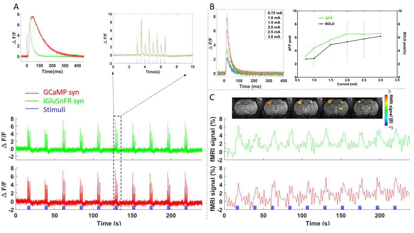
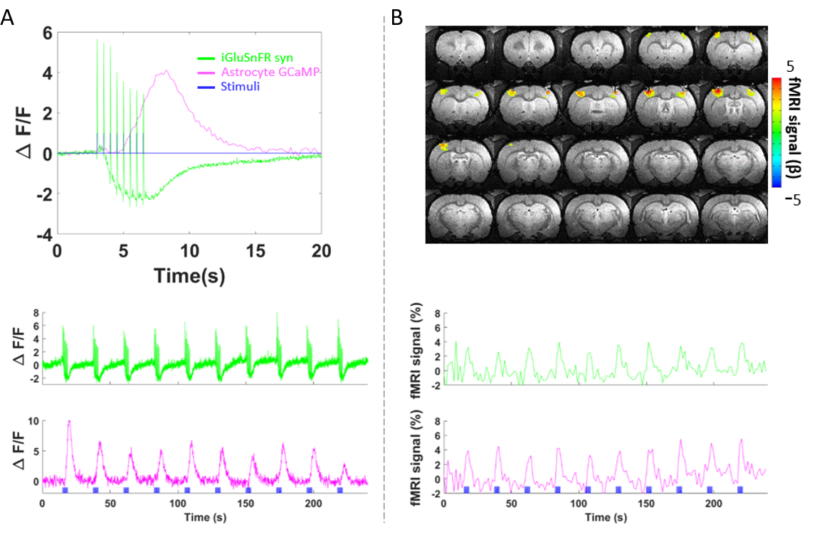
We first acquired evoked neuronal calcium and Glu signals with simultaneous fMRI from the FP-S1 of two hemispheres, respectively. Evoked neuronal calcium and Glu spikes detected from were shown to follow each electrical pulses (Fig. 1A). In contrast to the evoked neuronal calcium spikes, the evoked Glu spikes have earlier onset time (9.14±0.77 ms vs 12.7±0.42 ms, n=5 rats), and time to peak response (18.3±1.2 ms vs 41±2.96 ms), (Fig. 1 A). Also, amplitude of the evoked Glu spike increased proportionally to the amplitude of BOLD signals as the function of the stimulation intensity (0.75, 1, 1.5, 2, 2.5, and 3 mA, n=4 rats) (Fig. 1B). Fig. 1C shows the fMRI BOLD maps and the time course of BOLD signal from FP-S1 ROIs, which is simultaneous acquired with the neuronal calcium and Glu spikes. Besides the neuronal calcium, the evoked astrocytic calcium and Glu spikes were also acquired with fMRI simultaneously. Similar to previous study (Wang et al. ISMRM 2017), the astrocytic calcium signal is an integrated unitary spike, which has slower onset than the Glu spikes (Fig. 2A). Interestingly, we also observed the baseline drop of the Glu signal during the 4 s stimulation, which shows earlier onset with extended longer tail than the astrocytic signal. It remains unclear what contributes to the baseline drop of Glu signal. It may indicate fluorescent signal changes due to potential hemodynamic responses from vessels close to the fiber tip, similar to the intrinsic signal detection. Alternatively, it may implicate the extracellular clearance of Glu following synaptic glutamate release during stimulation. Also noteworthy is that the BOLD signals detected from both hemisphere are similar to each other (Fig. 2B). Future study will further clarify the neurovascular coupling events in the neuro-glial-vascular network and specify the source for the Glu baseline drop of during stimulation.Conclusion
Concurrent glutamate and calcium recording was established with the BOLD fMRI brain mapping in anesthetized rats. This platform would lead to a better understanding of neurovascular coupling through the neuro-glial-vascular network in the animal brain.Acknowledgements
This work was supported by the Max-Planck-Society.References
1. Schulz, Kristina, et al. Simultaneous BOLD fMRI and fiber-optic calcium recording in rat neocortex. Nat methods 9, 597-602, doi:10.1038/nmeth.2013 (2012).
2. Wang, M., He, Y., &Yu, X. 2017. A novel role of intrinsic astrocytic calcium spikes to mediate brain states through central/dorsal thalamic nuclei. ISMRM 2017.
3. Marvin, J. S., et al. An optimized fluorescent probe for visualizing glutamate neurotransmission. Nat methods 10, 162-170, doi:10.1038/nmeth.2333 (2013).
Figures