1000
High spatial and temporal free breathing T1 contrast enhanced imaging using a novel 4D variable density, elliptical centric radial stack-of-stars sharing approach1Philips Healthcare, Best, Netherlands, 2Philips Healthcare, Bangalore, India, 3Technical University Eindhoven, Eindhoven, Netherlands, 4Philips Research Europe, Hamburg, Germany
Synopsis
Non-Cartesian acquisitions combined with parallel imaging and compressed sensing have been proposed for high spatial and temporal resolution free breathing motion robust 4D contrast enhanced applications. While these approaches provide substantial reduction of contrast blurring and motion robustness, the sampling in itself may still provide limitations to capture the temporal image content. Our approach focuses on the optimization of the 4D sampling strategy with a unique 4D variable density, elliptical-centric radial stack of stars sharing approach. This approach reduces not only the minimum scan time of the 4D scan but also provide possibilties to optimally adjust and capture temporal information like motion and contrast enhancement.
Introduction
Non-Cartesian acquisitions based on radial or spiral trajectories combined with parallel imaging and compressed sensing [1], [2], [3], [4] have been proposed for high spatial and temporal resolution free breathing motion robust 4D contrast enhanced applications. While these approaches provide substantial reduction of contrast blurring and motion robustness, the sampling in itself may still provide limitations to capture the temporal image content. Furthermore, reconstruction is complex with long reconstruction times as a result. Our approach focuses on optimized radial 4D sampling strategies that allow to capture motion and contrast changes accurately. In this study, we provide the basic optimization steps, proof of concept and initial clinical results.Methods
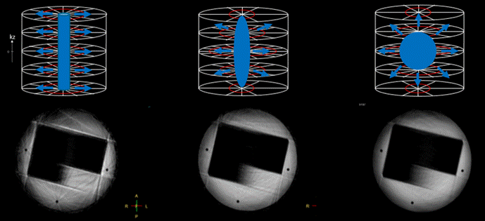
Non-Cartesian acquisitions require a higher reconstruction performance. Hence, minimizing the Non-Cartesian dimension using a robust 3D radial stack of stars (SOS) approach is advantageous. We propose a novel improvement to the standard SOS trajectory by using an elliptical, variable density sampling scheme (EVD) as shown in Figure 1 with a high-density radial grid [5] at central k-space and adjustable lower densities at peripheral k-space. To optimize 4D sampling, an elliptical centric, pseudo golden angle distributed sampling scheme is used. KWIC filtering [6] of the temporal closest radial spokes are used to sharpen the contrast phases over dynamics and standard gridding and FFT is applied.
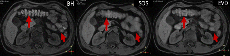
All examinations were performed on 1.5 and 3T clinical scanners (Ingenia, Philips Healthcare). Phantom experiments simulating contrast and motion changes were accomplished, comparing the improved elliptical VD sampling to the standard SOS. In an initial volunteer study of six subjects, the effects on free breathing and respiratory gated abdominal applications are shown and are compared to the standard state of the art breath hold (BH) and SOS sequences. Sequence parameters of these sequences are:
Sequence 1 (BH): 3D Dixon TFE, FOV = 400 x 352 mm2, resolution = 2.0 x 2.0 x 4.0 mm3, bandwidth = 555 Hz / pixel, flip angle = 15°, TR = 5.9 ms, TE 1 / TE 2 = 1.6 ms / 3.6 ms. SENSE Y, Z = 2.0, 1.3, halfscan Y, Z = 0.7, 0.85 leading to a scan time of 17 seconds.
Sequence 2 (SOS)/ 3 (EVD SOS): 3D radial Dixon TFE, FOV = 450 x 450 mm2, resolution = 1.5 x 1.5 x 3.0 mm3, bandwidth = 1086 Hz / pixel, flip angle = 10°, TR = 5.9 ms, TE 1 / TE 2 = 1.6 ms / 3.6 ms, radial density = 160 %, SENSE Y, Z = 1.0, 1.8, halfscan Y, Z = 1.0, 0.7 leading to scan time of 2:00 minutes / VD = 0.6, radial density = 110 % and scan time of 1:00 minute. Overall image quality and sharpness were compared by reviews.
Results
The phantom study shows that the information content is best preserved by the EVD approach. Streaking artifacts from k-space discontinuities are substantially reduced with the EVD approach over the standard SOS. In the volunteer study, the sharpness of intestines are greatly improved over the standard SOS demonstrating the potential to also capture temporal information like intestinal movement. In the six volunteers, the EVD approach was preferred by the reviewer compared to the conventional SOS.Discussion / Conclusion
We have shown a unique 4D elliptical centric variable density sampling and reconstruction approach. This approach reduces not only the minimum scan time of 4D contrast enhanced imaging (comparable to KOOSH ball) but also provide possibilties to optimally adjust and capture temporal information like motion and contrast enhancement.Acknowledgements
No acknowledgement found.References
[1] Xu et al., Fast 3D Contrast Enhanced MRI of the Liver using Temporal Resolution Acceleration with Constrained Evolution Reconstruction, MRM 2013
[2] Bi et al., Improving Respiratory Phase-resolved 3D Body Imaging Using Iterative Motion Correction and Average, ISMRM 2016
[3] Chen et. al., Free Breathing Liver Perfusion Imaging Using 3D Through-Time Spiral GRAPPA Acceleration, Invest Radiol. 2015
[4] Feng et. al., XD-GRASP: Golden-angle radial MRI with reconstruction of extra motion state dimensions using compressed sensing, MRM 2016
[5] Peters et al., Radial Undersampling That is Variable in Kz, ISMRM 2007
[6] Song et al., k-space weighted image contrast (KWIC) for contrast manipulation in projection reconstruction MRI, MRM 2000
Figures