0992
High-Resolution Multishot Diffusion-Weighted Body and Breast MRI using Locally Low-rank regularization1Department of Radiology, Stanford University, Stanford, CA, United States, 2Department of Electrical Engineering, Stanford University, Stanford, CA, United States, 3GE Healthcare, Menlo Park, CA, United States, 4Department of Bioengineering, Stanford University, Stanford, CA, United States
Synopsis
Multishot imaging has been shown to provide
Introduction
DWI in body is highly susceptible to motion artifacts due to the proximity to the lungs and heart, so the standard-of-care is often to use single-shot EPI. Multishot EPI could substantially improve both resolution and distortion due to off-resonance, but motion-induced phase variations must be corrected. Recently, methods such as MUSE or POCS-MUSE use SENSE reconstruction of each shot to estimate phase1-3, but may fail in areas of complex phase variations and are limited inherently by parallel imaging. Here we exploit the slowly-varying phase of each shot in a multishot acquisition and apply a locally-low rank penalty to more robustly correct for phase variations to enable high-resolution body and breast DWI.Theory
The spatial-shot locally low-rank matrix is constructed with images from all shots, similar to that introduced in CLEAR4,5. Based on the assumption that the motion-induced phase is spatially smooth, the rank of this matrix would be low. Intuitively, we can combine parallel imaging with a constraint on the rank of these matrices. The key trick of this method (“shot-LLR”) is to parameterize the image by multiple images rather than motion-induced phase and one single image, so that the phase estimation is not necessary and the reconstruction can be formulated as a convex optimization problem which is easy to solve and guaranteed to converge to the global minimum. Complex average or root sum of the square can be used to get one single DW image.Methods
The proposed method was tested in the pelvis and breast in a total of 4 subjects with a 3T GE MR750 scanner using a 2D DW EPI sequence.
Pelvis DWI data were acquired on a patient using an 18-channel coil with the following parameters: resolution = 1.1 x 1.1 x 4 mm3, b-value = 500 s/mm2, number of shots = 4/8, nex (number of averages) = 3/2.
Breast DWI data were acquired on two healthy volunteers and one patient with an invasive breast tumor using a 16-channel breast coil. The scan parameters for volunteers: resolution = 1.4 x 1.4 x 4mm3, number-of-shots = 4/8, b-value = 600 s/mm2, nex = 2, for the patient: resolution = 1 x 1 x 4 mm3, number-of-shots = 8, b-value = 600 s/mm2, nex = 2. For comparison, standard single-shot EPI data were also collected: reduction factor = 4, resolution = 1.4 x 1.4 x 4mm3, nex = 8/16 (to match the scan time), and all other parameters identical.
POCS-MUSE and POCS-ICE were implemented for comparison2-3. The proposed reconstruction was implemented based on BART6 with a block size of 8 x 8.
Results and Discussion
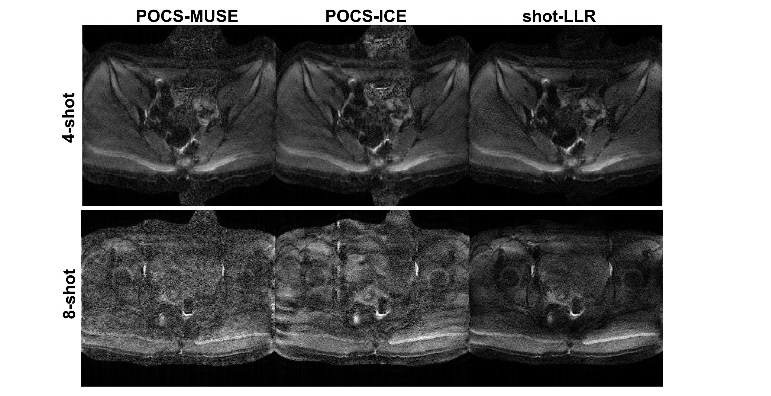
Figure 1 shows the 4-shot and 8-shot pelvis images and they demonstrate the capability of the proposed method to handle rapid phase variations. Residual aliasing artifacts exist in the 4-shot image reconstructed by POCS-MUSE and POCS-ICE, and they fail to reconstruct 8-shot data. Shot-LLR has significantly fewer artifacts, suggesting that shot-LLR can handle more complicated phase variations between shots.
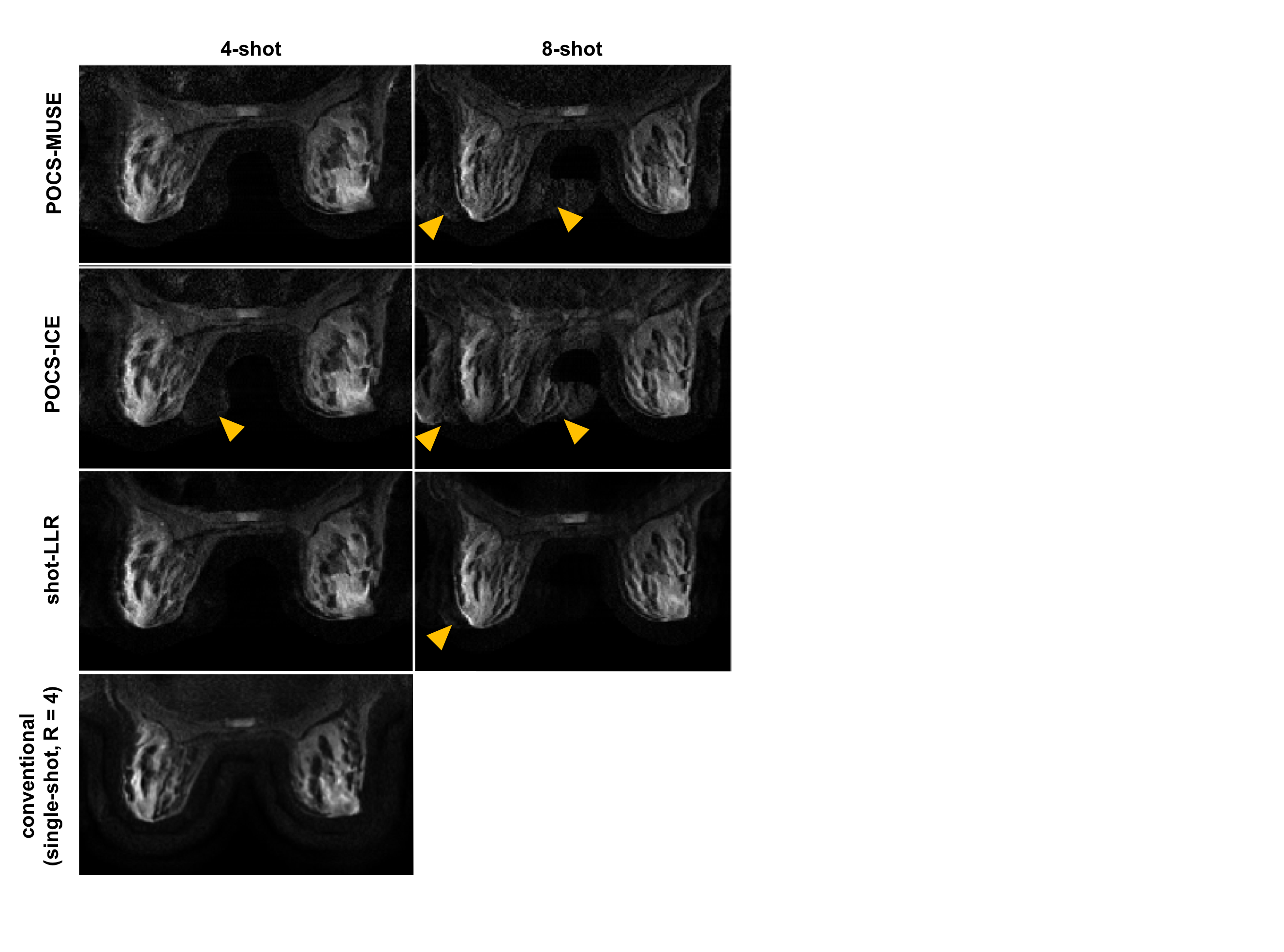
In the breast volunteer (Fig. 2), all 4-shot image reconstructions achieve comparable image quality with respect to the conventional single shot acquisition (R=4), and shot-LLR achieves higher SNR and reduced aliasing artifact in comparison to POCS-MUSE and POCS-ICE in the 8-shot acquisition.
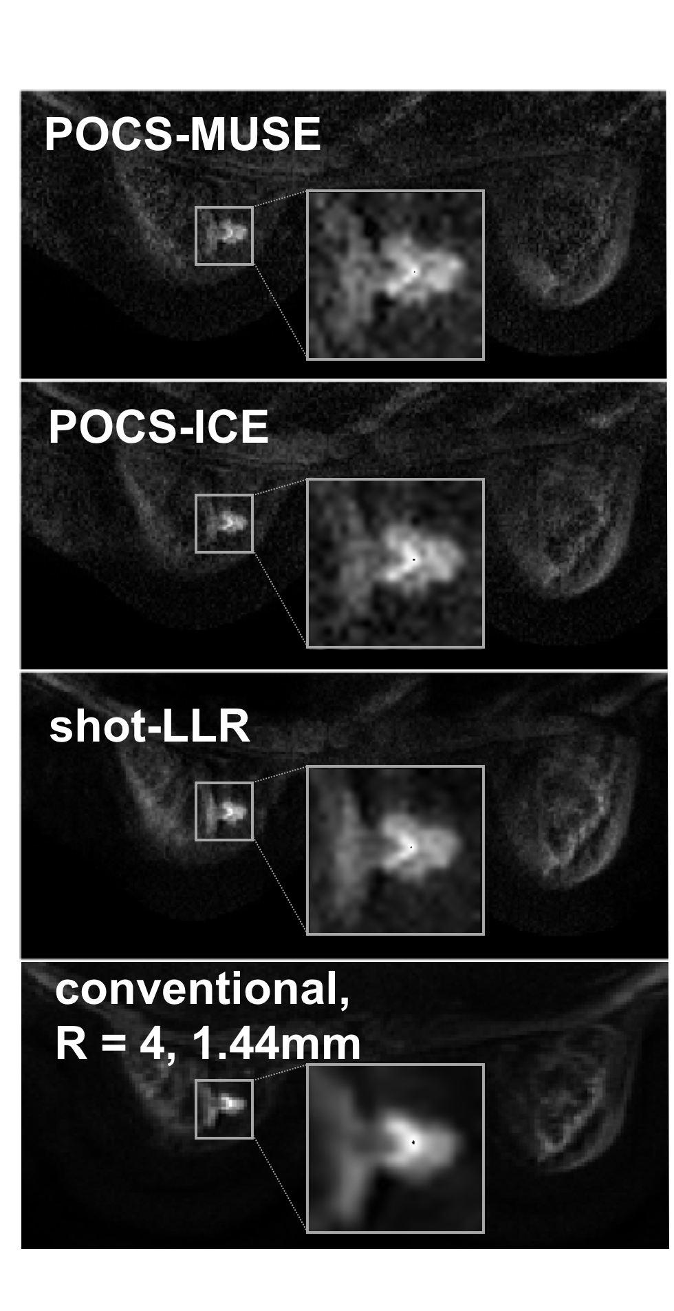
The increased SNR and reduced aliasing artifact achieved with shot-LLR is even more pronounced in Fig 3. with a higher in-plane resolution (1 mm). Diagnostically, in the breast, clear depiction of tumor morphology at 1 mm x 1 mm in-plane resolution is significant as it matches the standard in-plane resolution of DCE-MRI breast acquisitions. The diagnostic impact of improved image quality and respective higher resolution achieved with shot-LLR is demonstrated in the improved depiction of lesion morphology in the breast cancer patient.
Conclusion
We proposed shot-LLR: an alternative reconstruction method for multishot DWI that obviates the need for phase estimation. Proof-of-concept in-vivo experiments in the pelvis and breast demonstrate its capability to solve phase variations between shots and improved motion-robustness.Acknowledgements
Research support from R01-EB009055, P41-EB015891 and GE Healthcare.References
1. Chen, Nan-kuei, et al. "A robust multi-shot scan strategy for high-resolution diffusion weighted MRI enabled by multiplexed sensitivity-encoding (MUSE)." Neuroimage 72 (2013): 41-47.
2. Chu, Mei‐Lan, et al. "POCS‐based reconstruction of multiplexed sensitivity encoded MRI (POCSMUSE): A general algorithm for reducing motion‐related artifacts." Magnetic resonance in medicine 74.5 (2015): 1336-1348.
3. Guo, Hua, et al. "POCS‐enhanced inherent correction of motion‐induced phase errors (POCS‐ICE) for high‐resolution multishot diffusion MRI." Magnetic resonance in medicine 75.1 (2016): 169-180.
4. Liang ZP. Spatiotemporal imaging with partially separable functions. IEEE International Symposium on Biomedical Imaging: From Nano to Macro. 4th IEEE; 2007.
5. Trzasko, Joshua D., and Armando Manduca. "Calibrationless parallel MRI using CLEAR." Signals, Systems and Computers (ASILOMAR), 2011 Conference Record of the Forty Fifth Asilomar Conference on. IEEE, 2011.
6. BART Toolbox for Computational Magnetic Resonance Imaging, DOI: 10.5281/zenodo.592960
Figures