0980
Interaction of vascular and glymphatic systems in brain waste clearance after diabetesQuan Jiang1,2,3, Hiani Hu4, Guangliang Ding1, Esmaeil Davoodi-Bojd1, Yimin Shen4, Li Zhang1, Lian Li1, Qingjiang Li1, Michael Chopp1,2,3, and Zhenggang Zhang1,2,3
1Neurology, Henry Ford Health System, Detroit, MI, United States, 2Physics, Oakland University, Rochester, MI, United States, 3Neurology, Wayne State University, Detroit, MI, United States, 4Radiology, Wayne State University, Detroit, MI, United States
Synopsis
The recently discovered glymphatic system has become an exciting area of research because of it plays an important role in neurological diseases. However, the interaction between the vascular and the glymphatic systems in terms of waste clearance from the brain is not clear. In addition to the glymphatic system, our preliminary MRI results suggest that the venous system involves in waste removal and waste clearance increases after diabetes. Current study provide the first investigation of the interaction between the vascular and the glymphatic systems in brain waste clearance after diabetes.
Synopsis
The recently discovered glymphatic system has become an exciting area of research because of it plays an important role in neurological diseases. However, the interaction between the vascular and the glymphatic systems in terms of waste clearance from the brain is not clear. In addition to the glymphatic system, our preliminary MRI results suggest that the venous system involves in waste removal and waste clearance increases after diabetes. Current study provide the first investigation of the interaction between the vascular and the glymphatic systems in brain waste clearance after diabetes.PURPOSE:
Brain was long considered to be devoid of a conventional lymphatic system. Recent studies from Dr. Nedergaard’s group have fundamentally altered the traditional model of cerebrospinal fluid (CSF) hydrodynamics and discovered the brain glymphatic system which plays an important role in neurological diseases1-3. Despite many milestone achievements, conclusive findings on the efflux pathways are relatively limited. Consequently, the interaction between vascular and glymphatic systems on waste clearance, especially under condition of neurological diseases, is unclear. Here we present our first study on this important topic.METHODS
Male Wistar rats, 13 months of age, were either subjected to a streptozotocin and nicotinamide (STZ-NTM) induced type II diabetes mellitus (DM, n=26) or without induction (Non-DM, n=18). 3D dynamic T1-weighted MR images (T1WIs) were acquired continuously for 3 baseline scans followed by intra-cisterna magna (ICM) paramagnetic contrast delivery via the indwelling catheter while MRI acquisitions continued for 6 hours. The multi-echo SWI with ICM injection of FE dextran (40 k Da) was employed for detecting tracer changes in vascular system.RESULTS:
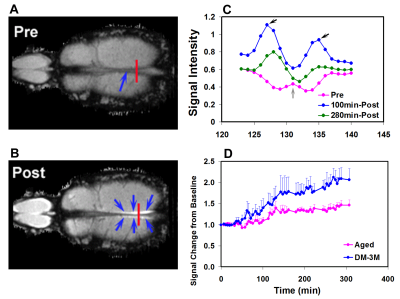
Our results demonstrated that the venous system also played a complementary role in waste clearance in addition to that of the glymphatic system, especially with DM. As demonstrated in Fig 1, the horizontal sections of T1WIs at the level of the superior sagittal (SS) sinus from a non-DM rat head before injection (A) and 100 min (B) after ICM infusion of Gd-DTPA showed bright signal in symmetric areas adjacent to the SS sinus (B, blue arrows), comparable to the pre-Gd (A). Fig 1C shows the changes of the signal intensity (SI) profiles measured from the red lines cross SS sinus in A and B. The center SI from venous blood (red arrow) is slightly increased with time but the symmetric peak SI from glymphatic fluid (solid black arrows) significant increase at 100 min and then decreased at 5 h. Fig 1D shows the SI changes with time from venous blood in non-DM and DM animals. Venous clearance of the tracer significantly increased in DM compared with non-DM animals. To test whether CSF drains through arachnoid granulations, directly into the major veins (sinuses) in the dura mater4, we performed a high resolution multi-echo SWI with ICM injection of FE dextran. The SWI-FE results exhibited that the venous not related to arachnoid granulations also directly participates in waste removal. However, the arterial did not involve in the waste removal. MRI glymphatic measurements revealed reduced glymphatic clearance rate leading to accumulation of the tracer in the brain. The Gd-DTPA clearance rate constant was 3.4 times slower (p=0.022) in DM than in non-DM rats. Also, The DM rats exhibited significantly increased residual intensity (signal intensity at the end of experiment minus baseline intensity, p=0.005, 772% of control) in the hippocampus compared with the non-DM rat.Discussion and conclusion:
This is the first MRI study of the interaction between glymphatic and vascular systems on waste clearance and exhibited that venous, but not arterial, also involved in brain waste clearance. Fully understanding the relationship between the venous and glymphatic systems in waste clearance is crucial for understanding mechanisms in the initiation or progression of neurological diseases. Whole brain MRI provides a sensitive, non-invasive tool to quantitatively evaluate waste clearance in glymphatic and vascular systems after DM and possibly in other neurological disorders, with potential clinical application.Acknowledgements
This work was supported in part by NIH grants, RF1 AG057494, RO1 NS097747 and R21 AG052735.References
1. Xie L, et al. Science. 2013;342:373-377; 2. Iliff JJ, et al. J Neurosci. 2013;33:18190-18199; 3. Iliff JJ, et al. Science translational medicine. 2012;4:147ra111; 4. Boulton M, et al. Neuropathology and applied neurobiology. 1996;22:325-333Figures

Fig 1. A&B Pre- and post-Gd horizontal
image through the SS sinus, para-ss sinus spaces are bright after
intracisternal injection of Gd-DTPA; C: profiles of SS sinus pre-Gd, post-Gd 100
and 280 minuts time points (the location of the profile measures is labeled in
red lines in A & B); D: MRI time evolution curve in SS sinus blood
indicate that the Gd-DTPA concentration increased in
DM compared with non-DM rats.