0975
Increased brain entropy in supplementary motor area and precuneus in amyotrophic lateral sclerosis1Huashan Hospital, Fudan University, Shanghai, China
Synopsis
Amyotrophic Lateral Sclerosis (ALS) is a fatal disease, but no fully validated and clinically specific biomarkers have been identified yet. We studied the brain entropy (BEN) of ALS patients using resting state functional magnetic resonance imaging (rs-fMRI) on fifty-six ALS patients without cognitive impairments and forty-six age- and sex-matched healthy controls. We found increased low frequency entropy in SMA/SMF and increased whole frequency entropy in precuneur/PCC regions in ALS patients. The results may improved our understanding of ALS and provide new biomarkers for diagnosis of ALS.
INTRODUCTION
Amyotrophic Lateral Sclerosis (ALS) is a fatal disease with mean survival time of 3-5 years and mean diagnosis delay of 1 year from symptom onset, which mainly affect lower and upper motor neurons1. Although neuroimaging studies on ALS in last decade have suggested some candidates, no fully validated and clinically specific biomarkers have been identified for diagnosis, stratification or therapeutic monitoring till now. We studied the brain entropy (BEN) of ALS patients using resting state functional magnetic resonance imaging (rs-fMRI), aiming to assess the alterations of brain functional complexity and provide new biomarker for ALS.METHODS
Fifty-six ALS patients (24 females. age: 26-75, mean 50.7) without cognitive impairments and forty-six age- and sex-matched healthy controls (NC: 21 females. age: 23-68, mean 50.3) were included in this study. Standard T1-weighted images and rs-fMRI were acquired using 3.0 T Siemens Trio with 8-channel head coil (Erlangen, Germany) at Huashan Hospital. Data analysis were performed using SPM12 (http://www.fil.ion.ucl.ac.uk/spm/), DPABI2 and BENtbx3. Generally, fMRI images were corrected for slice-timing and head movements, and nuisance covariates including movement-related parameters, white matter and cerebrospinal fluid mean signals were regressed. Then we calculated voxel-wise sample entropy (SampEn) on both whole-frequency (WF) and low-frequency (LF: 0.01-0.1Hz) smoothed images, with embedding dimension m = 3 and distance factor r = 0.6. The BEN maps were divided by its mean and subtracted by 1 to get rBEN maps. Individual T1 images were first co-registered with fMRI images and segmented, then nonlinearly registered with DARTEL and normalized to standard MNI space. The individual rBEN maps were warped with same warping parameters and smoothed. Then we conducted two-sample t-test on normalized and smoothed rBEN maps, with age and gender as covariates. Significant differences with voxel P < 0.005 and cluster P < 0.05 using GRF corrections for family-wise error (FWE) induced by multiple comparisons were reported. Volume of interest (VOI) were extracted from the significant results, and mean rBEN values of each VOI was calculated for each individual and illustrated.RESULTS
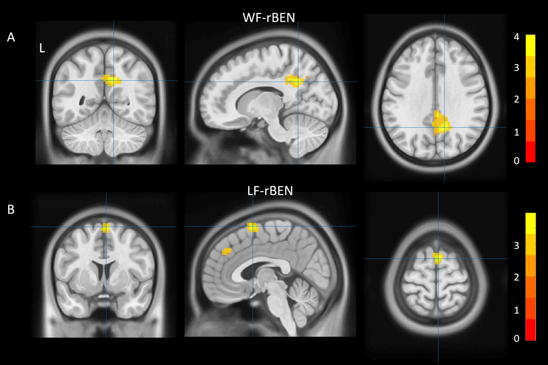
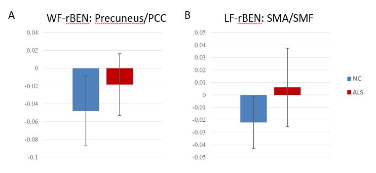
While comparing the whole frequency (WH) of resting state time series data (Figure 1A), the ALS patients showed significantly increased rBEN in Precuneus / posterior cingulate cortex (PCC) region (Number of Voxels: 269; peak MNI coordinate: [-12, -54, 33]; peak t-value: 4.05) compared with healthy controls (Figure 2A: ALS group, mean = -0.018, std = 0.039; NC group, mean = -0.048, std = 0.035). When Considering only the low frequency (LH) (Figure 1B), significally increased rBEN was found in a cluster centered in supplementary motor area (SMA) with extension to adjacent superior medial frontal cortex (SMF) (Number of Voxels: 119; peak MNI coordinate: [3, 6, 69]; peak t-value: 3.98) in ALS patients (Figure 2B: ALS group, mean = -0.006, std = 0.021; NC group, mean = -0.022, std = 0.032). No significantly decreased rBEN were found.DISCUSSION
Entropy is an important trait for indicating complexity or irregularity of time series. Compared with NC, we found significantly increased entropy in SMA/SMF of low frequency signals and Precuneus/PCC of whole frequency signals. As previous studies have reported white matter impairments mainly in corticospinal tract and decreased volume centered in primary motor regions, the increased low frequency entropy in SMA/SMF may indicate compensatory effect of neuronal activities in this area. And the increased whole frequency entropy in Precuneur/PCC region, which is the hub regions of default mode network in human brain, may indicate an overall functional reorganization of the ALS brain.CONCLUSION
We evaluated the resting state brain entropy, and found increased entropy in SMA/SMF and Precuneur/PCC regions in ALS patients. The results may improved our understanding of ALS and provide new biomarkers for diagnosis of ALS.Acknowledgements
We sincerely acknowledge the participants for their willingness to take part in this study.References
1.Menke, R. A. et al. Widespread grey matter pathology dominates the longitudinal cerebral MRI and clinical landscape of amyotrophic lateral sclerosis. Brain : a journal of neurology 137, 2546-2555, doi:10.1093/brain/awu162 (2014).
2.Yan, C. G., Wang, X. D., Zuo, X. N. & Zang, Y. F. DPABI: Data Processing & Analysis for (Resting-State) Brain Imaging. Neuroinformatics 14, 339-351, doi:10.1007/s12021-016-9299-4 (2016).
3.Wang, Z., Li, Y., Childress, A. R. & Detre, J. A. Brain entropy mapping using fMRI. PloS one 9, e89948, doi:10.1371/journal.pone.0089948 (2014).
Figures